Pulmonary atresia is a congenital malformation of the pulmonary valve with systemic-dependent pulmonary flow, classified into two main types according to the presence or absence of ventricular septal defect. Pulmonary atresia with an intact ventricular septum accounts for 2.5–4.0% of all congenital heart disease.Reference Freedom and Keith 1 , Reference Fricker and Zuberbulher 2 Pulmonary atresia with ventricular septum defect is less common – 0.2–0.4% – although it represents 25% of the cases of tetralogy of Fallot; it is also rarely associated with complete atrioventricular septal defects, intersegmental discordances, and univentricular hearts.Reference Kenna, Smithells and Fielding 3 , Reference Anderson, Macartney, Shinebourne and Tynan 4
The most frequently used classification of pulmonary circulation in pulmonary atresia patients comprises three types. Type A: all lobar branches are connected to the main branches, which are also confluent; Type B: some segments are connected to the main branches, whereas some are connected exclusively to the systemic-pulmonary collateral arteries; and Type C: absence of the main branches, with all intraparenchymal vessels being connected exclusively to collateral arteries.Reference Barbero-Marcial and Jatene 5 Types B and C are also denominated as non-confluent, and unifocalisation is required for surgical correction. The pulmonary circulation is defined as uni- or multifocal according to the number of collateral vessels supplying the pulmonary branches, and it basically originates from the descending aorta, or via a patent ductus arteriosus. A late teratogenic process during the embrionary period occurs in type A pulmonary circulation. In contrast, multifocal pulmonary circulation of types B and C, when multiple collaterals exist between the systemic and the pulmonary vessels, is an indication that the teratogenic process occured at the begining of foetal development, through the maintainance of communicant vessels from the sixth aortic archs to the pulmonary venous plexus.Reference Haworth and Macartney 6 The pulmonary circulation in pulmonary atresia with ventricular septum defect is unifocal and Type A, with rare exceptions.
The surgical correction of pulmonary atresia depends on the associated malformations, with the main decision being between uni- or biventricular correction. In the first case, the surgical alternative is a total cavopulmonary derivation because of the impossibility to obtain a circulation with two ventricles, one systemic and the other pulmonary. In the presence of two adequately formed ventricles, it is usually possible to establish a pulmonary circulation very close to normal.
The evaluation of pressures in pulmonary branches – main and/or intraparenchymal – is essential for surgical planning, particularly when the proposed intervention is univentricular, and a total cavopulmonary procedure is required. However, the selective catheterisation of pulmonary branches with conventional diagnostic catheters is frequently hindered by anatomical obstacles and the risk of jeopardising the critically low pulmonary perfusion.
Objective
The objective of this study was to test the safety, feasibility and the efficacy of the use of the pressure wire for the measurement of intravascular pulmonary pressures in the main branches, and/or lobar and segmental branches in successive infants and young children with pulmonary atresia and systemic-dependent pulmonary blood flow, in Type A, B, or C pulmonary circulations.
Materials and methods
This was a prospective observational registry. Signed informed consent approved by the local institutional ethics committee was obtained from the parents of all children included. We studied all sequential children with pulmonary atresia and congenital or surgical systemic-dependent pulmonary circulation referred for diagnostic catheterisation for evaluation of pulmonary pressures during the period from April, 2012 to April, 2013.
The catheterisation for the haemodynamic and angiographic study was performed under general anesthesia, with orotracheal intubation, and a fraction of inspired oxygen (FiO2) sufficient to maintain the peripheral oxygen saturation (SpO2) equal to or higher than that at the baseline condition. All patients received heparin at the doses of 100 U/kg of body weight. Monoplanar angiographic images were obtained with a non-rotational digital radiologic system. Through the femoral artery approach, a 4 or 5 Fr pigtail catheter was used for a contrast cine-aortography that allowed the opacification of the origin of systemic-pulmonary collateral circulation. A pressure guidewire, Certus™ or Aeris™, was attached to a console system RadiAnalyzer™, Saint Jude Medical, Saint Paul, Minnesota, United States of America, and introduced in the catheter. The pressure wire Certus™ is a mettalic wire, 0.014″ of outside diameter, with a pressure sensor located 30 mm proximal to the tip. The Aeris™ is basically the same wire with a hydrophilic coat. Although the tip of the pressure wire is less flexible than the majority of guidewires for coronary angioplasty, it has enough steerability to be distally advanced through tortuous arteries and stenotic lesions.
Calibration of the manometric system was performed assuring adequate equalisation between pressures obtained by the catheter and by the guidewire, when both were in the aorta, before the pressure wire was advanced through the collateral vessels into the pulmonary vessels and the calibration was checked again after the pressure wire was withdrawn from the pulmonary circulation. 4 or 5 Fr catheters of various design – for example, JR, LIMA, Amplatz – were positioned near the aortic ostia of the systemic-pulmonary collateral vessels, and the pressure wire was introduced to reach the main pulmonary artery, and/or lobar, and segmental branches supplied by these collaterals. In several cases, the distal tip of the pressure wire was manually bent and adapted to conform to the tortuous anatomy of the collateral vessels. Aortic and pulmonary pressures were simultaneously obtained with the diagnostic catheter and pressure wire, respectively. Pressures in the aorta and in the main pulmonary arteries, or in the lobar/segmental pulmonary vessels supplied by collaterals without critical stenoses at angiography, were simultaneously recorded. No vasodilator drugs were used in any patients.
In the cases of pulmonary atresia Types B or C, when it was necessary to measure the pressure in several sources of systemic–pulmonary collateral circulation, various catheters had to be used, sometimes requiring adequation of their tip, to ensure its appropriate positioning and allow the entrance of the pressure wire.
In specific, we evaluated whether in those patients with pulmonary atresia the pulmonary artery pressures could be successfully obtained by the pressure wire in the main branches, in Types A and B, and in the lobar and segmental arteries supplied by collaterals in Types B and C.
Results
From April, 2012 to April, 2013, we studied 10 patients with pulmonary atresia, age 21 days to 11 years, weight 3.1–33 kg (mean=9.7 kg), including six boys, and baseline SpO2 ranging from 65% to 85% (mean=76%). There was one patient who had a ventricular septal defect, Type B pulmonary circulation, and multifocal collaterals. In the patients with ventricular septal defect (n=9), a single ventricular-arterial connection with the aorta arising from the right ventricle was present in two patients, and one of them had an atrioventricular septal defect, featuring a non-aortic-related ventricular septal defect. Type A pulmonary circulation was present in five patients, Type B in two, and Type C also in two of the patients with ventricular septal defect. Unifocal collateral circulation was present in four patients and multifocal in five patients. There were four patients who had previously undergone a modified Blalock–Taussing procedure. The baseline clinical characteristics are displayed in Table 1.
Baseline Characteristics.

AAI=aortic arch interruption; AO-RV=aorta arising from the right ventricle; BGO=bi-directional Glenn operation; d=days; DP=during procedure; F=female; FiO2=fraction of inspired oxygen; IVS=intact ventricular septum; LBPAS=left branch pulmonary artery stenosis; Lig=ligadura cirúrgica; m=months; M=male; MBT=modified Blalock–Taussig; NC=no commited; PA=pulmonary atresia; PDA=patent ductus arteriosus; PP=pre procedure; SpO2=peripheral oxygen saturation; TAVSD=total atrioventricular septal defect; VSD=ventricular septal defect; y=years
In all 10 patients, the reliable manometric assessment of collateral vessels and the pulmonary circulation – main artery, and/or lobar, and segmental branches in the absence of significant stenosis detected by angiography – was successfully performed using the pressure wire.
The average measured systemic pressures for the group were: systolic 81 mmHg (68–96), diastolic 40 mmHg (30–64), and mean 64 mmHg (40–71). In cases of pulmonary circulation Type A, systolic, diastolic, and mean pulmonary branches had values from 20 to 35 mmHg, 15 to 28 mmHg, and 12 to 30 mmHg, respectively. One of these patients also showed a mild stenosis of the left pulmonary artery branch.
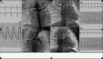
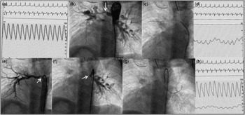
In patients with pulmonary circulation Types B (Fig 1) and C (Fig 2), the pressures recorded in the lobar or segmental branches supplied by collateral artery without an important angiographic stenosis were: systolic 15–37 mmHg, diastolic 8–15 mmHg, and mean 12–20 mmHg.
Patient 10, Type B MFPC and VSD. ( A ) Catheter and PW pressure equalisation at the aorta; ( B ) aortography demonstrating MAPCAs to pulmonary branches; ( C ) PW at the right superior lobar artery (arrow); ( D ) aortic and right superior lobar branch pressures. Despite the tortuosity, the pressure curve is totally appropriate and reliable; ( E ) bifurcated MAPCA: the right branch is not stenotic, it is connected to the pulmonary artery, and supplies the right upper and middle lobe pulmonary arteries, and the left upper lobe pulmonary artery; the left branch supplies the left lower lobe pulmonary artery and presents a subtotal stenosis (arrow); ( F ) PW at the right inferior lobe pulmonary artery (arrow); ( G ) aortic and RBPA curves. LBPA=left branch pulmonary artery; MAPCA=major aortic pulmonary collateral artery; MFPC=multifocal pulmonary circulation; PW=pressure wire; RBPA=right branch pulmonary artery; VSD=ventricular septal defect.

Patient 5, Type C, VSD and MFPC. ( A ) Catheter and PW equalised pressures; ( B ) stenotic MAPCA to SLRBPA; ( C ) PW at SLRBPA; ( D ) aortic and SLRBPA pressure curves; ( E ) MAPCA with a severe stenosis in the ILRBPA; ( F ) Stenotic MAPCA to LBPA; ( G ) PW at LBPA; ( H ) LBPA pressure curve. ILRBPA=inferior lobe right branch pulmonary artery; LBPA=left branch pulmonary artery; MAPCA=major aortic pulmonary collateral artery; MFPC=multifocal pulmonary circulation; PW=pressure wire; SLRBPA=superior lobe right branch pulmonary artery; VSD=ventricular septum defect.

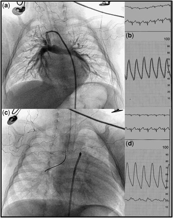
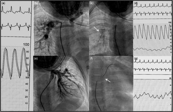
In patients 4 and 9, who were referred for assessing the feasibility of a total cavopulmonary shunt as the surgical approach, the diastolic pressure was 20 mmHg. Patient 4 (Fig 3) had unbalanced complete atrioventricular septal defects, single-outlet right ventricle, dextro-isomerism, and non-aortic-related ventricular septal defect. Patient 9 (Fig 4) had pulmonary atresia with ventricular septal defect, pulmonary circulation Type B, and right ventricle hypoplasia. (Table 2). In both cases, the elevation of the pulmonary pressures contraindicated the total cavopulmonary surgical correction.
Patient 4, Type A, UFPC, TAVSD, NC VSD and MBT. ( A ) MTB, without stenosis in the shunt conduit and pulmonary branches; ( B ) pressure equalisation; ( C ) PW at RBPA; ( D ) aortic and RBPA. MBT=modified Blalock–Taussig shunt; NC=not committed; PW: pressure wire; RBPA=right branch pulmonary artery; TAVSD=total atrioventricular septal defect; UFPC=unifocal pulmonary circulation; VSD=ventricular septal defect.

Patient 9, Type B, IVS and MFPC. ( A ) Catheter and PW equalised pressures; ( B ) non-stenotic MAPCA to RBPA, connected through a hypoplasic main pulmonary artery to LBPA that supplies only the superior lobe; ( C ) PW at RBPA; ( D ) aortic and RBPA pressures curves; ( E ) MAPCA with a moderate stenosis to the LBPA; ( F ) pressure wire at the LBPA; ( G ) LBPA pressure curve. IVS=intact ventricular septum; LBPA=left branch pulmonary artery; MAPCA=major aortic pulmonary collateral artery; MFPC=multifocal pulmonary circulation; PW=pressure wire; RBPA=right branch pulmonary artery.

Haemodynamic parameters.

AAI=aortic arch interruption; DEXTROISOM=dextroisomerismo; IVS=intact ventricular septum; LBPA=left branch pulmonary artery; LBPAS=left branch pulmonary artery stenosis, MAPCA=major aortic pulmonary collateral artery; MBT=modified Blalock–Taussig; NC=not committed; PA=pulmonary atresia; PDA=patent ductus arteriosus; RBPA=right branch pulmonary artery; TAVSD=total atrioventricular septum defect; TCPA=total cavopulmonary anastomosis; Unifoc=unifocalisation; VSD=ventricular septal defect
* In lobar or segmental artery pressures supplied by non-severely stenotic anastomosis as defined by angiography the higher values were considered
No significant changes occurred in arterial oxygen saturation during selective catheterisation of the pulmonary collateral arteries with the pressure wire, and no haemodynamic instability, arrhythmias, or death supervened during these manoeuvres.
The final diagnosis of pulmonary circulation characteristics, with their related source of collaterals from the systemic circulation, and the presence of surgical anastomoses as defined by angiography is displayed in Table 2. This table also shows the manometric data obtained with the catheter and pressure wire, respectively, in the systemic and pulmonary circulation, including the pressure in the lobar or segmental arteries supplied by non-stenotic anastomosis as defined by angiography. The type of surgical correction proposed for each of those complex congenital heart diseases, as defined on the basis of the catheterisation data, is also shown in Table 2.
Discussion
The anatomical diagnosis of complex congenital heart diseases is currently performed in most cases by echocardiography and magnetic resonance imaging. The recently described method of rotational angiography has demonstrated that because of the eccentricity of vascular stenosis frequently found in congenital heart disease, the accurate assessment of stenosis severity is usually not possible using conventional angiographic projections.Reference Berman, Khan, Gutierrez and Zahn 7 Hence, cardiac catheterisation remains essential for accurate evaluation of pulmonary intravascular pressures and rotational angiography characteristics. However, there are technical difficulties and limitations when conventional diagnostic catheters are used, because of obstructed and tortuous collateral arteries, in addition to the risk of vascular injury and consequently impairment of pulmonary blood flow.Reference Lloyd, Donnerstein and Shirazi 8 The technique of wedging catheters in the pulmonary veins may underestimate the pulmonary artery pressures when they are low, and it is also not accurate when there is pulmonary venous hypertension.Reference Mullins 9
The pressure wire method of assessing coronary fractional flow reserve is now in current use since its original description by Piljs et al.Reference Pijls, Van Gelder and Van der Voort 10 There are few reports in the literature of its application in evaluating blood pressure in other adult heart diseases.
Our review of the literature revealed few articles that reported the use of the pressure wire system in congenital heart disease. Everett et alReference Everett and Matherne 11 used the pressure wire to reach the pulmonary artery in 11 patients, but always through the linear conduct of a modified surgical systemic–pulmonary shunt. Hamid et alReference Hamid, Hadi, Clarke and Mahadevan 12 described its use in three adult patients with pulmonary atresia where it was not possible to advance the conventional catheters through the collateral vessels into the pulmonary artery. Zampi et alReference Zampi, Hirsch, Goldstein and Armstrong 13 evaluated the effectiveness of pulmonary artery banding, intra-operatively, during the stage I of the Norwood surgical procedure in eight patients. Goldstein et alReference Goldstein, Fifer and Armstrong 14 evaluated the gradient pre and post balloon dilation in a foetus at 216/7 weeks of gestation with critical aortic stenosis.
In our prospectively designed experience of 10 patients with pulmonary atresia, the use of pressure wires allowed the adequate haemodynamic evaluation by reaching the main and distal pulmonary arteries through the collateral vessels, even in cases of severe tortuosity or stenosis. Its inherent very low profile (0.014′′) allows for a more accurate measurement of pulmonary pressures, as it does not artificially increase the degree of stenosis and does not induce the appearance of damped curves, as usually happens when diagnostic catheters are negotiated through collateral vessels, which have prominent bends and tortuousity. The pressure wire also minimised the risk of vascular injury, or of haemodynamic instability caused by the presence of catheters in the collateral vessels leading to the pulmonary arteries.
The selective instrumentation of intraparenchymal arteries with the pressure wire allowed the pressure measurement in all lobar and segmental arteries in Types B and C pulmonary circulations.
Although less flexible than a regular 0.014″ coronary angioplasty guidewire, the distal end of the pressure wire can be shaped, to overcome obstructions or tortuousity and reach the pulmonary vessels. The repeated shaping of the distal end does not affect the micromanometer that lies 30 mm proximal to the tip of the guide.
This distance could be a limitation in small children. However, in our series the pressure measurement was feasible even in these small patients, because the technical difficulty is overcome with the excellent steerabiliy of the wire that allows its positioning in distal regions.
In this series of patients, we observed four cases of a significant gradient in collateral arteries or pulmonary branches, as detected with the pressure wire technique. This was a clear evidence for the occurrence of stenoses not shown by non-rotational angiography. This information was nevertheless essential in determining the surgery approach in those cases. It is important to emphasise that stenoses in collateral vessels frequently evolve with time, and thus in older children it is possible to see pulmonary segments or even lobar territories supplied by stenotic vessels at the time of the catheterization. Distal vascular lesions can be found in such cases because the vessels have been exposed to a high-pressure regimen in the past, before the development of more severe stenosis. On the other hand, in patients with critical or sub-total stenoses, the measurement of distal pressures is not mandatory, as in this setting a regimen of very low-flow and low pressure occurs.
Finally, in a short-term analysis the procedure has an additional cost of around US$650.00 to pay for the pressure wire. Thus, the cost-effectiveness of this method to evaluate pulmonary pressures in congenital heart disease should be evaluated in future studies. In the scenario of coronary heart disease, pressure wire-guided percutaneous coronary intervention improves outcomes and saves costs in 1-year follow-up.Reference Fearon, Bornschein and Tonino 15
Study limitations
Our series is small, and the absence of any complications in our study may be related to this limitation. A more extensive number of cases would be necessary to validate the method as a gold standard, by a study directly comparing the pressure wire method with the use of standard catheters to measure pulmonary pressures in patients with pulmonary atresia. The low prevalence of this condition would warrant a multi-centre design of such study.
Follow-up measurement of pulmonary pressures, in the operating room or during a post-surgery catheterisation, was not performed in our trial. The analysis of these parameters would add clinical basis to the utilisation of pressure wire in this specific scenario.
Conclusion
Surgical planning of pulmonary atresia correction requires a thorough evaluation of the anatomy and haemodynamics in all segments of the pulmonary circulation. In these patients, the measurement of pulmonary pressures using diagnostic catheters is frequently hindered by the presence of bends, excessive tortuosity and stenoses, and by artefacts arising from the mere presence of the catheter in these small collateral vessels. In addition, there is always the risk of vascular injury and consequent pulmonary hypoperfusion. The pressure wire system is a reliable, safe, and effective method of measurement of pulmonary pressures, permitting an adequate surgical planning.
Acknowledgement
The authors are thankful for the expert assistance of the nurses and technicians staff of our Hemodynamics and Interventional Cardiology Laboratory.
Financial Support
This research received no specific grant from any funding agency, commercial or not-for-profit sectors.
Conflicts of Interest
None.