The American physiologist Walter Cannon proposed and developed the concept of homeostasis, building on Claude Bernard's concept of the “milieu interieur” (Cannon, Reference Cannon1932). Cannon enunciated four principles: (a) our bodies require mechanisms that act to maintain constancy. (b) Steady-state conditions require that any tendency toward change automatically meets with factors that resist change. For example, an increase in blood sugar results in thirst as the body attempts to dilute the concentration of sugar in the extracellular fluid. (c) The regulating system that determines the homeostatic state consists of a number of cooperating mechanisms acting simultaneously or successively. For example, blood sugar is regulated by insulin, glucagons, and other hormones that control its release from the liver or its uptake by the tissues. (d) Homeostasis does not occur by chance but is instead the result of organized self-government.
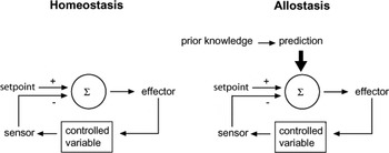
The concept of homeostasis has been interpreted literally to mean that the purpose of physiological regulation is to clamp each internal parameter at a “set point” by sensing errors and correcting them with negative feedback (see Figure 1). Based on this model, physicians and scientists reason that some internal mechanism must be broken when a parameter deviates from its set point value. Consequently, clinicians, including those in mental health, design therapies to restore the “inappropriate” value to “normal.”
Alternative models of regulation. Homeostasis describes mechanisms that hold constant a controlled variable by sensing its deviation from a “set point” and feeding back to correct the error. Allostasis describes mechanisms that change the controlled variable by predicting what level will be needed and overriding local feedback to meet anticipated demand.

Allostasis
The homeostasis model has contributed immeasurably to the theory and practice of scientific medicine. Yet, all scientific models eventually encounter new facts that do not fit, and it has been argued that this is now the case for homeostasis. In physiology, evidence accumulates that parameters are not constant, and their variations, rather than signifying error, are designed to reduce error. Psychiatric illnesses have causes that the homeostasis model cannot explain. In contrast to the hypertension caused by a constricted renal artery and the diabetes caused by immune destruction of insulin-secreting cells, no mental illnesses present as yet with an obviously defective neural mechanism, and treating these diseases with drugs or psychological therapies to fix low-level mechanisms that are not “broken” turns out not to work particularly well. For example, the most effective therapies for the treatment of acute depressions in adolescents are associated with up to 30% nonresponse even by 36 weeks posttreatment (Goodyer et al., Reference Goodyer, Dubicka, Wilkinson, Kelvin, Roberts and Byford2007; Kennard et al., Reference Kennard, Silva, Tonev, Rohde, Hughes and Vitiello2009).
The second model, allostasis (“stability through change”), appears to take virtually the opposite view (Sterling & Eyer, Reference Sterling, Eyer, Fisher and Reason1988). It suggests that the goal of regulation is not constancy, but rather fitness under natural selection. Fitness implies preventing errors and minimizing costs to the organism. Both needs are best accomplished by using prior information to predict demand and then adjusting all parameters to meet it (Figure 1). Thus, allostasis considers an unusual parameter value not as a failure to defend a set point, but rather as a response to some prediction, and the model attributes diseases such as depression to sustained neural signals that arise from unsatisfactory social interactions. Consequently, the allostasis model would redirect therapy away from manipulating low-level mechanisms toward improving higher levels in order to restore predictive fluctuation, which, under this model, is the hallmark of health (Sterling & Eyer, Reference Sterling, Eyer, Fisher and Reason1988).
A key distinction of allostasis over homeostasis is therefore the ability of the organism to predict response and minimize prediction errors, in order to retain behavioral fitness in the presence of an alteration (good or bad) in the environment either within itself or from without.
Allostasis provides a conceptual window for mechanistic studies to determine how individual differences develop in metabolic systems including neurocognitive systems through, in part, experience-dependent learning. From the physiological perspective, real or interpreted threats from the environment rapidly (seconds) initiate the sympathetic–adrenal–medullary (SAM) axis release of catecholamines and more slowly (minutes) the hypothalamic–pituitary–adrenal axis (HPAA) secretion of glucocorticoids that mobilize energy necessary for fight-or-flight responses (Sapolsky, Romero, & Munck, Reference Sapolsky, Romero and Munck2000). A substantial proportion of these initiations come from bottom-up neural reactions in emotion perception areas of the brain (amygdala–hippocampal activations). Programming and coordination of allostasis depends on the brain's evaluation of bottom-up physiological signals regarding perceived threat and the top down execution (frontal and prefrontal neural systems) of behavioral and physiological regulatory response. Such top down control leading to a coordinated reaction to social threats was recently demonstrated in rhesus monkeys, showing association between cortisol response and metabolism in the subgenual prefrontal cortex, regardless of the context in which brain activity and cortisol were assessed (Jahn et al., Reference Jahn, Fox, Abercrombie, Shelton, Oakes and Davidson2010). This primate data is likely to be one of many such integrative research investigations we are likely to see over the coming decade, as researchers look for relations between peripheral physiological markers and controlling neural systems (that may reflect allostatic regulation).
In our view, allostasis does not replace homeostasis but expands the latter concept. In particular, allostasis provides a model for understanding how prediction error can result in disrupted outcomes. Experience-dependent learning, if efficient, will minimize prediction error for the future management of adverse experiences. Poor learning may impair this ability, and error risk will increase when individuals engage in future social activities that require adaptation and response. Thus, set points in resting state parameters themselves may show individual differences within a population (e.g., variations in blood pressure, peripheral glucose levels, or cognitive rumination style). What allostasis opens up conceptually is the ability to investigate the mediators that effect change in the system that may reveal mechanistically how an individual retains well-being or develops deviance or illness in the face of adversities. Of key importance for successful allostasis is that the individual has a response strategy and can predict from the environment when this strategy should be employed with the minimum of error to ensure that change results in continued stability and well-being. Researchers in this field have over the past decade turned their attention to how allostatic failures may compromise well-being and promote ill health.
Allostatic Load and Overload
Maintaining physiological stability by changing parameters of the internal milieu by matching them appropriately to environmental demands is not always successful. That is, there may be faulty learning from events, resulting in an increase in prediction error when considering future experiences (a very simple cognitive illustration is: I had a bad experience the last time something like this happened and I will again). In particular, exposure to recurrent or chronic adversities may accrue a repeatedly learned response whose effects become deleterious. Pathological models can flow from this concept, grouped collectively as allostatic failures. How might they come about? The terms allostatic load and overload were introduced to describe how chronic over or under activity of mediators whose effects are designed to retain fitness may lead to wear and tear within metabolic systems and eventually impaired functioning (McEwen, Reference McEwen1998). Compromising brain function in this manner will inevitable raise the risk for psychopathology (McEwen & Gianaros, Reference McEwen and Gianaros2010).
There are a number of putative pathological processes that may arise from allostatic failure as follows: (a) not predicting the need to evoke a response, (b) predicting the need but not turning on an adequate response when required, (c) not turning off a normative response when it is no longer needed, and (d) not habituating to the recurrence of the same stressor and thus not dampening the allostatic response.
Our own group has shown that allostatic failure exists in males with conduct disorder. They do not show the expected HPAA response to threat which, although recognized cognitively, does not result in the appropriate rise in peripheral cortisol during a provoking video task (Fairchild, van Goozen, Stollery, & Goodyer, Reference Goodyer2008). Furthermore, male and female adolescents with conduct disorder do not habituate to fear stimuli, decreasing the likelihood of an adaptive behavior response to threats in the environment (Fairchild et al., Reference Fairchild, Van Goozen, Stollery and Goodyer2008; Fairchild, Stobbe, van Goozen, Calder, & Goodyer, Reference Fairchild, Stobbe, van Goozen, Calder and Goodyer2010). These constitute examples of allostatic failures associated with a behavioral psychopathology. They do not reveal whether they arise as a cause or a consequence of the antisocial behavior.
What of those conditions where there is an overactivity of a putative allostatic mediator, such as the frequently reported HPAA overactivity (with loss of circulating cortisol diurnal variation) in around 50% of unipolar depressions? Although adaptive acutely, chronic overproduction of the SAM and HPA axis products induces a “domino effect” on interconnected biological systems that include inflammatory processes, other endocrine systems (thyroid, dehydroepiandrosterone), as well as a concatenation of putative effects within the cell. The allostatic overload is that metabolic systems overcompensate and eventually function suboptimally, leaving the organism susceptible to stress-related diseases that may occur at different points in an individual's life. This gives an important developmental life course trajectory to the impact of allostatic failures (Lupien, McEwen, Gunnar, & Heim, Reference Lupien, McEwen, Gunnar and Heim2009).
The rest of this review focuses on how allostatic failures may arise to increase the liability for depressive disorders over the life course. We have previously outlined some of the mechanisms that may be operating in increasing the emergence of these mental disorders in the first two decades of life (Goodyer, Reference Goodyer2008). In this review we outline how exposure to chronic or recurrent environmental adversities occurring in infancy and the preschool years leads to inappropriate HPAA activity and thereby to allostatic overload. There is a decoupling of adaptive responses, leading to further persistent HPAA overactivity, even after the environmental adversities are no longer present. This leads to increased risks for affective illnesses.
We start by presenting evidence that early environmental adversity increases the risk for depressions and then describe the normal physiology and allostatic regulation of the HPAA. We provide evidence from animal and human studies that early adversity leads to allostatic overload of the HPAA and discuss potential intermediate mechanisms. Finally, we demonstrate that HPAA dysregulation does increase vulnerability to depression and again discuss potential intermediate mechanisms.
Early Adversities and Risk for Depression Episode Onset
There has been over a century of theory, conjecture, observations, and scientific study asserting that a child exposed to relatively persistent adverse parenting styles in the infancy period and the preschool years is at increased risk for mental illness over the first four decades of the life course. For example, proven child maltreatment (physical, sexual, or emotional abuse) of sufficiently negative quality to require removal into the custody of the State is associated with depression episode onset through to the end of the fourth decade (Widom, DuMont, & Czaja, Reference Widom, DuMont and Czaja2007). Epidemiological studies have substantially supported the association between adverse early experiences of various types and later psychopathology. For example, a community survey of over 9000 adults in San Diego demonstrated that multiple types of childhood adversity each increased the risk for lifetime and recent depressive symptoms in adults. Adversities measured included emotional abuse, physical abuse, sexual abuse, mother being battered, household substance abuse, parental separation, and a criminal household member (Chapman et al., Reference Chapman, Whitfield, Felitti, Dube, Edwards and Anda2004). Such associations were significant for both men and women; remained significant when adjusting for living with a mentally ill parent (thus removing the effects of a gene–environment correlation; it may be the sharing of depressogenic genes with a parent that leads to depression, not the environmental adversity); and showed a dose–response relationship, with a greater number of types of adversity associated with a greater risk for depression.
Many studies have found a similar strong association (with a clear dose–response relationship where measured) between childhood adversity and adult depressive symptoms (Goldberg, Reference Goldberg1994; Surtees et al., Reference Surtees, Wainwright, Willis-Owen, Luben, Day and Flint2006) and adult depressive disorder (Caspi et al., Reference Caspi, Sugden, Moffitt, Taylor, Craig and Harrington2003; Kendler et al., Reference Kendler, Bulik, Silberg, Hettema, Myers and Prescott2000; Parker, Reference Parker1979). Recent findings from the National Comorbidity Survey Replication have shown that multiple types of childhood adversity are associated with first-episode onset and persistence of a wide range of psychiatric disorders (including depression; Green et al., Reference Green, McLaughlin, Berglund, Gruber, Sampson and Zaslavsky2010; McLaughlin et al., Reference McLaughlin, Green, Gruber, Sampson, Zaslavsky and Kessler2010). Again, there was an additive dose–response relationship between number of adversities and risk of disorder.
The association between early environmental adversity and later depressions is not, however, straightforward. Statistical modeling in large epidemiological studies has demonstrated mediation by multiple intermediate variables, potentially acting with different degrees of effects at varying life stages. These include personality (neuroticism), low self-esteem, conduct disorder, increased risk of adverse life events, low social support, substance misuse, and difficulties in interpersonal relationships (Brown, Craig, & Harris, Reference Brown, Craig and Harris2008; Kendler, Gardner, & Prescott, Reference Kendler, Gardner and Prescott2002, Reference Kendler, Gardner and Prescott2006). Classical studies of the social origins of depression in adult females showed that both the quality and timing of adverse experiences are important in determining the risk of depression episode onset (Brown & Harris, Reference Brown and Harris1978). Longitudinal findings suggest that common pathways exist within individuals with differing clinical phenotypes over time (Kessler et al., Reference Kessler, Ormel, Petukhova, McLaughlin, Green and Russo2010). These common pathways should be the focus of future research. These studies have not reported measurements of HPAA activity, so we are unable to establish whether HPAA abnormalities mediate the early adversity–adult depression association, nor whether they mediate specific etiological pathways. Therefore, this review will rely on a two-stage indirect approach: is early adversity associated with HPAA abnormalities, and are HPAA abnormalities associated with subsequent depression?
Physiology of the HPAA
As part of the body's generic, nonspecific response to acute threatening or demanding events, there is a transient rise in cortisol levels. Cortisol regulates the body's specific response to stressors. Variations in cortisol at the cellular level alter gene transcription, thereby changing metabolism, mobilizing the body's energy resources, and increases cardiovascular tone (Argyropoulos et al., Reference Argyropoulos, Bailey, Hood, Kendrick, Rich and Laszlo2002; Herbert et al., Reference Herbert, Goodyer, Grossman, Hastings, de Kloet and Lightman2006).
The HPAA provides allostatic regulation of cortisol levels. Neurones from the parvocellular nucleus (PVN) of the hypothalamus secrete corticotrophin-releasing hormone (a 41-amino acid peptide, sometimes known as corticotrophin-releasing factor [CRF]). Corticotrophin-releasing hormone [CRH] is secreted into the hypothalamohypophyseal portal venous system and is transported to the anterior pituitary gland. CRH activates CRH1 receptors of pituitary corticoprophic cells, which leads to release of ACTH into the systemic circulation. ACTH stimulates production and release of the steroid hormone cortisol (cortisone in rodents) by the cortex of the adrenal glands (Gillespie, Phifer, Bradley, & Ressler, Reference Gillespie, Phifer, Bradley and Ressler2009).
Most cortisol in the blood is bound to cortisol binding globulin (CBG). CBG acts as a high-affinity, low-capacity reservoir, buffers natural fluctuations in cortisol, and in particular, reduces the amount of cortisol entering the brain (cortisol is very lipophilic, and so would otherwise freely enter the brain). In the brain, cortisol is detected by cytoplasmic glucocorticoid receptors (GRs) and mineralocorticoid receptors (MRs). GRs are widely expressed, particularly in the hippocampus. Expression of MRs is more restricted, with highest concentrations found in the hippocampus, amygdala, and lateral septum. MRs have a 10-fold greater affinity for cortisol than GRs (Herbert et al., Reference Herbert, Goodyer, Grossman, Hastings, de Kloet and Lightman2006).
Cortisol is detected by GR and MR at the hippocampus, PVN, and pituitary, and this leads to reduced CRH and ACTH secretion, acting as a negative feedback mechanism and damping down the stress response (Gillespie et al., Reference Gillespie, Phifer, Bradley and Ressler2009). In view of the lower affinity of GR compared with MRs, GR-mediated negative feedback occurs at higher levels of cortisol. It is clear here that if GR receptors do not respond optimally to changes in cortisol level, then negative feedback from high levels will not function optimally. In these circumstances this leads to continued high levels of CRH, ACTH, and cortisol, thereby risking allostatic overload. This is crucial in the context of this review, which will argue that impaired MR/GR functioning may be a key mediator between early environmental adversity and later depression.
Cortisol levels vary greatly both within and between individuals. In particular, there is a diurnal pattern of cortisol regulation. In humans, cortisol levels rise in the later part of the night; there is a further increase until about 30 min after waking (cortisol awakening response [CAR]), followed by a gradual reduction through the waking day (Herbert et al., Reference Herbert, Goodyer, Grossman, Hastings, de Kloet and Lightman2006). Cortisol may be higher in the morning to encourage the organism to search for more carbohydrate and novel experiences (Gunnar & Cheatham, Reference Gunnar and Cheatham2003).
Young humans do not show the same patterns of diurnal cortisol variation as adults, partly because of the pattern of irregular sleep and daytime naps in babies. Newborns show two peaks in cortisol, 12 hr apart, not related to time of day (Gunnar & Donzella, Reference Gunnar and Donzella2002). The single early morning peak is not reliably established until 12 weeks (Price, Close, & Fielding, Reference Price, Close and Fielding1983). Only after 4 years is there significantly higher morning compared with afternoon cortisol (Gunnar & Donzella, Reference Gunnar and Donzella2002).
Cortisol and ACTH are present in the systemic circulation. Levels can therefore be measured from blood. Cortisol is present in bound and free form. It is the free form that is active and that crosses the blood–brain barrier. Consideration needs to be made as to whether free or total cortisol is measured. Cortisol is also released into the saliva. In humans, salivary levels accurately reflect free cortisol levels that cross the blood–brain barrier. Salivary levels are about 5% of the blood levels and positively correlate with levels in the cerebrospinal fluid (Guazzo, Kirkpatrick, Goodyer, Shiers, & Herbert, Reference Guazzo, Kirkpatrick, Goodyer, Shiers and Herbert1996). Measurement in saliva is more acceptable to patients, and can also be done in their own homes, meaning collection can occur at multiple times of day in large number of participants. CRH is present in the hypophyseal portal vein. This can be directly accessed in animal, but not human, studies. In human studies, cerebral spinal fluid (CSF) CRH (from lumbar puncture) is used to estimate portal CRH. However, it has been noted that CRH in the CSF may come from a variety of sources, such as the prefrontal cortex and limbic system, which are not part of the HPAA. Therefore, CSF CRH may not accurately reflect HPAA CRH (Stetler & Miller, Reference Stetler and Miller2011).
Hormone levels can be measured at single or at multiple times of day. Careful consideration is needed as to the best time(s) to collect cortisol. Urinary cortisol and metabolites can be collected over 24 hr to estimate total daily production. We can measure HPAA reactivity to stress. In animals, multiple methods are used to elicit stress including restraint (Coplan et al., Reference Coplan, Andrews, Rosenblum, Owens, Friedman and Gorman1996; Liu et al., Reference Liu, Diorio, Tannenbaum, Caldji, Francis and Freedman1997; Weaver et al., Reference Weaver, Cervoni, Champagne, D'Alessio, Sharma and Seckl2004), separation from peers (Levine & Mody, Reference Levine and Mody2003), strong puffs of air onto the cheek (Plotsky et al., Reference Plotsky, Thrivikraman, Nemeroff, Caldji, Sharma and Meaney2005), and novel environments (McCormack, Newman, Higley, Maestripieri, & Sanchez, Reference McCormack, Newman, Higley, Maestripieri and Sanchez2009). In humans, it has been demonstrated that a social stressor with both social evaluation and uncontrollability is most likely to raise cortisol (Dickerson & Kemeny, Reference Dickerson and Kemeny2004). A widely used social stress test in humans is the Trier Social Stress Test, in which participants are given a mock job interview followed by a mental arithmetic task in front of a panel of “experts,” which is videotaped for “later analysis” (Kirschbaum, Pirke, & Hellhammer, Reference Kirschbaum, Pirke and Hellhammer1993). HPAA response to this test is mediated by how much the participant believes it and their own natural ability in/anxiety toward such tasks, not just HPAA reactivity. A physiological stress test has been developed that is less likely to be confounded by such cognitive/emotional factors: participants take in a single tidal breath of 35% carbon dioxide (Kaye et al., Reference Kaye, Buchanan, Kendrick, Johnson, Lowry and Bailey2004). This task is generally safe, with the most common adverse event being panic attacks. Both the Trier Social Stress Test and the 35% CO2 test lead to significant rises in cortisol in many, but not all, participants. Their relative effectiveness in inducing changes in the HPA axis has yet to be reported.
We may want to investigate GR functioning. The first widely used experimental method to test receptor activity indirectly in humans was the Dexamethasone Suppression Test (DST; Pfohl, Sherman, Schlechte, & Stone, Reference Pfohl, Sherman, Schlechte and Stone1985). A total of 1–2 mg of oral dexamethasone (DEX) is given the night of the test. The following day, peripheral cortisol, generally blood, is measured. If central GR function well, they are activated by the DEX, leading to negative feedback of the HPAA and reduced cortisol production the next day. High cortisol suggests impaired GR sensitivity to cortisol, leading to loss of negative feedback. The development of the DEX/CRH test (Heuser, Yassouridis, & Holsboer, Reference Heuser, Yassouridis and Holsboer1994) adds an additional physiological challenge by stimulating the system to produce cortisol from the adrenal glands. A total of 1.5 mg of oral DEX is given in the evening. The following afternoon, 100 µg CRH is infused intravenously. Cortisol levels are subsequently measured; again, high cortisol levels indicating nonsuppression of the HPAA, suggesting impaired GR functioning and escape from negative feedback. The DEX/CRH test better discriminates depressed from nondepressed participants than the DST (Heuser et al., Reference Heuser, Yassouridis and Holsboer1994), but neither has sufficient sensitivity and specificity to be of clinical utility. This indicates the difficulties of clinical heterogeneity, not a fundamental flaw in testing the effectiveness of the HPAA.
There is growing evidence for a genetic effect on the integrity of the HPAA. Polymorphisms of the GR gene are associated with higher basal cortisol levels, in the morning, at bedtime, and over the whole day (Rautanen et al., Reference Rautanen, Eriksson, Kere, Andersson, Osmond and Tienari2006; Rosmond et al., Reference Rosmond, Chagnon, Chagnon, Perusse, Bouchard and Bjorntorp2000). Polymorphisms of the GR gene and FKBP5 (an immunophilin involved in the regulation of GR) are associated with increased cortisol secretion in the DST (Derijk & de Kloet, Reference Derijk and de Kloet2008; van Rossum et al., Reference van Rossum, Koper, Huizenga, Uitterlinden, Janssen and Brinkmann2002) and increased cortisol response to stress (Wust et al., Reference Wust, Van Rossum, Federenko, Koper, Kumsta and Hellhammer2004). Two intronic single nucleotide polymorphisms in the CRH1 receptor gene (CRHR1) are associated with lower cortisol response to the DEX/CRH test in healthy adults, but only in individuals with a history of childhood adversity (Tyrka et al., Reference Tyrka, Price, Gelernter, Schepker, Anderson and Carpenter2009). Therefore, variation in the genes controlling the sensitivity of various components of the HPAA are likely to contribute to individual differences in cortisol levels both at rest and at times of demand.
Early Adversity and Dysregulation of the HPAA
Rodent studies
Laboratory studies in rats have shown that prolonged (>3 hr) separation from the mother leads to raised ACTH and corticosterone in offspring (Stanton, Gutierrez, & Levine, Reference Stanton, Gutierrez and Levine1988; van Oers, de Kloet, Whelan, & Levine, Reference van Oers, de Kloet, Whelan and Levine1998). Of importance, studies have demonstrated that the effects of this undesirable separation lasted beyond the separation period itself. Thus, if prolonged separation occurs in the first 10 days of life, there is greater ACTH and corticosterone response to stress when pups are mature adults, implying a negative effect of early adverse environment on the programming of the HPAA (Plotsky et al., Reference Plotsky, Thrivikraman, Nemeroff, Caldji, Sharma and Meaney2005). Of interest, pups not separated from the mothers at all have adult HPAA reactivity intermediate between those with prolonged separation and pups who undergo a brief 15-min separation each day (Plotsky et al., Reference Plotsky, Thrivikraman, Nemeroff, Caldji, Sharma and Meaney2005; Vallee et al., Reference Vallee, Mayo, Dellu, Le Moal, Simon and Maccari1997). This latter observation indicates a potentially positive programming effect of intermittent separation in infancy on mature rat HPAA functions. It has been demonstrated that after rats are briefly separated from their mothers there is greater positive maternal behavior: licking and grooming of pups and arched-back nursing (LG-ABN; Liu et al., Reference Liu, Diorio, Tannenbaum, Caldji, Francis and Freedman1997). Higher frequency of LG-ABN by mothers is correlated with lower subsequent ACTH and corticosterone reactivity to stress when their pups are adults (Liu et al., Reference Liu, Diorio, Tannenbaum, Caldji, Francis and Freedman1997; Weaver et al., Reference Weaver, Cervoni, Champagne, D'Alessio, Sharma and Seckl2004). In other words, active maternal care is implicated in the development of individual differences in rodent HPAA axis sensitivity and perhaps in contributing to setting the “resting state level” of the HPAA.
It is interesting that there is a stress hyporesponsive period (SHRP) in the first 2 weeks of a rat's life, where basal plasma corticosterone levels and HPAA response to stress are relatively low. This may be to protect the immature brain from the effects of high corticosterone levels (Lupien et al., Reference Lupien, McEwen, Gunnar and Heim2009; Sanchez, Reference Sanchez2006). Presence of the mother seems to maintain the SHRP, partly by licking and grooming behavior (Lupien et al., Reference Lupien, McEwen, Gunnar and Heim2009; Sanchez, Reference Sanchez2006; Sanchez, Ladd, & Plotsky, Reference Sanchez, Ladd and Plotsky2001; van Oers et al., Reference van Oers, de Kloet, Whelan and Levine1998). LG-ABN may be effective at reducing HPAA response to stress via two mechanisms: maternal licking modulates ACTH activity and effective nursing leads to more milk entering the gut, which leads to hypoactivity of the adrenal gland (Gunnar & Cheatham, Reference Gunnar and Cheatham2003). It is therefore possible that a lack of LG-ABN reduces the buffering from the SHRP, increasing corticosterone and/or the developing brain's vulnerability to corticosterone. This is an example in rodent development of putative allostatic overload with poor maternal care leading to long-term HPAA hyperactivity.
Effects of low LG-ABN during infancy on the adult HPAA in the rodent are likely to be mediated through a number of different mechanisms both in the brain and in the periphery. Rats exposed to either low LG-ABN or no handling (and by implication absent LG-ABN) show multiple HPAA abnormalities including: greater ACTH following acute restraint stress after pretreatment with corticosterone (hence, reduced negative feedback from GR; Liu et al., Reference Liu, Diorio, Tannenbaum, Caldji, Francis and Freedman1997); lower hippocampal GR mRNA and greater CRH mRNA expression in the paraventricular nucleus of the hypothalamus (PVNh; Liu et al., Reference Liu, Diorio, Tannenbaum, Caldji, Francis and Freedman1997); greater CSF CRH (Plotsky et al., Reference Plotsky, Thrivikraman, Nemeroff, Caldji, Sharma and Meaney2005); higher CRH1 receptor binding and mRNA together with greater CRH mRNA and immunoreactivity at the PVNh, central nucleus of the amygdala, bed nucleus of the stria terminalis, and the locus coeruleus/parabrachial nucleus (Liu et al., Reference Liu, Diorio, Tannenbaum, Caldji, Francis and Freedman1997; Plotsky et al., Reference Plotsky, Thrivikraman, Nemeroff, Caldji, Sharma and Meaney2005). This suggests that the mechanisms whereby a suboptimal early environment leads to impaired GR sensitivity is due in part to reduced GR levels in the hippocampus, and both higher brain CRH levels and CRH1 receptor upregulation across brain regions.
Combining a randomization procedure within an experimental design demonstrated that rats born to low LG-ABN but cross-fostered and raised by high LG-ABN mothers resemble pups of high LG-ABN rather than low LG-ABN mothers in terms of fearfulness under conditions of novelty (Francis, Diorio, Liu, & Meaney, Reference Francis, Diorio, Liu and Meaney1999) and methylation of GR (Weaver et al., Reference Weaver, Cervoni, Champagne, D'Alessio, Sharma and Seckl2004). A strain of mice with more reactive HPAA has HPAA response to stress (and behavioral impairments) attenuated by being fostered by a more resilient strain (Anisman, Zaharia, Meaney, & Merali, Reference Anisman, Zaharia, Meaney and Merali1998). Rats gently brushed for 15 min each day had lower corticosterone and greater GR gene expression than an unbrushed control group (Jutapakdeegul, Casalotti, Govitrapong, & Kotchabhakdi, Reference Jutapakdeegul, Casalotti, Govitrapong and Kotchabhakdi2003). These studies demonstrate that mechanistic effects on offspring HPAA are indeed likely to be due in part to the environment rather than gene–environment correlations. This demonstrates a dynamic allostatic process within the infant that may have life-long impact.
Several important caveats need to be made when trying to extrapolate the results of the above studies in rats to humans: first, patterns of rearing and maternal behavior are clearly very different between rats and humans; results from primate studies may greater resemble what happens in humans. Second, rats are born with much more immature brains and HPAA than humans, and so stress in the early days of a rat's life may model prenatal stress in humans (Lupien et al., Reference Lupien, McEwen, Gunnar and Heim2009; Owen, Andrews, & Matthews, Reference Owen, Andrews and Matthews2005). Third, and of importance, the above studies model lack of positive nurturing, not the introduction of maltreatment experiences such as physical abuse. Human applications are therefore better considered in terms of the effects of emotional neglect rather than physical or sexual abuse as is often assumed.
Primate studies
Primates more closely resemble humans both in terms of maternal behavior and maturity at birth. Caregiving is very involved, both in infancy and also into the primate equivalent of adolescence. As well as complex parent–offspring relationships, there are important effects on development from the wider social environment including relatives and the peer group (Sanchez, Reference Sanchez2006).
In primates, separation from the mother leads to greater screaming and coo calls than in nonseparated controls (Dettling, Feldon, & Pryce, Reference Dettling, Feldon and Pryce2002; Sanchez et al., Reference Sanchez, Noble, Lyon, Plotsky, Davis and Nemeroff2005). It also leads to higher cortisol levels in plasma (Bayart, Hayashi, Faull, Barchas, & Levine, Reference Bayart, Hayashi, Faull, Barchas and Levine1990; Levine, Reference Levine1993; Levine & Wiener, Reference Levine and Wiener1988; Lyons, Martel, Levine, Risch, & Schatzberg, Reference Lyons, Martel, Levine, Risch and Schatzberg1999; Sanchez et al., Reference Sanchez, Noble, Lyon, Plotsky, Davis and Nemeroff2005) and urine (Dettling et al., Reference Dettling, Feldon and Pryce2002). There may be a stress hyporesponsive period in primates as with rodents, with less of a rise in cortisol in response to stressful situations if the mother is present, compared with if the infant and mother are separated (Sanchez, Reference Sanchez2006). The separated-control difference in plasma cortisol was present in female, but not male, monkeys in one sex-differentiated study (Sanchez et al., Reference Sanchez, Noble, Lyon, Plotsky, Davis and Nemeroff2005). The authors suggested that sex steroids may moderate the response of the HPAA to stress but there was no measurement of sex steroids to test this postulate.
Primate infants separated from their mothers and raised with peers have greater HPAA activity in adulthood in response to separation stress (Heim & Nemeroff, Reference Heim and Nemeroff1999). A small number of primate mothers are actively abusive to their offspring, with behaviors that include dragging, crushing, hitting, and biting the infant (Maestripieri, Reference Maestripieri1998). When infants of abusive and nonabusive mothers had basal cortisol/ACTH and hormone response to CRH challenge tested every 6 months up to 3 years of age, there were no effects of abuse on basal levels. However, following CRH challenge, offspring of abusive mothers had greater cortisol increase, but also a greater reduction in ACTH, than nonabused comparison offspring (Sanchez et al., Reference Sanchez, McCormack, Grand, Fulks, Graff and Maestripieri2010). Cortisol rise was also greater in abused female than male offspring. The authors postulated that ACTH fell more in abused monkeys due to the effects of negative feedback from the rapid rise in cortisol.
In addition, early adversity has been shown to exert effects on the integrity of GR sensitivity in primates: infants with or without repeated separation from their mothers were given a modified cortisol/CRH test in adulthood. Monkeys who were separated in infancy showed greater suppression by pretreatment with cortisol of the ACTH response to CRH, suggesting greater GC receptor sensitivity (Lyons, Yang, Mobley, Nickerson, & Schatzberg, Reference Lyons, Yang, Mobley, Nickerson and Schatzberg2000).
Serotonin is a key monoamine system that modulates emotion, including inhibiting responses to mood in rodents and primates (Cools, Roberts, & Robbins, Reference Cools, Roberts and Robbins2008). There is considerable evidence in rodents and primates for a neurobiological interaction between HPAA and serotonergic systems of relevance to psychopathology (Goodyer, Reference Goodyer2008; Goodyer, Bacon, Ban, Croudace, & Herbert, Reference Goodyer, Bacon, Ban, Croudace and Herbert2009). The serotonergic system may influence allostasis and the risk for allostatic overload, via the HPAA in primates: infants of abusive mothers show a greater cortisol response to stress when 6 months old than infants of nonabusive mothers. This increase is moderated by variations in the serotonin transporter linked polymorphic repeat (5-HTTLPR) genotype, with long/short (l/s) abused infants showing a greater rise in cortisol than l/l abused infants (McCormack et al., Reference McCormack, Newman, Higley, Maestripieri and Sanchez2009). The l/s peer-raised macaques have greater ACTH response to separation stress at 6 months than l/l peer-raised and all mother-raised macaques (Barr et al., Reference Barr, Newman, Shannon, Parker, Dvoskin and Becker2004). Overall, these studies point to the notion that in some primate species there are genetic influences on the liability for offspring to respond adversely to maternal separations and abusive maternal events. Thus homeostasis, allostasis, and the putative effects of allostatic overload are likely to vary with gene variants within offspring. This suggests that individual differences in HPAA function, particularly at times of demand, are moderated by genes.
The notion that early adversities in infancy are associated with HPAA deficits expressed via cortisol hypersecretion are not consistently reported across all studies. Some authors have reported opposite findings, with early trauma being associated with reduced adult HPAA activity. Thus, Sanchez and colleagues (Reference Sanchez, Noble, Lyon, Plotsky, Davis and Nemeroff2005) demonstrated that separated monkeys showed flatter diurnal cortisol rhythm 6 months later than controls, accounted for by lower morning cortisol in separated monkeys. The authors concluded that this study demonstrated that repeated rises in cortisol in early life led to downregulation of components of the HPAA.
Mothers with inconsistent requirements on how hard they must work to obtain food (variable foraging demand [VFD]) show inconsistent, erratic, sometimes dismissive rearing behavior to their infants, similar to the poor attachment behavior of some human mothers (Rosenblum & Andrews, Reference Rosenblum and Andrews1994). When these monkeys were later put under restraint stress as adolescents, those whose mothers who had VFD had higher CSF CRH, but lower CSF cortisol than control monkeys reared by mothers with stable FD (Coplan et al., Reference Coplan, Andrews, Rosenblum, Owens, Friedman and Gorman1996). Similar results were found by other groups: monkeys removed from their mothers at 6 months of age had lower cortisol response to peer-separation stress at 2 years and 3 years than nonseparated controls (Levine & Mody, Reference Levine and Mody2003); VFD-reared macaques had lower CSF CRH than controls two years after differential rearing (Mathew et al., Reference Mathew, Coplan, Smith, Scharf, Owens and Nemeroff2002).
Reasons for these apparently contradictory findings are unclear. Timing of stress may be important: when VFD begins at 10–12 weeks, there are increases in adult CSF CRH; when VFD begins after 18 weeks, there is subsequently lower CSF CRH (Carpenter et al., Reference Carpenter, Tyrka, McDougle, Malison, Owens and Nemeroff2004). Genotype may moderate HPAA response to stress (McCormack et al., Reference McCormack, Newman, Higley, Maestripieri and Sanchez2009). In small studies, genotypes may not be balanced between groups and so may confound results.
Human studies
Exposure to stress in the first week of human life leads to a rise in cortisol. The rise in cortisol is positively associated with the personal impact of the situation, with circumcision leading to a larger rise than a medical examination (Gunnar, Reference Gunnar1992; Gunnar, Fisch, Korsvik, & Donhowe, Reference Gunnar, Fisch, Korsvik and Donhowe1981). At 12 weeks of age, being taken out of the bath leads to an increase in cortisol and maternal behavior does not influence this response. However, infants with mothers who were more sensitive and less interfering in their behavioral style had a more rapid reduction of cortisol back toward normal levels after this initial rise (Albers, Riksen-Walraven, Sweep, & de Weerth, Reference Albers, Riksen-Walraven, Sweep and de Weerth2008). Over the first year of life, the extent of cortisol rise in response to stress in general reduces in offspring of both sexes (Gunnar & Cheatham, Reference Gunnar and Cheatham2003). However, this seems to be moderated by parenting quality: cortisol response to stress only reduces with age if caregivers are responsive, and/or the infant has secure attachment (Gunnar & Cheatham, Reference Gunnar and Cheatham2003). Leaving 9-month-olds with a sensitive and attentive babysitter has no effect on cortisol levels; however, leaving an infant with a babysitter who is not attentive when the child is not distressed leads to a rise in cortisol (Gunnar, Larson, Hertsgaard, Harris, & Brodersen, Reference Gunnar, Larson, Hertsgaard, Harris and Brodersen1992). This seems to suggest that HPAA function in normal infants is sensitive to the style of caring more than who delivers that care, at least with regard to brief separations and substitutions.
A common frequent separation that children are exposed to is preschool care at a nursery. Three- to 4-year-old children show the normal fall in cortisol from morning to afternoon if they are at home, but a rise from morning to afternoon if they are in daycare (Dettling, Gunnar, & Donzella, Reference Dettling, Gunnar and Donzella1999; Tout, de Haan, Campbell, & Gunnar, Reference Tout, de Haan, Campbell and Gunnar1998). The rise is cortisol is greater in daycare centers with lower quality of care (Tout et al., Reference Tout, de Haan, Campbell and Gunnar1998); in addition, there is a greater rise in cortisol in children having home-based nonparent care if quality of that care is poor (Dettling, Parker, Lane, Sebanc, & Gunnar, Reference Dettling, Parker, Lane, Sebanc and Gunnar2000). Such a dose–response effect of poor quality day care makes it more likely that it is the stress of separation from parents and receiving poorer quality daycare that leads to an elevation of cortisol, rather than simply being separated.
Daycare leads to a greater rise in cortisol in children with higher negative temperament, but this effect is only seen in low quality childcare (Dettling et al., Reference Dettling, Parker, Lane, Sebanc and Gunnar2000). As temperament is under strong genetic influence (Plomin et al., Reference Plomin, Emde, Braungart, Campos, Corley and Fulker1993), this suggests a possible Gene × Environment interaction. Other research has also demonstrated negative temperament to be associated with higher cortisol in response to stress (Kagan, Reznick, & Snidman, Reference Kagan, Reznick and Snidman1987).
Depressed children living in current conditions of environmental adversity have higher ACTH response to CRH than depressed children with past abuse but current stable environment (Kaufman et al., Reference Kaufman, Birmaher, Perel, Dahl, Moreci and Nelson1997). It is not just quality of care from adults that is associated with HPAA activity: rejection by peers at preschool is also correlated with higher cortisol levels (Gunnar & Donzella, Reference Gunnar and Donzella2002). Current environment influences morning cortisol levels of 6–10 years olds: lower socioeconomic status and higher maternal depressive symptoms are correlated with higher cortisol (Lupien, King, Meaney, & McEwen, Reference Lupien, King, Meaney and McEwen2000).
Such effects of early environmental adversity may last after the adversity has finished. Postnatal depression, itself associated with poorer quality maternal care, is associated with higher cortisol at the age of 3; and this association is strongest for depression in the first year of the infant's life (Gunnar & Donzella, Reference Gunnar and Donzella2002). Romanian orphans who had lived in an orphanage for more than 8 months of their life had higher cortisol levels throughout the day than early-adopted Romanians or Canadian parent-reared children 6 years after adoption. There was a clear dose–response effect, with higher time in an orphanage correlated with higher cortisol (Gunnar, Morison, Chisholm, & Schuder, Reference Gunnar, Morison, Chisholm and Schuder2001). Prepubertal children with past maltreatment and current posttraumatic stress disorder had higher 24-hr urinary cortisol than nonmaltreated comparisons, with duration of maltreatment again correlated with cortisol (De Bellis et al., Reference De Bellis, Baum, Birmaher, Keshavan, Eccard and Boring1999). Thirteen-year-olds exposed to maternal postnatal depression had higher and more variable morning cortisol than comparisons who were not exposed to maternal postnatal depression, a finding not confounded by current maternal depressive symptoms (Halligan, Herbert, Goodyer, & Murray, Reference Halligan, Herbert, Goodyer and Murray2004). Adult women who had been sexually or physically abused as children had higher ACTH and cortisol responses to a psychosocial stress test when adults compared with nonabused comparisons (Heim et al., Reference Heim, Newport, Heit, Graham, Wilcox and Bonsall2000). The type of maltreatment may be relevant to HPAA dysregulation: in one study that carefully delineated the type of maltreatment, only children with both sexual and physical abuse had higher (morning) cortisol than nonmaltreated comparisons there was no significant maltreated comparison difference if maltreatment was physical or sexual alone, or emotional/neglect (Cicchetti & Rogosch, Reference Cicchetti and Rogosch2001a).
There may be direct effects of the early environment on the control mechanisms of the HPAA axis. For example, early adversity may increase HPAA activity by directly promoting persistently high central CRH secretion, leading to increases in cortisol production from the adrenal glands. High CSF CRH in adults (healthy, with depression and with personality disorders) has been shown to be associated with a history of childhood abuse (Heim, Newport, Mletzko, Miller, & Nemeroff, Reference Heim, Newport, Mletzko, Miller and Nemeroff2008; Lee, Geracioti, Kasckow, & Coccaro, Reference Lee, Geracioti, Kasckow and Coccaro2005), general environmental adversity (Carpenter et al., Reference Carpenter, Tyrka, McDougle, Malison, Owens and Nemeroff2004), and low parental care (Lee, Gollan, Kasckow, Geracioti, & Coccaro, Reference Lee, Gollan, Kasckow, Geracioti and Coccaro2006). High CRH itself may arise due to GR downregulation, as evidenced by the finding that childhood abuse (Heim, Mletzko, Purselle, Musselman, & Nemeroff, Reference Heim, Mletzko, Purselle, Musselman and Nemeroff2008; Rinne et al., Reference Rinne, de Kloet, Wouters, Goekoop, DeRijk and van den Brink2002) and parental loss (Tyrka et al., Reference Tyrka, Wier, Price, Ross, Anderson and Wilkinson2008) are associated with higher cortisol response to the DEX/CRH challenge test in adults with and without depression. Abused women with major depression have lower GR binding than healthy comparisons (Heim, Newport, et al., Reference Heim, Mletzko, Purselle, Musselman and Nemeroff2008).
However, some studies have shown opposite findings, with early adversity associated with lower HPAA activity. Children living in neglected environments either at home (Gunnar & Cheatham, Reference Gunnar and Cheatham2003) or in Romanian orphanages (living in conditions of great emotional and physical privation; Carlson & Earls, Reference Carlson and Earls1997) have been demonstrated to have flattened cortisol circadian rhythm, with lower morning cortisol, and hence, a much smaller fall in levels across the day. Armenian adolescents living closest to the epicentre of the 1988 earthquake had lower morning salivary cortisol levels and greater suppression of cortisol from DEX 5 years after the earthquake than those living further away (Goenjian et al., Reference Goenjian, Yehuda, Pynoos, Steinberg, Tashjian and Yang1996). Twelve- to 16-year-old girls with a history of prior abuse but no history of depression had lower cortisol response to a psychosocial stress test than nonabused comparisons (MacMillan et al., Reference MacMillan, Georgiades, Duku, Shea, Steiner and Niec2009).
De Bellis has hypothesized that a possible reason for lower basal cortisol several years after trauma is that at the time of trauma, there is a great increase in central CRH, and therefore peripheral cortisol. There is subsequent compensatory pituitary CRH receptor downregulation, and thus lower ACTH (and cortisol). However, the HPAA is primed to be hyperresponsive to later stress (De Bellis, Reference De Bellis2002). Partial evidence for this theory comes from blunted ACTH response (alongside normal cortisol response) to CRH in people several years after abuse (De Bellis et al., Reference De Bellis, Chrousos, Dorn, Burke, Helmers and Kling1994).
Age of adversity exposure may influence direction of effects, with adult CSF CRH positively correlated with environmental adversity before the age of six but negatively correlated with environmental adversity both in the perinatal period and between the ages of 6 and 13 (Carpenter et al., Reference Carpenter, Tyrka, McDougle, Malison, Owens and Nemeroff2004). Conversely, CSF CRH was found to be higher in adult women who had childhood physical or sexual abuse after the age of 6 compared with before the age of 6 (Heim, Newport, et al., Reference Heim, Mletzko, Purselle, Musselman and Nemeroff2008). When multiple age groups were tested, there was an association between low socioeconomic status and high morning cortisol only in elementary school (before 11 years) and not in high school (after 11 years) children (Lupien, King, Meaney, & McEwen, Reference Lupien, King, Meaney and McEwen2001). Pubertal stage at the time of testing may influence direction of results, possibly due to the differing hormonal environment (MacMillan et al., Reference MacMillan, Georgiades, Duku, Shea, Steiner and Niec2009).
A significant limitation of comparing the effects of early adversity on cortisol in studies recruiting participants from different ages is the cross-sectional nature of such studies, and the fact that other differences between studies may account for different results at different ages. One longitudinal study has followed up cohorts of sexually abused 6- to 16-year-old females and nonabused comparisons over 16 years, and compared resting cortisol levels in midmorning (9:00–10:00 a.m.) between groups at multiple time points (Trickett, Noll, Susman, Shenk, & Putnam, Reference Trickett, Noll, Susman, Shenk and Putnam2010). There were two important findings: cortisol was higher in abused females than nonabused comparisons up to when they were aged 15, and lower in abused females than comparisons at older ages; cortisol was higher in abused females up to 6 years after the abuse was reported but lower in abused females more than 6 years later. This supports De Bellis' theory of compensatory CRH receptor downregulation in the years following trauma (De Bellis, Reference De Bellis2002).
Although these studies are demonstrating associations between early adversity and associated changes in the HPAA, there is as yet no clear data to support a main effects model.
Several studies have shown that HPAA activity is different in people with histories of depression and abuse compared with people with past abuse but no depression. This may reflect either the effects of the depressed state on the HPAA; or effects of biological risk factors that both increase the risk for depression and influence the HPAA. The physiological impact of adversity may be mediated via effects on psychological function. For example, in a factorial design, 49 healthy women were split into four groups, categorized by the presence/absence of childhood abuse and presence/absence of current depression, and underwent a psychosocial stress challenge (Heim et al., Reference Heim, Newport, Heit, Graham, Wilcox and Bonsall2000). Rise in ACTH was significantly higher in the two abused groups compared with the nonabused groups. However, on reanalysis, cortisol rise was significantly higher in the depressed/abused group than all three other groups (which were not significantly different from each other), suggesting that environmental adversity on its own may not be enough to lead to greater cortisol reactivity to stress; other factors (depression itself, or factors that increase risk for depression) are also needed.
The same group used a similar factorial design to investigate the effects of early abuse and current depression on HPAA response to hormonal challenge in adult females (Heim, Newport, Bonsall, Miller, & Nemeroff, Reference Heim, Newport, Bonsall, Miller and Nemeroff2001). Women with a history of abuse but no depression had higher ACTH response to CRH than nondepressed, nonabused comparisons. However, both depressed groups (with and without abuse) had lower ACTH response to CRH than comparisons. Both depressed groups (in particular, the group with prior abuse) had significantly blunted cortisol response to both CRH and ACTH. This suggests that early abuse may lead to greater sensitization of the pituitary to CRH; however, this mechanism does not take place during depressive episodes: either state-related features of the depressive state or other biological variables associated with risk for depression dampen down such hypersensitivity. However, there may still be downregulation of the GR in depression + abuse, as demonstrated by the fact that adult men with a history of childhood abuse have higher cortisol response to the DEX/CRH challenge test than nonabused comparisons, and this effect is strongest for men with abuse and current depression (Heim, Mletzko, et al., Reference Heim, Mletzko, Purselle, Musselman and Nemeroff2008).
Similar results have been found in preadolescent children. An advantage of recruiting children is that samples can be recruited with objective evidence of abuse, as documented by child protection services. This is likely to define abuse history more objectively and accurately than adulthood memories. Cichetti's group have recruited serial samples of abused children and nonabused comparison children, matched for social deprivation, to summer camps and collected cortisol in controlled conditions, at exact times of day, on multiple days. In one study, they found that children with histories of maltreatment and current high internalizing symptoms had higher morning and afternoon cortisol than those with maltreatment alone, current internalizing symptoms alone or neither; and these latter three groups were not significantly different (Cicchetti & Rogosch, Reference Cicchetti and Rogosch2001b). However, contrasting results have been found, even by the same research group using similar methodology: one study demonstrated that children with both physical and/or sexual abuse and high internalizing symptoms had a flatter diurnal cortisol slope (i.e., less of a reduction in cortisol between morning and afternoon, partly due to lower morning cortisol) than children in other groups; however, this was only present in those children who were physically/sexually abused in the preschool period (Cicchetti, Rogosch, Gunnar, & Toth, Reference Cicchetti, Rogosch, Gunnar and Toth2010). An earlier study suggested that morning cortisol was lower in maltreated children with high depressive symptoms compared with maltreated children with lower depressive symptoms, although the difference was not statistically significant (Hart, Gunnar, & Cicchetti, Reference Hart, Gunnar and Cicchetti1996). This group has conceded that a limitation of their methods is that cortisol sampling at 9:00 a.m. misses the awakening cortisol peak and so may not accurately estimate this key measure. An independent team also found that morning cortisol was lower in depressed maltreated children than those in other groups (Kaufman, Reference Kaufman1991). These studies suggest a complex interplay between adversity, and its timing, and symptoms in the etiology of HPAA dysfunction.
These studies do demonstrate the clear possibility that the physiological consequences of severe recurrent and/or chronic early adversities may indeed be mediated through psychological processes, with the HPAA changes arising secondarily to the activation of psychopathology. It is also possible that risk factors that increase the risk of psychopathology (possibly genetic) also increase HPAA dysregulation. From the psychiatric perspective, we need better confirmation from prospective designs that there are HPAA vulnerabilities that exert effects on activation of an episode of mental illness.
A key limitation of most of the above studies is that they simply measure resting cortisol. This may not be the best marker of the HPAA dysfunction that is induced by environmental adversity, and which may increase the risk of depression. As stated by De Bellis (Reference De Bellis2002), there may be compensation in the years after abuse to make sure that resting cortisol levels are normal, or even lower. However, at times of stress, or immediately after awakening, there may be a greater rise in cortisol, possibly as a result of GR downregulation. Differences between samples, including age, years since abuse, and presence of psychiatric illness, may lead to differences in degree of compensation of resting cortisol levels, rather than underlying GR function.
Genetic moderation of the influence of early adversities on HPAA function
In the 1990s, Lesch and colleagues suggested that genetic variation in the 5-HTTLPR gene, which partially moderates the rate of serotonin transporter synthesis) may reveal individual differences in mood states, including depression and anxiety (Collier et al., Reference Collier, Stober, Li, Heils, Catalano and Di Bella1996). In recent years there has been considerable interest in characterizing molecular genetic vulnerability for psychopathology, in particular, whether genotype moderates the effects of environmental adversity on psychopathology. For example, being a carrier of one or both short form alleles of 5-HTTLPR is strongly positively moderates the association between depression and childhood maltreatment (p = .00007). However, there is only marginal evidence that 5-HTTLPR moderates the association between depression and acute stressful life events (p = .03; Karg, Burmeister, Shedden, & Sen, Reference Karg, Burmeister, Shedden and Sen2011). This risk is particularly apparent when the environmental effects are assessed utilizing qualitatively comprehensive interview methods rather than brief life event checklists (Uher & McGuffin, Reference Uher and McGuffin2010). This is unlikely to be a “bad” gene (Belsky et al., Reference Belsky, Jonassaint, Pluess, Stanton, Brummett and Williams2009; Homberg & Lesch, Reference Homberg and Lesch2011); rather, this short form conveys greater differential sensitivity for response to environmental demands than the wild type long form. The hypothesis therefore is that following exposure to adverse environments, the short carriers will show a dose response effect on risk for psychopathology with homozygous s/s being most susceptible and homozygous l/l least. Whether there is a main effect of gene variant on a physiological system such as HPAA, and therefore indicating a putative variation in allostasis and therefore allostatic overload, has only recently begun to be investigated.
To date there have been only a few reports of genetic moderation by serotonergic genes on the HPAA. What evidence there is suggests that in adolescents at known high psychosocial risk for psychopathology, there is a dose response effect of the 5-HTTLPR gene variant on circulating morning cortisol, with levels being incrementally higher with none, one, or both copies of the short form of the allele in both sexes (Goodyer et al., Reference Goodyer, Bacon, Ban, Croudace and Herbert2009). A positive association between peripheral cortisol levels and s/s genotype has also been reported for older adults (O'Hara et al., Reference O'Hara, Schroder, Mahadevan, Schatzberg, Lindley and Fox2007). Using an experimental stress paradigm, it has been demonstrated that 9- to 14-year-old girls with s/s 5-HTTLPR genotype have greater cortisol response to a psychosocial stress test than girls with l/l or l/s variant (Gotlib, Joormann, Minor, & Hallmayer, Reference Gotlib, Joormann, Minor and Hallmayer2008). Alexander and colleagues found no main effects of either 5-HTTLPR genotype or earlier severe stressful life events on HPAA (Alexander et al., Reference Alexander, Kuepper, Schmitz, Osinsky, Kozyra and Hennig2009). However, there was a significant Gene × Environment interaction: men with the combination of s/s and prior stressful life events had significantly higher cortisol rise after stress than the other groups. This somewhat mirrors the primate study described earlier that demonstrated that l/s abused infants had a greater rise in cortisol than l/l abused infants (McCormack et al., Reference McCormack, Newman, Higley, Maestripieri and Sanchez2009), implying a moderating effect of the 5-HTTLPR short allele.
The evidence such as it is to date supports the notion that gene variants in 5-HTTLPR moderate the effects of the social environment on HPAA activity, probably over the life course. This makes theoretical sense and is biologically plausible given the key role of serotonergic mechanisms in the brain for modulating behavior, both in resting state and under stressful circumstances.
There is no reason to assume any specificity in genetic moderation of HPAA. For example, gene variants in brain-derived neurotrophic factor (BDNF) have recently been shown to exert differential effects on the liability of subsequent depression via an interaction with cortisol. Well adolescents of either sex carrying the common wild-type Val/66/Val BDNF variant show increased levels of subsequent unipolar depression, but only in the presence of higher levels of morning cortisol (Goodyer, Croudace, Dudbridge, Ban, & Herbert, Reference Goodyer, Croudace, Dudbridge, Ban and Herbert2010). This BDNF × Cortisol interaction was independent of the known interaction between 5-HTTLPR short carriers and morning cortisol in the same sample. BDNF is a developmentally sensitive peptide with a key role in promoting brain development and neural plasticity. These findings need replication before any substantive implications for HPAA integrity and allostatic mechanisms can be drawn. Nevertheless, the findings illustrate the importance of examining the contribution and effects of biological systems that are involved in brain development and behavior in influencing the integrity and function of the HPAA.
Overall these findings suggest that allostatic overload and failure may be partially dependent on gene variants outside of the genetic variation that is directly responsible for the control, production, and release of cortisol. These findings should also inform future research on HPAA function and psychopathology which should become more genetically sensitive than hitherto.
Early adversity and HPAA dysregulation: Possible mechanisms
Randomized experimental studies, with manipulation of early environment, provide the best evidence for a causal link. The clearest evidence is found in cross-fostering studies, where neonates are randomly allocated to be raised by their genetic mother or a foster mother. Such randomized experiments are, of course, not possible in humans. However, quantitative genetic (QG) studies of twins can partition environmental and genetic influences on variation (Plomin et al., Reference Plomin, Emde, Braungart, Campos, Corley and Fulker1993). Monozygotic twins originate from a common fertilized egg. They therefore have identical DNA sequences (and epigenetic methylation) before the embryo splits. Any difference between monozygotic twins is therefore due to differences in environment (pre- or postnatal). Some relatively small QG studies have demonstrated that there are both environmental and genetic influences on morning cortisol (Bartels, Van den Berg, Sluyter, Boomsma, & de Geus, Reference Bartels, Van den Berg, Sluyter, Boomsma and de Geus2003). However, such studies do not disentangle whether it is current or past environment that is influencing HPAA activity.
Methylation of CpG islands within promoter regions of genes is associated with reduced gene transcription through two epigenetic mechanisms: first, methylation of DNA makes it more difficult for transcription factors to bind to DNA; second, methylated cystosine attracts methylated-DNA binding proteins, attracting repressor complexes and reducing transcription, partly by deacetylation of histones (Meaney, Reference Meaney2010). Such methylation may be permanent, thus being a mechanism by which early adversity leads to permanent changes of the HPAA.
Rats are born with virtually no methylation of the transcription factor (nerve factor-inducible protein A [NGFI-A]) binding site of the promoter region (exon 17) of the hippocampal GR. Low quality of maternal care, in particular lack of LG-ABN in the first week of life, is associated with greater methylation at this promoter region (Weaver et al., Reference Weaver, Cervoni, Champagne, D'Alessio, Sharma and Seckl2004). Cross-fostering demonstrates that this is not due to a gene–environment correlation: pups with low LG-ABN genetic mothers who are fostered to high LG-ABN mothers have low methylation and pups with high LG-ABN genetic mothers who are fostered to low LG-ABN mothers have high methylation (Weaver et al., Reference Weaver, Cervoni, Champagne, D'Alessio, Sharma and Seckl2004). Methylation at this site inhibits NGFI-A binding (Weaver et al., Reference Weaver, D'Alessio, Brown, Hellstrom, Dymov and Sharma2007). High maternal LG is associated with greater hippocampal GR mRNA in both infancy and adulthood (Weaver et al., Reference Weaver, D'Alessio, Brown, Hellstrom, Dymov and Sharma2007) and lower corticosterone response to stress in adulthood (Weaver et al., Reference Weaver, Cervoni, Champagne, D'Alessio, Sharma and Seckl2004). The histone deacetylase inhibitor trichostatin A reverses demethylation. Rats raised by low LG-ABN dams but treated with trichostatin A have GR mRNA expression and corticosterone response to stress in adulthood similar to rats raised by high LG-ABN dams, but significantly different to rats raised by low LG-ABN dams but treated with inactive vehicle (Weaver et al., Reference Weaver, Cervoni, Champagne, D'Alessio, Sharma and Seckl2004). This experimental evidence suggests that in rats at least, early environmental adversity leads to (reversible) methylation of the GR promoter region, leading to reduced GR expression and therefore reduced GR negative feedback and greater corticosterone response to stress in adult life.
Serotonin has a significant role in this epigenetic modification of GR. Licking–grooming increases hippocampal 5-HT turnover, leading to greater 5-HT7 receptor stimulation, which causes greater NGFI-A activity, leading to demethylation at GR exon 17 (Meaney, Reference Meaney2010). This may explain why polymorphisms of serotonergic genes may moderate the effects of early adversity on later HPAA activity and depression risk (Alexander et al., Reference Alexander, Kuepper, Schmitz, Osinsky, Kozyra and Hennig2009; Uher & McGuffin, Reference Uher and McGuffin2010).
One human study has demonstrated that early adversity is associated with epigenetic modification of the GR: the postmortem hippocampus was compared between suicide victims with and without a history of childhood abuse and nonsuicide comparison subjects. Abused suicide victims differed from the other groups in: reduced GR mRNA; greater methylation at the NR3C1 promotor region of GR exon 1F (equivalent to rat exon 17); and reduced NGFI-A-induced gene expression through NR3C1. There was no difference in NR3C1 DNA sequence between groups; there were no effects of psychiatric disorder; and nonabused suicide victims did not differ from comparisons (McGowan et al., Reference McGowan, Sasaki, D'Alessio, Dymov, Labonte and Szyf2009). Childhood adversity may also lead to epigenetic modification of genes that have an indirect effect on the HPAA: childhood abuse is associated with greater methylation of lymphoblast serotonin transporter promoter polymorphic region in adulthood (Beach, Brody, Todorov, Gunter, & Philibert, Reference Beach, Brody, Todorov, Gunter and Philibert2010).
However, some geneticists believe that early environmental adversity does not truly lead to postnatal epigenetic modification (Buchen, Reference Buchen2010; Meaney & Ferguson-Smith, Reference Meaney and Ferguson-Smith2010), partly because there is no robust demonstration of an enzyme that removes methyl groups from DNA; for example, mice lacking a putative Mbd2 demethylase gene have normal patterns of DNA methylation (Hendrich, Guy, Ramsahoye, Wilson, & Bird, Reference Hendrich, Guy, Ramsahoye, Wilson and Bird2001). The implication is that behavioral science is at risk of treating correlation as causation. It is important to recognize that we do not as yet have a fundamental mechanistic understanding as to how the adverse environment influences molecular and cellular processes (Meaney & Ferguson-Smith, Reference Meaney and Ferguson-Smith2010). Nevertheless, we believe that the current correlational evidence is positive enough to state that epigenetic strategies should be pursued vigorously at this time until and unless it becomes definitively proven that a mechanistic effect is not possible. The experimental evidence detailed above demonstrates that methylation clearly occurs after birth and demethylation can be experimentally manipulated (Weaver et al., Reference Weaver, Cervoni, Champagne, D'Alessio, Sharma and Seckl2004). In humans, methylation differences have been demonstrated at multiple loci on the genome between monozygous twins, who originate from a single fertilized egg and who therefore start life with identical DNA sequences and methylation (Ollikainen et al., Reference Ollikainen, Smith, Joo, Ng, Andronikos and Novakovic2010); any variation in methylation must therefore be due to differences in (pre- or postnatal) environment.
Conclusions on early adversity and HPAA dysregulation
In mammals, environmental adversity in early life leads to short-term activation of the HPAA, which is an adaptive allostatic response to stress. However, there is likely to be allostatic overload, possibly arising from repeated cycles of HPAA activation: rodents, primates, and humans with early environmental adversity seem to have a dysregulated HPAA in adulthood. The evidence is made harder to interpret by the fact that some studies seem to show opposite effects, with early adversity leading to both lower and higher HPAA activity. Such results may be due to different timings of abuse, differing measurements of the HPAA and different levels of confounding factors, such as genes and age, in different studies. Apparently contradictory findings may also occur because many studies measure resting cortisol, which may normalize, or become lower, as a result of compensation to the underlying HPAA dysfunction.
Overall, studies seem to point toward adversity in the first few years of life being associated with greater central CRH and systemic cortisol levels emerging in childhood and adolescence and being persistently revealed in adulthood. The elevation in cortisol is most marked at times when cortisol is naturally high: after awakening and after acute stress. This may be mediated by a core deficit of downregulation of the GR, leading to reduced negative feedback from high peripheral cortisol, leading to higher central CRH levels. The elevation in cortisol levels is at its greatest at times of peak cortisol because it is then that the GR is overloaded and is unable to provide adequate negative feedback. This downregulation may occur because early adversity leads to permanent methylation, and hence epigenetic modification, of the GR.
Dysregulation of the HPAA and Risk for Major Depressive Disorder
Pathophysiological effects of HPAA dysfunction
Elevated levels of cortisol in plasma, urine, and CSF (within the normal physiological range) have long been known to be present during episodes of depression (Carroll, Curtis, Davies, Mendels, & Sugerman, Reference Carroll, Curtis, Davies, Mendels and Sugerman1976; Carroll, Curtis, & Mendels, Reference Carroll, Curtis and Mendels1976). A recent meta-analysis has confirmed these findings in adults, adolescents and children (Stetler & Miller, Reference Stetler and Miller2011).
Intraventricular (but not peripheral) administration of CRH to rodents causes behavior that is similar to both depression and anxiety: reduced feeding, increased (lower dose) and decreased (higher dose) motor activity in a novel environment, reduced sexual activity (in males and females), enhanced startle response, more cautious behavior in an elevated plus maze, increased freezing behavior after a shock (Dunn & Berridge, Reference Dunn and Berridge1990). In primates, intraventricular administration of CRH leads to increased vigilance and withdrawal behavior (Dunn & Berridge, Reference Dunn and Berridge1990). Transgenic mice overexpressing central CRH show increased anxietylike behavior. This continues after adrenalectomy and normalization of cortisone levels, demonstrating effects are likely to be due to central CRH, not cortisone (Keck, Holsboer, & Muller, Reference Keck, Holsboer and Muller2004). Mice with CRH1 receptor knockout have reduced anxiety behavior (Keck et al., Reference Keck, Holsboer and Muller2004). Anxiogenic effects are mainly mediated by the CRH1 receptor, but are moderated by the CRH2 receptor, with reduced CRH2 activation associated with greater anxiety (Keck et al., Reference Keck, Holsboer and Muller2004). These preclinical studies clearly demonstrate that high central CRH levels are associated with depressive and anxiety symptoms. However, caution is needed: all of these symptoms could be due to anxiety, and so there may be no relevance to depression per se. It is possible that elevated CRH in humans leads to greater “emotionality,” with environmental variables determining whether people experience depressive disorders, anxiety disorders, or both.
Studies do suggest that increased central CRH is relevant to human depression. Some studies have demonstrated that CSF CRH is higher in patients with depressive disorder than comparison subjects with other psychiatric disorders or no disorders (Banki, Bissette, Arato, O'Connor, & Nemeroff, Reference Banki, Bissette, Arato, O'Connor and Nemeroff1987; Nemeroff et al., Reference Nemeroff, Widerlov, Bissette, Walleus, Karlsson and Eklund1984). However, a recent meta-analysis has demonstrated no depressed–nondepressed difference in CSF CRF (Stetler & Miller, Reference Stetler and Miller2011). It is possible that heterogeneity of results between studies is due to heterogeneity of samples. Adults with depression and early abuse have greater dysregulation of the HPAA (in particular, elevated HPAA response to stress; Heim et al., Reference Heim, Newport, Heit, Graham, Wilcox and Bonsall2000) and so it may be that only adults with depression and early abuse have greater CRH; depressed comparison differences would then be diluted within a nonselected depression sample. Another source of heterogeneity could be the presence of anxiety disorders (a very common comorbid disorder in depression). As stated earlier, animal models suggest CRH may lead to anxiety rather than depressive symptoms, so any depressed comparison differences in CRH in human studies may be due to high levels of anxiety disorders in those studies. Of course, genetic heterogeneity may lead to differing results; it may be that only subtypes of depression with a certain genetic profile have raised CRH.
As stated earlier, CSF CRH only leads to a crude estimation of CRH levels in the HPAA, and indeed at the receptor level in the whole brain. The best methodology to test human CRH neuronal neurotransmission is postmortem studies. There are higher levels of both CRH and CRH-expressing neurones in the brain in the postmortems of depressed patients than nondepressed comparison subjects (Austin, Janosky, & Murphy, Reference Austin, Janosky and Murphy2003; Bissette, Klimek, Pan, Stockmeier, & Ordway, Reference Bissette, Klimek, Pan, Stockmeier and Ordway2003; Raadsheer, Hoogendijk, Stam, Tilders, & Swaab, Reference Raadsheer, Hoogendijk, Stam, Tilders and Swaab1994). CRH1 (but not CRH2) receptors/binding sites are reduced in postmortem brains of people who have committed suicide (many of whom were depressed in the studies; Merali et al., Reference Merali, Du, Hrdina, Palkovits, Faludi and Poulter2004; Nemeroff, Owens, Bissette, Andorn, & Stanley, Reference Nemeroff, Owens, Bissette, Andorn and Stanley1988). This is likely to be in response to high levels of CRH.
Support for the hypothesis of raised CRH in depression is provided by the fact that CSF CRH is reduced by successful treatment of depression, by antidepressants (De Bellis, Gold, Geracioti, Listwak, & Kling, Reference De Bellis, Gold, Geracioti, Listwak and Kling1993; Heuser et al., Reference Heuser, Bissette, Dettling, Schweiger, Gotthardt and Schmider1998) and ECT (Nemeroff, Bissette, Akil, & Fink, Reference Nemeroff, Bissette, Akil and Fink1991). Tricyclic antidepressants also lead to a reduction in CSF CRH in healthy individuals (Heuser et al., Reference Heuser, Bissette, Dettling, Schweiger, Gotthardt and Schmider1998), suggesting that they directly reduce CRH as a mechanism of action, rather than this being an epiphenomenon of depression improvement. High CSF CRH after antidepressant treatment is associated with greater risk of relapse (Banki, Karmacsi, Bissette, & Nemeroff, Reference Banki, Karmacsi, Bissette and Nemeroff1992).
ACTH increases less after administration of exogenous CRH in depressed patients than healthy comparisons, although there is no difference in cortisol levels following CRH (Amsterdam, Maislin, Winokur, Kling, & Gold, Reference Amsterdam, Maislin, Winokur, Kling and Gold1987; Gold et al., Reference Gold, Chrousos, Kellner, Post, Roy and Augerinos1984). This suggests reduced pituitary sensitivity to CRH, supporting a theory of CRH receptor downregulation secondary to chronically high CRH. Cortisol levels rise more after ACTH administration and adrenal glands are larger in depressed patients than nondepressed comparison subjects (Amsterdam, Marinelli, Arger, & Winokur, Reference Amsterdam, Marinelli, Arger and Winokur1987). This may compensate for or be in direct response to the lower ACTH; or may be an independent source of HPAA overactivity in depresion.
Cortisol and ACTH levels are higher after both the DST (Pfohl et al., Reference Pfohl, Sherman, Schlechte and Stone1985; Rubin, Poland, Lesser, Winston, & Blodgett, Reference Rubin, Poland, Lesser, Winston and Blodgett1987; Stetler & Miller, Reference Stetler and Miller2011) and the more sensitive DEX/CRH test (Heuser et al., Reference Heuser, Yassouridis and Holsboer1994; Stetler & Miller, Reference Stetler and Miller2011) in depressed patients than healthy comparisons. This suggests downregulation of GRs in depression, which leads to higher cortisol through escape from negative feedback. High baseline cortisol and reduced cortisol diurnal variation are correlated with high cortisol after the DST, suggesting that GR downregulation partly causes these baseline cortisol level abnormalities in depression (Pfohl et al., Reference Pfohl, Sherman, Schlechte and Stone1985; Poland, Rubin, Lesser, Lane, & Hart, Reference Poland, Rubin, Lesser, Lane and Hart1987). Cortisol levels after the DEX/CRH test in depressed patients do reduce significantly after successful treatment, suggesting that GC downregulation is at least in part a depressive state-related phenomenon (Baghai et al., Reference Baghai, Schule, Zwanzger, Minov, Holme and Padberg2002; Rybakowski & Twardowska, Reference Rybakowski and Twardowska1999).
Overall, these studies imply that allostatic failure of the HPAA is prevalent in depressive disorders, with a putative mechanism that involves a central deficit in feedback mechanisms. Measuring cortisol may not of itself provide sufficient precision in understanding the mechanistic aspects of escape from negative feedback. Such work requires a detailed assessment of the axis at the level of at least CRH and ACTH and a greater understanding of the cellular and molecular events that control negative feedback effectiveness within the cell. This is clearly not a disease model as such given that the associations with depressive disorders refer to higher levels within the normal range for these dysregulated components (cortisol, ACTH, CRH). It would be misleading to equate allostatic overload and failure with pathophysiological mechanisms per se.
Genetic variants within the HPAA and risk for depression
Genetic polymorphisms of the HPAA influence the risk for depressive disorder, adding further evidence for the role of the HPAA in risk for depression. As stated earlier, polymorphisms of the GR gene and FKBP5 (an immunophilin involved in the regulation of GR) are associated with increased cortisol secretion at rest, after DST and after psychosocial stress (Derijk & de Kloet, Reference Derijk and de Kloet2008). These (and other GR) polymorphisms are significantly more common in depressed patients than nondepressed comparison subjects (van Rossum et al., Reference van Rossum, Binder, Majer, Koper, Ising and Modell2006; van West et al., Reference van West, Van Den Eede, Del-Favero, Souery, Norrback and Van Duijn2006; Zobel et al., Reference Zobel, Jessen, von Widdern, Schuhmacher, Hofels and Metten2008, Reference Zobel, Schuhmacher, Jessen, Hofels, von Widdern and Metten2010). This FKBP5 genotype also predicts differential risk for suicidal events in treatment resistant depressed adolescents (Brent et al., Reference Brent, Melhem, Ferrell, Emslie, Wagner and Ryan2010). A polymorphism of the CRH binding protein is associated with both higher plasma ACTH and poorer response to antidepressant treatment (Binder et al., Reference Binder, Owens, Liu, Deveau, Rush and Trivedi2010).
Two intronic single nucleotide polymorphisms (SNPs) in the CRH1 receptor gene (CRHR1) are associated with lower cortisol response to the DEX/CRH test in healthy adults, but only in individuals with a history of childhood adversity (Tyrka et al., Reference Tyrka, Price, Gelernter, Schepker, Anderson and Carpenter2009). A haplotype of these two SNPs and a further intronic SNP in CRHR1 also reduced the effects of retrospectively reported childhood/adolescent adversity on risk for adult depressive symptoms and disorder in three independent samples (Bradley et al., Reference Bradley, Binder, Epstein, Tang, Nair and Liu2008; Polanczyk et al., Reference Polanczyk, Caspi, Williams, Price, Danese and Sugden2009). However, no such Gene × Environment association was found in a fourth sample (Polanczyk et al., Reference Polanczyk, Caspi, Williams, Price, Danese and Sugden2009). The authors of this study hypothesized that the difference in results may be due to differences in ages at which adversity was measured, or differences in the measures. The studies with positive results used a measure that particularly identifies adversity that leads to strong emotional memories, whereas the negative study used a measure that enquired about trauma in a more emotionally neutral way and also measured some trauma prospectively. CRHR1 polymorphisms may therefore moderate the effects of emotional memories of abuse, possibly due to effects on the amygdala, which has a high density of CRH1 receptors and a strong role in emotional memory consolidation (Polanczyk et al., Reference Polanczyk, Caspi, Williams, Price, Danese and Sugden2009). One study has now demonstrated that genetic polymorphisms of the GR (23/23EK and 9beta) moderate the effects of childhood adversity on depressive symptoms in adulthood (Bet et al., Reference Bet, Penninx, Bochdanovits, Uitterlinden, Beekman and van Schoor2009).
HPAA and the clinical phenotype of depression
HPAA dysregulation is greater in some depressed patients than others. The rise in ACTH after CRH administration is more attenuated in patients with melancholic depression than nonmelancholic depression (Amsterdam, Maislin, et al., Reference Amsterdam, Maislin, Winokur, Kling and Gold1987). Cortisol response to ACTH and after the DST is higher in patients with melancholic than nonmelancholic depression (Amsterdam, Maislin, Abelman, Berwish, & Winokur, Reference Amsterdam, Maislin, Abelman, Berwish and Winokur1986; Carroll et al., Reference Carroll, Feinberg, Greden, Tarika, Albala and Haskett1981; Stetler & Miller, Reference Stetler and Miller2011) and in psychotic as opposed to nonpsychotic depression (Nelson & Davis, Reference Nelson and Davis1997; Stetler & Miller, Reference Stetler and Miller2011). High evening cortisol is associated with greater risk for depressive episodes persisting (Goodyer, Park, & Herbert, Reference Goodyer, Park and Herbert2001), complementing results stated earlier that high CSF CRH after antidepressant treatment is associated with greater risk of relapse (Banki et al., Reference Banki, Karmacsi, Bissette and Nemeroff1992). As described in an earlier section, adults with depression and a history of childhood abuse have greater HPAA abnormalities than adults with depression and no history of abuse, including greater cortisol in response to both a psychosocial stress test and the DEX/CRH test (Heim, Mletzko, et al., Reference Heim, Mletzko, Purselle, Musselman and Nemeroff2008; Heim et al., Reference Heim, Newport, Heit, Graham, Wilcox and Bonsall2000). Cortisol has actually been found to be lower in nonpsychotically depressed patients than healthy comparison subjects, whereas patients with psychotic depression had higher cortisol than healthy comparisons (Posener et al., Reference Posener, DeBattista, Williams, Chmura Kraemer, Kalehzan and Schatzberg2000). Patients with atypical depression have downregulated HPAA and lower central CRH than healthy comparisons (Gold & Chrousos, Reference Gold and Chrousos2002; Kasckow, Baker, & Geracioti, Reference Kasckow, Baker and Geracioti2001).
These studies indicate the dangers of lumping together all patients with depression into one single homogenous disorder; there are likely to be multiple forms of depression with different biological profiles. It may be that people with depression and a history of abuse are characterized by a more abnormal HPAA. This may be associated with poorer prognosis (Goodyer et al., Reference Goodyer, Park and Herbert2001). Different subtypes of depression, delineated by HPAA abnormalities, may respond best to different treatments. Indeed, a large randomized controlled trial of depressed adults demonstrated that psychological therapy is more effective than antidepressants alone for depressed adults with childhood trauma, whereas there is no difference for adults without early trauma (Nemeroff et al., Reference Nemeroff, Heim, Thase, Klein, Rush and Schatzberg2003). However, there was no measurement of HPAA activity in this study. Further research is needed to test these hypotheses.
HPAA abnormalities and depression: Causal or a state-dependent epiphenomenon?
The above studies indicate that the HPAA is more active in depressed individuals (particularly those with melancholic and psychotic depression) than healthy comparisons. However, HPAA abnormalities do improve with treatment (Baghai et al., Reference Baghai, Schule, Zwanzger, Minov, Holme and Padberg2002; Heuser et al., Reference Heuser, Bissette, Dettling, Schweiger, Gotthardt and Schmider1998). Therefore, it is possible that raised activity and loss of negative feedback regulation is simply a consequence of the depressed state. Evidence that high HPAA activity is associated with future risk of depression onset would increase the probability that allostatic load of the HPAA mediates the association between early environmental adversity and depression.
The CAR is higher in patients recovered from depression than healthy comparisons (Bhagwagar, Hafizi, & Cowen, Reference Bhagwagar, Hafizi and Cowen2003), suggesting a degree of biological independence between illness and HPAA abnormalities. Although this may indicate that the high CAR was present before depression, it may of course be an effect of scarring from the depressive episode. In a prospective study, CAR has been shown to predict new episodes of major depression (Adam et al., Reference Adam, Doane, Zinbarg, Mineka, Craske and Griffith2010). High morning cortisol measured at 8:00 a.m. is also associated with future risk of depression onset (Goodyer et al., Reference Goodyer, Bacon, Ban, Croudace and Herbert2009; Goodyer, Herbert, Tamplin, & Altham, Reference Goodyer, Herbert, Tamplin and Altham2000; Harris et al., Reference Harris, Borsanyi, Messari, Stanford, Cleary and Shiers2000). In contrast higher cortisol at other time points during the 24-hr cycle is not associated with future depression onset.
Of note, high CAR is also found in healthy relatives of depressed patients (Mannie, Harmer, & Cowen, Reference Mannie, Harmer and Cowen2007). The implications of this finding are not straightforward. High CAR in first-degree relatives may be the result of depression susceptibility genes, exposure to the negative environmental impact of being related to a depressed patient, or environmental adversity shared with the depressed proband. In addition, the genes increasing the CAR may act by moderating the effects of early environment on the HPAA, as described earlier (Alexander et al., Reference Alexander, Kuepper, Schmitz, Osinsky, Kozyra and Hennig2009).
If actively intervening to improve HPAA overactivity led to an improvement in depressive symptoms, we could be more confident that there is a causal link between the two. Medications that directly target the HPAA have been shown to be effective antidepressants. The CRH1 antagonist drug, R121919, leads to improvement in depressive symptoms, which is reversed when the drug is stopped (Kunzel et al., Reference Kunzel, Zobel, Nickel, Ackl, Uhr and Sonntag2003; Zobel et al., Reference Zobel, Nickel, Kunzel, Ackl, Sonntag and Ising2000). This occurs at doses that do not affect peripheral cortisol levels, suggesting the effects are directly due to reduced central CRH effects rather than reduced effects of cortisol. However, a subsequent randomized controlled trial of a different CRH1 antagonist, CP-316,311, was terminated early due to nonefficacy (Binneman et al., Reference Binneman, Feltner, Kolluri, Shi, Qiu and Stiger2008). The lack of significant results here suggests that improvement in earlier noncontrolled studies may have been a placebo effect or regression to the mean. However, it remains possible that R121919, but not CP-316,311, is an effective antidepressant, or that clinical heterogeneity between samples accounted for the differences in results, and CRH1 antagonists are effective for a subtype of depression.
The steroid dehydroepiandrosterone (DHEA) may act as a natural antagonist to cortisol. The cortisol to DHEA ratio is higher in depressed patients than healthy comparisons (Young, Gallagher, & Porter, Reference Young, Gallagher and Porter2002). High cortisol to DHEA ratio is associated with longer duration of depressive episode, both retrospectively up until sampling (Young et al., Reference Young, Gallagher and Porter2002) and persistence of episode after sampling (Goodyer, Herbert, & Altham, Reference Goodyer, Herbert and Altham1998). DHEA has been shown in randomized controlled trials to be significantly better than placebo at treating depression, either as a monotherapy (Schmidt et al., Reference Schmidt, Daly, Bloch, Smith, Danaceau and St Clair2005) or combined with antidepressants (Wolkowitz et al., Reference Wolkowitz, Reus, Keebler, Nelson, Friedland and Brizendine1999). The steroid synthesis inhibitor metyrapone is more effective than placebo as an adjunctive therapy in treating depression (Jahn et al., Reference Jahn, Schick, Kiefer, Kellner, Yassouridis and Wiedemann2004). The corticosteroid antagonist mifepristone is more effective than placebo in the treatment of bipolar and psychotic depression (Belanoff, Flores, Kalezhan, Sund, & Schatzberg, Reference Belanoff, Flores, Kalezhan, Sund and Schatzberg2001; Young et al., Reference Young, Gallagher, Watson, Del-Estal, Owen and Ferrier2004). The fact that these studies used randomized designs means that it is less likely that confounders led to the differences between groups, making it more likely that there is a true causal link between HPAA overactivity and depression.
The above studies suggest that HPAA abnormalities are associated with risk of depression onset and persistence, rather than just being state-dependent epiphenomena. However, it is also possible that early adversity leads to both HPAA abnormalities and depression through independent mechanisms, and that the HPAA abnormalities that are associated with depression onset are simply markers of the earlier adversity, and themselves are not causal for depression onset. Studies are needed that demonstrate in the same sample that early adversity is associated with HPAA dysfunction, that is itself then associated with future depression risk, and that there is true statistical mediation.
Possible mechanisms for HPAA dysregulation and depression
Biological plausibility for the link between HPAA abnormalities and risk for depression would increase our confidence that there is a true causal association. We have argued that early environmental adversity leads to high secretion of CRH within the brain and high production of cortisol by the adrenal glands, some of which enters the brain. We shall now argue that both high CRH and cortisol may increase the risk for depression.
Evidence for the depressogenic effects of CRH mainly comes from the animal studies cited above, that demonstrate that direct intraventricular infusion of CRH leads to depressive and anxiety symptoms, even if peripheral cortisol levels are normal (Dunn & Berridge, Reference Dunn and Berridge1990) and that CRH1 receptor knockout reduces anxiety behavior (Keck et al., Reference Keck, Holsboer and Muller2004). This suggests that CRH neural pathways may directly mediate depressive symptoms, and so greater activity in these pathways may lead to greater depressive symptoms. However, intervention studies of CRH1 antagonists in humans have had contradictory results, as described earlier (Binneman et al., Reference Binneman, Feltner, Kolluri, Shi, Qiu and Stiger2008; Kunzel et al., Reference Kunzel, Zobel, Nickel, Ackl, Uhr and Sonntag2003; Zobel et al., Reference Zobel, Nickel, Kunzel, Ackl, Sonntag and Ising2000).
Increased cortisol (in animals and humans, exogenously administered or as an endogenous result of stress) mediates and facilitates consolidation, but impairs recall of, new memories (Buchanan, Tranel, & Adolphs, Reference Buchanan, Tranel and Adolphs2006; Kuhlmann & Wolf, Reference Kuhlmann and Wolf2005; Wolf, Reference Wolf2008). Some studies have demonstrated that high levels of arousal are needed for these effects of cortisol to be found (Herbert et al., Reference Herbert, Goodyer, Grossman, Hastings, de Kloet and Lightman2006; Wolf, Reference Wolf2008). This suggests an interaction between the HPAA and the amygdala; in particular, cortisol may increase noradrenergic stimulation of the amygdala (Herbert et al., Reference Herbert, Goodyer, Grossman, Hastings, de Kloet and Lightman2006; Wolf, Reference Wolf2008). The hippocampus plays an important role in episodic memory. Chronic stress, and chronic exposure to glucocorticoids, leads to atrophy of hippocampal cells and reduced hippocampal neurogenesis, which is reversed by SSRI antidepressants (Herbert et al., Reference Herbert, Goodyer, Grossman, Hastings, de Kloet and Lightman2006); and reduced hippocampal volume (Herbert et al., Reference Herbert, Goodyer, Grossman, Hastings, de Kloet and Lightman2006). Of interest, adults with childhood abuse and current depression have lower hippocampal volume than depressed adults with no abuse or healthy comparisons (Vythilingam et al., Reference Vythilingam, Heim, Newport, Miller, Anderson and Bronen2002).
However, the effects of cortisol on memory are not simply global. There is a mnemonic bias to impaired recall memory: in healthy individuals, there is less of a deterioration in recall for negative compared to neutral or positive words after cortisol administration (Domes, Heinrichs, Rimmele, Reichwald, & Hautzinger, Reference Domes, Heinrichs, Rimmele, Reichwald and Hautzinger2004; Wolf, Reference Wolf2008). A consistent cognitive correlate of depression is overgeneral autobiographical memory: providing a summary of prior experience rather than a specific event when prompted to recall a specific event in response to a cue word (Williams et al., Reference Williams, Barnhofer, Crane, Herman, Raes and Watkins2007). In adolescents, overgeneral memories are associated with more severe depressions and somewhat lower general intelligence but not with comorbid anxiety or conduct disorder (Park, Goodyer, & Teasdale, Reference Park, Goodyer and Teasdale2004). Exogenous cortisol reduces the specificity of memories in the autobiographical memory test (Buss, Wolf, Witt, & Hellhammer, Reference Buss, Wolf, Witt and Hellhammer2004). A mood-related ruminative response style is defined as “repetitive and passive thinking about one's symptoms of depression and the possible causes and consequences of these symptoms” (Nolen-Hoeksema, Reference Nolen-Hoeksema, Papageorgiou and Wells2004). Mood-related ruminative response is associated with the onset and persistence of major depression in young people and adults (Goodyer, Herbert, & Tamplin, Reference Goodyer, Herbert and Tamplin2003; Nolen-Hoeksema & Morrow, Reference Nolen-Hoeksema and Morrow1991; Spasojevic & Alloy, Reference Spasojevic and Alloy2001). Ruminating on negative cognitions about the self increases the use of over general autobiographical memory and lowers mood and has a deleterious effect on memory retrieval processing.
Prolonged higher cortisol levels within the normal range are likely to block retrieval of consolidated positive memories so it may be difficult when depressed to recall positive experiences and distract working memory from ruminating on negative thoughts. This mnemonic block hypothesis may account in part for persisting cognitive features and treatment resistance in depressed cases. Higher morning cortisol may also interfere with emotion perception and consolidation: perhaps events and experiences are perceived, and consolidated, as more negative in such individuals. They are later retrieved at times of stress preferentially simply because there are more negatively stored memories even if they were misperceived at the time of real experience. This is a theoretical a example based on the presented evidence of how allostatic overload may occur within a physiological system, the HPAA, and exert pathological effects on a cognitive process that would result in clinical symptoms.
There is a need to further test the effects of variations in cortisol levels both at rest and at times of stress on affective and cognitive processing. Such experimental studies would likely aid in unraveling the corticoid mediated mechanisms that lead to cognitive vulnerabilities for depressions (Goodyer, Reference Goodyer2008).
Conclusions on HPAA dysregulation and depression
Human adults have greater HPAA activity during episodes of depression than healthy comparisons. In particular, they produce more cortisol over the day and may have higher levels of CRH in the brain. These may be caused by reduced sensitivity of GRs. HPAA abnormalities are also associated with increased risk of future onset of depression and experimental manipulation of the HPAA leads to an improvement in depressive symptoms, suggesting that these are not simply depressed state effects. However, this HPAA dysregulation is not seen in all depressed patients, and is more likely to be found in a putative depressive subtype with past childhood environmental adversity and greater risk for future persistence.
Conclusions
From all the available evidence we can conclude that environmental adversities that occur in the preschool and perhaps the early school-age years lead to increased risk for onset and persistence of depressive disorder in humans. The mechanisms by which such statistical risks translate to causal mechanisms within an individual remain poorly characterized. Early environmental adversity is associated with both short-term adaptive activation of the HPAA (allostasis) and long-term dysregulation of the HPAA (allostatic overload). HPAA abnormalities present in people who have suffered early adversity have been demonstrated in people with current depressive disorder and crucially with risk for future depression onset in healthy individuals. There is indirect evidence that getting environmental factors “under the skin” is partially mediated by allostatic overload of the HPPA. This is likely to lead to alterations in central neural control of resting state cortisol levels and differential responses to subsequent environmental demands. It is crucial to remember that the risk for activating a depressive episode is associated with higher resting morning cortisol levels within the normal physiological range. This indicates a biomarker effect with a latent mechanism that remains to be fully revealed. This is likely to involve variations in genetic control of HPAA in concert with tonic changes in serotonergic and neurotrophic genes and their protein products arising in part as a consequence of early childhood adversities. In contrast, cortisol response at times of acute demand reveals high levels that may lie outside the expected normal range (Gotlib et al., Reference Gotlib, Joormann, Minor and Hallmayer2008) in genetically vulnerable individuals, indicating perhaps a peripheral component of a central neural mechanism likely to involve neural systems subserving emotion processing, memory encoding and retrieval, and reward and punishment mechanisms (Goodyer, Reference Goodyer2008).
These preliminary conclusions reflect an early phase in the investigations and understanding of how the HPAA contributes to the onset and recurrence of affective disorders and may inform treatment decisions. Although this review has focused exclusively on the HPAA it is crucial to bear in mind that environmental pathways and other physiological systems may influence the risk for affective disorders, and corticoid-mediated pathways are unlikely to be the only source of metabolic risk. For example, early adversities may exert their effects through a different biological pathway that is independent of the HPAA, such as contributing to suboptimal inflammatory and immune systems (Danese et al., Reference Danese, Moffitt, Harrington, Milne, Polanczyk and Pariante2009).
Overall, however, we believe that the evidence presented is in favor of a causal contribution with at least two plausible biological mechanisms: increased central CRH neurotransmission and neurotoxicity in limbic–frontotocortical circuits; and a cortisol-induced negative mnemonic bias to cognitive processing, which may occur due to atypical neurogenesis in the hippocampus. These mechanisms may operate at basal levels and/or at time of stress, as HPAA activity is elevated in both. There is experimental evidence that reducing HPAA overactivity treats depression, providing important clinical proof of principle that there is a pathophysiology involving the HPAA that is amenable to intervention.
More evidence is clearly needed. First, prospective research is needed to demonstrate that HPAA dysregulation truly mediates the association between early adversity and adult depression within the same individuals. Second, neuroimaging studies should examine in developmentally sensitive designs whether growth in white matter tracts in neural systems that subserve emotion and cognitive processing vary by individual differences in childhood adverse experiences and if these are mediated by variations in HPAA.
Could this be clinically useful? When we go to a physician with shortness of breath, the professional approach is to take a history, examine us, and perform some investigations. The clinical diagnosis is underpinned by physical investigations that tell the physician whether this is heart failure, asthma, pneumonia, or renal failure (or another diagnosis). All of these will lead to very different treatment plans. When we go to a mental health professional such as psychiatrist or clinical psychologist with feelings of sadness and loss of concentration, they would ask us about all of our symptoms and see if they meet DSM-IV criteria and make a diagnosis with no recourse to independent validation through further investigations. If so, we would get the simple diagnosis of depression. The professional may then decide to offer us antidepressants, or psychotherapy, or both, or neither, depending on local treatment availability and personal opinions. However, there is virtually no evidence to guide her as to which type of depression this is, and which treatment will work best.
It is possible that a deeper understanding of the role of the HPAA in depression will help us to better delineate clinical subtypes and so help clinicians to provide appropriate personalized treatment for our depressed patients. One study has demonstrated that depressed adults who were abused are more likely to need psychological therapy (Nemeroff et al., Reference Nemeroff, Heim, Thase, Klein, Rush and Schatzberg2003). However, there is a three way association within depressed individuals between early adversity, HPAA dysregulation, and persistence of depression. As well as early adversity being associated with HPAA dysregulation (Heim, Mletzko, et al., Reference Heim, Newport, Mletzko, Miller and Nemeroff2008), both HPAA dysregulation and early adversity are associated with greater persistence of depression (Brown et al., Reference Brown, Craig and Harris2008; Goodyer et al., Reference Goodyer, Park and Herbert2001).
Is it the HPAA dysregulation or the early abuse that mean people are more likely to need psychological therapy? Does high cortisol lead to negative cognitive biases that require psychological therapy? Or is HPAA dysregulation again simply a marker for abuse? Alternatively, would depressed patients with HPAA dysregulation benefit most from antidepressants that directly target the HPAA, such as cortisol or CRH antagonists? To answer these questions, we require large-scale randomized controlled treatment trials that incorporate measures of HPAA activity and early environment, with adequate power to test whether early environment or HPAA dysregulation is a stronger moderator of treatment.
In summary, we believe that allostatic overload of the HPPA partially probably does partially mediate the association between childhood environmental adversity and adult depression. Further research is needed to prove such a causal link. It is possible that further research into the HPAA will help us to better identify subtypes of depression that respond best to different treatments.