INTRODUCTION
Squid in the family Onychoteuthidae Gray, 1847 have been reported from every ocean but the Arctic, and are important in the diets of many apex marine predators (e.g. Clarke, Reference Clarke1980; Clarke & MacLeod, Reference Clarke and MacLeod1982a, Reference Clarke and MacLeodb; Piatkowski et al., Reference Piatkowski, Heinemann and Pütz2001; Cherel & Duhamel, Reference Cherel and Duhamel2004). However, onychoteuthid life history remains poorly understood; several of the ~25 species remain known only from a single early- or late-life stage (Bolstad, Reference Bolstad2008a), preventing even full morphological descriptions throughout ontogeny. Reports on biology or ecology have generally focused on either the minor commercial species Onychoteuthis borealijaponica Okada, 1927 (Murata & Ishii, Reference Murata and Ishii1977; Murata et al., Reference Murata, Ishii and Osako1982; Okutani & Murata, Reference Okutani and Murata1983), Onychoteuthis ‘banksii’ (Arkhipkin & Nigmatullin, Reference Arkhipkin and Nigmatullin1997, likely comprising a number of recently disambiguated taxa—see Bolstad, Reference Bolstad2008b), or the common notalian by-catch species Onykia (Moroteuthopsis) ingens Smith, 1881 (e.g. Jackson, Reference Jackson1993, Reference Jackson1995, Reference Jackson1997, Reference Jackson2001). Spent females from an indeterminate number of Onychoteuthis species have also been reported, under the name ‘Chaunoteuthis mollis’ Appellöf, Reference Appellöf1891 (Grimpe, Reference Grimpe1921; Torchio, Reference Torchio1967; Nesis, Reference Nesis1970; Adam, Reference Adam1972; Okutani & Ida, Reference Okutani and Ida1986; Bello, Reference Bello1998).
Reproduction, in particular, remains largely unreported in onychoteuthids. In general, males lack a hectocotylus and transfer spermatophores directly from the terminal organ to the female, and both sexes undergo late-life degeneration of the musculature (females to a greater extent), frequently losing the tentacles (e.g. Nesis, Reference Nesis1995; Arkhipkin & Nigmatullin, Reference Arkhipkin and Nigmatullin1997). Additional piecemeal information on physical reproductive structures has been reported for six species, mostly from the genera Onychoteuthis Lichtenstein, 1818, and Onykia Lesueur, 1821. Spent female morphology and spermatangium implantation have been reported in Onychoteuthis borealijaponica (Murata et al., Reference Murata, Ishii and Osako1982), Onykia (Onykia) robsoni Adam, 1962 (Clarke, Reference Clarke1980), and Onykia (M.) ingens (Lipinski & Linkowski, Reference Lipinski and Linkowski1986; Jackson & Jackson, Reference Jackson and Jackson2004; Hoving & Laptikhovsky, Reference Hoving and Laptikhovsky2007; Laptikhovsky et al., Reference Laptikhovsky, Arkhipkin and Hoving2007), and Hess (Reference Hess1987) reported spermatophore morphology for Onychoteuthis compacta Berry, 1913 (his Onychoteuthis ‘banksi’ [sic]), O. borealijaponica, Onykia (Onykia) robusta Verrill, 1876, Onykia (Onykia) robsoni (his ‘Ancistroteuthis lichtensteinii’), and briefly Kondakovia longimana Filippova, 1972. Spawning and mating strategies have been reported and discussed only in Onykia (M.) ingens (Jackson & Mladenov, Reference Jackson and Mladenov1994; Jackson & Jackson, Reference Jackson and Jackson2004; Jackson et al., Reference Jackson, Semmens, Phillips and Jackson2004; Bolstad, Reference Bolstad2006; Laptikhovsky et al., Reference Laptikhovsky, Arkhipkin and Hoving2007).
From these reports, spermatangium implantation strategies appear to differ between onychoteuthid taxa, e.g. Onykia (M.) ingens and Onychoteuthis borealijaponica (non-localized deposition of individual spermatangia in the former, infliction of wounds with concentrations of spermatangia in the latter). In order to provide a more complete overview of spermatophore transfer strategies in the family, we provide new information on spermatangium morphologies and implantation sites for several species of Onykia and Onychoteuthis. Observations are also made on spent females of Notonykia Nesis, Roeleveld & Nikitina, 1998, and Callimachus Bolstad, 2010 for which no aspect of reproduction has been previously reported.
MATERIALS AND METHODS
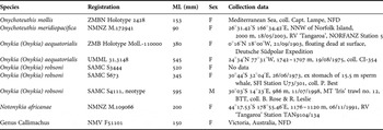
Onychoteuthid specimens were loaned from, or examined at, the Museum of New Zealand Te Papa Tongarewa (NMNZ), Wellington, New Zealand; Museum Victoria, Melbourne, Australia (NMV); South African Museum, Cape Town, South Africa (SAMC); Rosenstiel School of Marine and Atmospheric Science, University of Miami, Florida, USA (UMML); Zoologisches Museum, Museum für Naturkunde der Humboldt-Universität, Berlin, Germany (ZMB); and Zoological Museum, Universitetet i Bergen, Norway (ZMBN). Details of all material examined are given in Table 1, with incomplete collection data indicated ‘NFD’ (no further data) and collection dates given as dd/mm/yyyy. Collection acronyms include: BTT, bottom trawl; MT, motorized trawler; NORFANZ, New Zealand and Australia Norfolk Ridge-Lord Howe Rise Biodiversity Voyage; SFI, Sea Fish Institute, South Africa; RV, research vessel.
Onychoteuthid material examined herein.

Additional morphological acronyms used in the text are: ML, dorsal mantle length; maxW, maximum width (of spermatophore or spermatangium). The term ‘terminal organ’ is used for the narrowed extension of Needham's sac through which individual spermatophores travel and exit during mating. This portion is often improperly referred to as the ‘penis’; however, a true penis is used for internal fertilization, as in mammals.
Although Arkhipkin & Nigmatullin (Reference Arkhipkin and Nigmatullin1997) analysed the relationship between Onychoteuthis ‘banksii’ Leach, 1817, and two ‘Chaunoteuthis mollis’ specimens from the south-eastern Pacific, and concluded that the latter name was a junior synonym of the former, their O. ‘banksii’ specimens appear to have included a number of recently disambiguated taxa (see Bolstad, Reference Bolstad2008b). The C. mollis holotype is distinct from each of these (Bolstad, Reference Bolstad2008a, 2010), and Onychoteuthis mollis (sensu stricto) is accordingly treated herein as a taxonomically valid species.
More complete systematic descriptions of the material reported below are given in Bolstad (Reference Bolstad2008a). Subgeneric distinctions in the genus Onykia follow Bolstad (Reference Bolstad2008a) and Bolstad (2010).
RESULTS
Genus Onychoteuthis
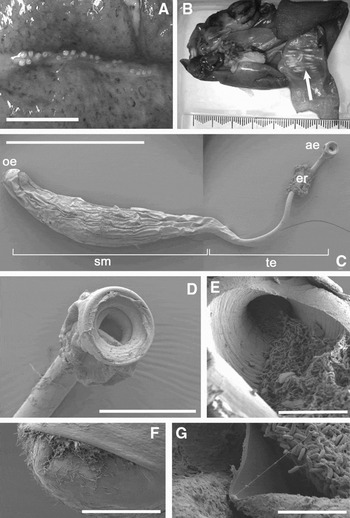
One spent female each of Onychoteuthis mollis (sensu stricto) and O. meridiopacifica was observed. The tissues of both were thin and flaccid, with the relative proportions of the mantle and fins greatly distorted, although the fin margins were whole; the tentacles were reduced to short stumps and the arms appeared shrunken, with many suckers lost. The external mantle surface of each specimen bore two longitudinal cuts (incisions part-way into the mantle musculature, with clear-cut edges), one on each side of the ventral midline. Spermatangia were implanted along the full length of each cut, with their aboral ends protruding externally and the oral portion (sperm mass) enclosed within the mantle musculature, oriented perpendicularly to the cuts in either direction (Figure 1B). Particulars of implantation for each specimen follow.
Onychoteuthis meridiopacifica (NMNZ M.172941, ♀, ML 90 mm). (A) External view of left-hand cut with protruding spermatangium bulbs; (B) internal view of implanted spermatangia embedded in mantle wall on either side of cut (arrow); (C–G) scanning electron micrographs of spermatangia extracted from mantle wall: (C) entire spermatangium (ae, aboral end, with ‘bulb’; er, epidermal remnant; oe, oral end; sm, sperm mass; te, trailing end); (D) aboral bulb; (E) spermatozoa emerging from bulb; (F) oral invagination in middle membrane; (G) breach in spermatangium wall, showing sperm mass within. Scale bars: A, 5 mm; B (major increments), 10 mm; C, 2 mm; D, 250 µm; E, 50 µm; F, 100 µm; G, 25 µm.

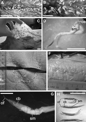
Onychoteuthis mollis (ZMBN Holotype 2428, Figure 2A): two ventral cuts, approximately parallel to ventral midline, each ~20% ML, near ventral anterior margin; one incision Y-shaped (with stem parallel to midline), with ~80 spermatangia in total; the other incision a single straight cut, with ~50 embedded spermatangia. Aboral, protruding end of each spermatangium with rounded, bulbous opening (hereafter ‘bulb’) (Figure 2B, C), through which spermatozoa are likely to exit spermatangium. Spermatangia not extracted (to avoid damaging holotype); approximate length of each 10 mm.
(A–B) Onychoteuthis mollis (ZMBN Holotype 2428, ♀, ML 153 mm), cut with implanted spermatangia, with trailing ends protruding externally (B, courtesy of R. Young). (C–D) Onykia (Onykia) aequatorialis: (C) ZMB Holotype Moll.-110000, ♀, ML 380 mm preserved, backlit mantle wall, showing silhouettes of individually implanted spermatangia; (D) UMML 31.3148, ♀, ML 545 mm, extracted spermatangium from mantle wall (oe, oral end with filament; sm, sperm mass); (E–H) Onykia (Onykia) robsoni (E,G,H) SAMC S3957, ♂, ML 595 mm: (E) scratches on ventral surface of mantle (incision along ventral midline); (G, H) extracted spermatangia from terminal organ (cb, convoluted cement body; of, oral filament; sm, convoluted sperm mass); (F) SAMC S3444, ♀, ML 520 mm, implanted spermatangia visible on exterior mantle surface of specimen. Scale bars: A, C, E, F (major increments), 10 mm; B, D, G, 5 mm; H, 20 mm.

Onychoteuthis meridiopacifica (NMNZ M.172941): two cuts midway along length of mantle, with anterior end of each cut slightly closer to midline; each cut ~9 mm long (~15% ML), with 25–30 embedded spermatangia (Figure 1A, B). Spermatangium maxW 1 mm; middle membrane of oral end with invagination (Figure 1F); trailing end of spermatangium equal in length to sperm mass, terminating in a ‘bulb’ (Figure 1C, D) with emerging spermatozoa (Figure 1E); heads of spermatozoa (Figure 1G) 4.3–5.1 µm long and 1.2–1.7 µm wide (N = 10). Length of entire spermatangium ~8 mm (preserved).
Genus Onykia
Two spent females each of Onykia (Onykia) aequatorialis and O. (O.) robsoni, and one mature male of O. (O.) robsoni were examined. The tissues of both O. (O.) aequatorialis specimens were so thin as to be translucent, with silhouettes of implanted spermatangia clearly visible when backlit (Figure 2C), while those of the O. (O.) robsoni remained more robust, in spite of one specimen having been collected from a sperm whale stomach. In most cases spermatangia were implanted individually into the exterior mantle surface at various positions. Particulars of implantation for each species follow.
Onykia (Onykia) aequatorialis: mantle of holotype (ZMB Holotype Moll.-110000) studded with ~80 individually implanted spermatangia in middle third of left side, dorsally and ventrally; implantation sites characterized by transverse puncture in epidermis (length 1–5 mm), dark red-purple and puckered epidermal tissue, and hard subcutaneous knot of implanted spermatangium (Figure 2C). Second specimen (UMML 31.3148) with individually implanted spermatangia over dorsal mantle surface, occasionally clustered in small groups but each separately implanted; spermatangium length ~12 mm, maxW 1.5 mm, with slender oral filamentous extension, forming a loop (Figure 2D); free trailing end of spermatangium may protrude from implantation site.
Onykia (Onykia) robsoni: females (SAMC S3444, SAMC S673) with spermatangia implanted on ventral mantle surface between fins (300–330 mm from ventral anterior mantle margin in larger specimen (ML 520 mm)), mostly individually, although groups of up to ten were observed (Figure 2E); small punctures (maximum diameter ~7 mm) overlying implanted spermatangia apparent over most external mantle surfaces; free trailing end of spermatangium may protrude from implantation site. Male (SAMC S4111, neotype) with implanted spermatangium observed in terminal organ; intact spermatophores, partly and completely everted spermatangia also removed from terminal organ (Figure 2G, H); spermatangia elongate, length 30–40 mm, maxW 1.5–2 mm, with narrow oral extension/filament; cement body visible in oral part of spermatangium, followed by sperm mass; spermatangium convoluted (Figure 2G). Large scratches observed on ventral mantle surface, 90–145 mm from anterior mantle margin (Figure 2E), and on dorso-lateral right side, 90–170 mm from anterior mantle margin.
Genus Notonykia
One spent female Notonykia africanae was observed (NMNZ M.109066). Numerous white sucker-ring imprints over all external surfaces of mantle, fin, head and arms. Single remaining fin greatly enlarged relative to mantle and brachial crown; tentacles absent, ~1 cm long stumps remaining; some arm tips and arm suckers missing; tissues loose and flaccid in general; some residual mature eggs present in ovary, but nidamental glands and other viscera shrunken. No implanted spermatangia apparent.
Genus Callimacus
One spent female of Callimacus rancureli was observed (NMV F51101). Fins greatly enlarged relative to mantle and brachial crown; tentacles absent, ~1 cm long stumps remaining; some arm tips and arm suckers missing; some residual mature eggs present in ovary, but nidamental glands and other viscera shrunken. No implanted spermatangia apparent.
DISCUSSION
What little is known of mating in oceanic and deep-sea squid has been extrapolated almost exclusively from dead specimens. To date, only one potential mating event has been observed between live deep-sea squid (Brachioteuthis beanii, see Roper & Vecchione, Reference Roper and Vecchione1996). However, our body of knowledge is slowly increasing; descriptions of many mated female deep-sea squid are available, and Nesis (Reference Nesis1995) published an overview of then-known sperm transferral and storage strategies, recently augmented by Hoving (Reference Hoving2008). Several trends in squid reproductive morphology are apparent, including the presence of seminal receptacles or seminal tissues (a specialized organ or modified tissue for the reception of sperm) in some taxa. In species lacking seminal receptacles (including all presently known Onychoteuthidae), spermatangia are implanted directly and deeply into the female's tissues (for overviews, see Nesis, Reference Nesis1995 and Hoving, Reference Hoving2008). More specific strategies for sperm transfer and storage vary between, but also within squid families, as observed in the present study.
Onychoteuthids employ at least two different strategies for spermatangium implantation and storage. At present, these strategies appear generally consistent within, yet specific to, the two most speciose onychoteuthid genera: males of Onychoteuthis species make longitudinal cuts in the female's exterior mantle surface, and implant rows of spermatangia along the length of the cuts, while males of Onykia species implant spermatangia individually on various parts of the female's mantle externally or internally; wounds may be inflicted in the process but do not appear required for/associated with spermatangium implantation in Onykia. (One apparent exception was a female of Onykia (Onykia) robsoni reported by Clarke (Reference Clarke1980, p. 118) with ‘large longitudinal cuts in the anterior mantle with spermatophores embedded in them;’ he also reported one female with embedded spermatophores that were apparently not associated with cuts.) Ventral longitudinal cuts with embedded spermatangia have been reported for Onychoteuthis borealijaponica (Murata et al., Reference Murata, Ishii and Osako1982) and ‘Chaunoteuthis mollis’ (=Onychoteuthis spp.: Grimpe, Reference Grimpe1921; Torchio, Reference Torchio1967; Nesis, Reference Nesis1970; Adam, Reference Adam1972; Okutani & Ida, Reference Okutani and Ida1986; Bello, Reference Bello1998; not Appellöf, Reference Appellöf1891). Jackson & Jackson (Reference Jackson and Jackson2004) reported scattered, individual spermatangia implanted externally and internally in the mantles of female Onykia (M.) ingens. Thus, the patterns observed on specimens herein are consistent with those previously reported in congeners, although some variability in the use of cuts is possible, given those observed by Clarke (Reference Clarke1980) in O. (O.) robsoni.
The specific function of (or advantage conferred by) cuts into the mantle remains unknown. The external mantle surface certainly differs between genera, being smooth in Onychoteuthis and rugose or longitudinally ridged in all species of Onykia where adults are known. The chemistry of the mantle musculature is also believed to vary by genus, with Onykia tissues containing higher levels of ammonia than those of Onychoteuthis (Nesis, Reference Nesis2000). It is possible that the cuts therefore overcome some not-yet-understood physical or chemical barrier to implantation, but in the absence of evidence it is unwise to speculate further.
Hoving & Laptikhovsky (Reference Hoving and Laptikhovsky2007) demonstrated that spermatophores of Onykia (M.) ingens implant autonomously into squid tissue, once the spermatophoric reaction is initiated by the tip of the terminal organ (as each spermatophore leaves the terminal organ, the oral end is pressed, triggering eversion). Given that all reported mature male onychoteuthids have a long terminal organ for spermatophore storage and transfer (in O. (M.) ingens, terminal organ length may exceed mantle length—see Bolstad, Reference Bolstad2006; Arkhipkin & Laptikhovsky, Reference Arkhipkin and Laptikhovsky2010), and that the sperm mass portions of all implanted spermatangia observed herein were fully embedded within, and difficult to extract from, the female mantle musculature, the mechanism reported by Hoving & Laptikhovsky (Reference Hoving and Laptikhovsky2007) is likely consistent throughout the family (although it is not necessary in all squid—see Drew, Reference Drew1919). Autonomous spermatangium implantation would permit rapid deployment of many spermatophores, whether in individual locations or within purpose-inflicted cuts.
Several insights into the more specific mechanics of spermatangium implantation and subsequent deployment in Onychoteuthis and Onykia are suggested by the morphology of structures reported herein for Onychoteuthis meridiopacifica (Figure 1C–G), and Onykia (Onykia) aequatorialis and robsoni (Figure 2E–H). In Onychoteuthis meridiopacifica, the oral part of the spermatangium is smooth, as in Rossia moelleri (a sepiolid). The cement body is orally secreted during implantation in the latter (Hoving et al., Reference Hoving, Nauwelerts, Van Genne, Stamhuis and Zumholz2009), and probably also in O. meridiopacifica, through the oral invagination of the middle membrane on the spermatangium (Figure 1F), facilitating implantation into the female's tissues. In contrast, the oral part of the spermatangium is typified by a filamentous extension in Onykia. Oral extensions of the spermatangia probably also facilitate implantation and originate from the cement body in the intact spermatophore; although morphologically different, oral extensions of the spermatangium have also been described for Todarodes pacificus (a spike-like extension (Takahama et al., Reference Takahama, Kinoshita, Sato and Sasaki1991)) and Octopoteuthis sicula (a tubular extension with an oral broadening, which extends forward during implantation but adheres closely along the spermatangium in the final implanted spermatangium (Hoving et al., Reference Hoving, Lipinksi and Videler2008)).
In other morphological aspects, similarities were observed between the spermatangia of Onychoteuthis and Onykia. The mid-section of the implanted spermatangium (encasing the sperm mass) is firmly attached and difficult to extract intact in both genera. The trailing end of the spermatophore protrudes through a small window in the tissue, through which the spermatangium would have entered during implantation (Figure 1C). This trailing end quite evidently forms the exit route for spermatozoa, at least in Onychoteuthis, since hundreds of spermatozoa were observed within each in O. meridiopacifica (Figure 1E). In other (non-onychoteuthid) taxa, spermatozoa presumably also exit the spermatangium through the trailing end during fertilization (Architeuthis sp., Hoving et al., Reference Hoving, Roeleveld, Lipinski and Melo2004; Octopoteuthis sicula, Hoving et al., Reference Hoving, Lipinksi and Videler2008; Rossia moelleri, Hoving et al., Reference Hoving, Nauwelerts, Van Genne, Stamhuis and Zumholz2009). The ‘bulb’ observed in Onychoteuthis spermatangia has not been described for any other squid; it may serve to prevent the trailing end of the spermatangium from becoming completely implanted into the tissue, where the spermatozoa would be unable to exit. However, the bulb does not sit flush with the female's external tissues, and the same risk of over-implantation would affect other taxa, so the bulb may serve some additional function that is not apparent at present.
Spermatangium implantation was not observed in Notonykia or Callimachus. Given the degenerate state of the reproductive systems in these specimens, it is possible that spermatangia had been implanted and used in spawning, leaving no evidence. The general state of these specimens was consistent with spent females of other onychoteuthid genera, notably the thinned, loosened mantle musculature, lost tentacles, and distorted mantle and fin proportions. Arkhipkin & Nigmatullin (Reference Arkhipkin and Nigmatullin1997) suggested that evolution of this late-life ‘Chaunoteuthis’ stage in onychoteuthids permitted mature females to ‘escape from the ecologically active epipelagic zone […] into a deep water refuge characterized by low predator densities and low temperatures,’ and to utilize earlier accumulated energy to fuel the final stages of maturation. Of the five spent females examined herein with known collection depths, four were taken in relatively deep water (986–2000 m), so conclusion of the life cycle at these depths may be a strategy common to most onychoteuthid genera.
ACKNOWLEDGEMENTS
Thanks to the following persons and institutions for facilitating collection visits and specimen loans: the Museum of New Zealand Te Papa Tongarewa; Museum Victoria; South African Museum; Rosenstiel School of Marine and Atmospheric Science; Zoologisches Museum, Museum für Naturkunde der Humboldt-Universität, Berlin; and Zoological Museum, Universitetet i Bergen; B. Marshall (NMNZ), M. Norman and C. Rowley (NMV); L. Hoenson and M. Cochrane (SAMC), N. Voss (UMML); F. Köhler and M. Glaubrecht (ZMB); E. Willassen and J. Kongsrud (ZMBN). S. O'Shea (AUT), S. Donati (University of Groningen), V. Laptikhovsky (Falkland Islands Government Fisheries Department), A. Turner and B. James (University of Auckland) provided additional assistance in the preparation of this manuscript. This research was supported by the following institutions (K.S.R.B.): Auckland University of Technology, the American Museum of Natural History (Lerner–Gray Fund for Marine Research); the Malacological Society of London (Research Grant), and the Harvard Museum of Comparative Zoology (Ernst Mayr Travel Grant in Animal Systematics).