There are several approaches to the categorisation of the holes, or channels, that permit shunting between the ventricles. The system now used by most centres in the United Kingdom depends on assessing, as the primary feature of the defect, its phenotype. This is determined by examination of the boundaries of the defect as seen from the aspect of the right ventricle. Attention is then paid to the fashion in which the defect opens to the right ventricle, with further details provided of the presence of malalignment of any septal components.Reference Soto, Becker and Moulaert 1 This is the system that was used to provide the codes currently existing in the European Paediatric Cardiac Code. They also provided the basis for its successor, known as the Association for European Paediatric Cardiology-derived, or European, version of the International Paediatric and Congenital Cardiac Code (www.ipccc.net).Reference Franklin 2 The approach favoured by many centres in North America, in contrast, used the location of the defect as the primary focus, basing the categorisation on a concept for development of the normal ventricular septum.Reference Van Praagh, Geva and Kreutzer 3 It is this system that underscores some of the molecules found in the coding system established by the Society of Thoracic Surgeons and the European Association of Cardiothoracic Surgeons.Reference Mavroudis and Jacobs 4 A long-standing approach, also used in the construction of the system favoured by the paediatric cardiac surgeons, had used a simple numerical classification.Reference Wells and Lindesmith 5 Of the sets identified in this system, three correspond broadly with the phenotypes identified in the European system. The so-called inlet defect, however, is defined exclusively on the basis of its location.Reference Wells and Lindesmith 5 Paradoxically, this category has no specific phenotype, since holes, or channels, with markedly different anatomic features can open into the inlet of the morphologically right ventricle.Reference Spicer, Anderson and Backer 6 If these lesions are coded simply on the basis of their phenotypic features, the category for the “inlet defect” becomes redundant. With these ongoing differences in mind, we have taken the opportunity afforded us of examining specimens stored within the archive of Birmingham Children’s Hospital to assess, first, whether all cases can adequately be coded within the European system, and second, if it is possible to correlate the current different approaches.
Materials and Methods
We have examined 147 hearts found in the morphological archive of Birmingham Children’s Hospital. The archive has been collected over a period extending from 1939 to the present time, and contains approximately 2,000 specimens. It is currently segregated alphabetically, with so-called “isolated” ventricular septal defects making up Category C. Specimens having ventricular septal defect associated with other lesions, such as transposition, or tetralogy of Fallot, are catalogued in different alphabetical sets. We have not included most of these latter specimens within our current analysis. We have, however, included hearts coded primarily on the basis of aortic coarctation or interruption, including these hearts when the ventriculo-arterial connections were concordant (Category F). In some these hearts, there was a degree of overriding of the aortic root relative to the apical muscular ventricular septum. In the hearts examined, we did not consider this sufficient to warrant making the diagnosis of double outlet ventriculo-arterial connection. In all the hearts, we then established the nature of the borders of the communication providing the potential for shunting between the ventricles as seen from the aspect of the right ventricle. This permitted us to distinguish between phenotypes according to whether these borders were exclusively muscular (Figure 1), or made up in part of fibrous continuity between the hinges of the leaflets of the cardiac valves (Figures 2–5). These specific phenotypic features were in keeping with the anatomic arrangements already established by our study of the archive of Lurie Children’s Hospital in Chicago.Reference Spicer, Anderson and Backer 6 The phenotypes also correspond with the definitions used to provide the categories existing in the European Paediatric Cardiac Code.Reference Franklin 2
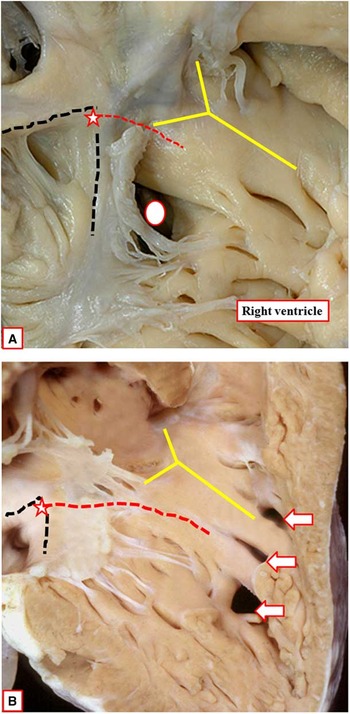
The images show defects with exclusively muscular rims, as viewed from the right ventricle. In each heart, the atrioventricular node (star) is at the apex of the triangle of Koch (dashed black lines). The defect in panel A (white oval) opens to the inlet of the right ventricle, and is inferior to the septomarginal trabeculation (yellow Y). The conduction axis runs superiorly relative to the defect. In the heart shown in panel B, there is a solitary defect as viewed from the left ventricle, opening anteriorly in the right ventricle relative to the septomarginal trabeculation (yellow Y), but it is crossed by two septoparietal trabeculations, so that three holes are seen from the right ventricle (arrows). The conduction axis (red dashed line) is located between an intact membranous septum and the crest of the muscular ventricular septum.

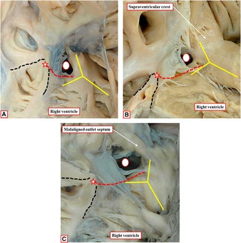
The images show the right ventricular aspect of holes or channels between the ventricles (white ovals) having fibrous continuity between the leaflets of the aortic and tricuspid valves (see Figure 5). Each defect opens to the right ventricle between the limbs of the septomarginal trabeculation, or septal band (yellow Y). In each heart, the atrioventricular node (star) is at the apex of the triangle of Koch (dashed black lines), and the atrioventricular conduction axis (red dashed lines) runs inferiorly relative to the defects. The defect shown in panel A opens to the inlet of the right ventricle, the defect in panel B opens centrally, and the one shown in panel C opens to the outlet, with malalignment of the muscular outlet septum into the cavity of the right ventricle. In the latter defect, the curved surface patched by the surgeon to restore septal integrity will not correspond with the relatively planar geometric interventricular communication, the latter defined on the basis of the cranial continuation of the long axis of the muscular ventricular septum.

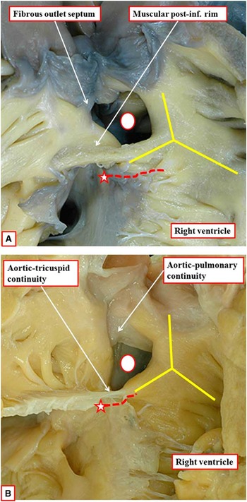
The images show doubly committed and juxta-arterial ventricular septal defects (white ovals), which exist because of failure of formation of the muscular subpulmonary infundibulum. The defects open to the outlet of the right ventricle, between the limbs of the septomarginal trabeculation, or septal band (yellow Y). The phenotypic feature is fibrous continuity between the leaflets of the aortic and pulmonary valves, reinforced in the heart shown in panel A by a fibrous outlet septum. In the heart shown in panel A, there is a muscular postero-inferior rim to the defect, which protects the atrioventricular conduction axis (red dashed line). In the heart shown in panel B, in contrast, the defect extends to become bordered by an area of fibrous continuity between the leaflets of the aortic and tricuspid valve, with the conduction axis then being exposed in the postero-inferior quadrant of the defect.

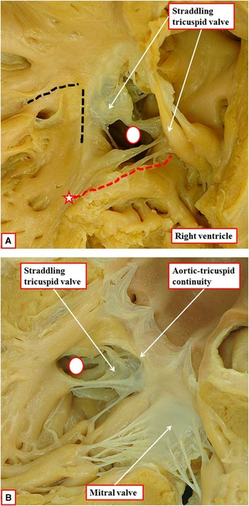
The phenotypic feature of the heart shown in these images, as seen from the right ventricle in panel A, and the left ventricle in panel B, is malalignment between the atrial septum and the muscular ventricular septum, with the muscular ventricular septum deviated inferiorly so as to be attached to the right atrioventricular junction, away from the cardiac crux. Because of this, the atrioventricular conduction axis (red dashed line) no longer arises from the apex of the triangle of Koch (black dashed lines). Instead, it originates from an anomalous inferiorly located atrioventricular node (star). The left ventricular view (panel B) shows the separate atrioventricular junction supporting the leaflets of the mitral valve, so that the heart does not exhibit an atrioventricular canal, the phenotypic feature of this being a common atrioventricular junction. Note that the area of fibrous continuity between the leaflets of the tricuspid and aortic valves, by virtue of the overriding of the tricuspid valve, is within the cavity of the left ventricle. If, during surgical repair, the surgeon attached the patch to the septal hinge of the tricuspid valve, then the curved surface closed by the patch will again differ from the virtual plane representing the geometric interventricular communication.

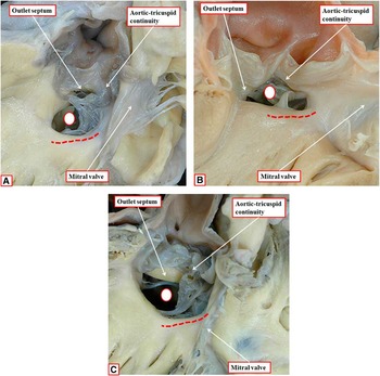
The images show the left ventricular aspects of the three defects (white ovals) shown in Figure 1. In each heart, the phenotypic feature is fibrous continuity between the leaflets of the aortic and tricuspid valves. Note that, in Figure 5A, the defect opens to the inlet of the right ventricle, so that there is also an extensive area of fibrous continuity between the leaflets of the tricuspid and mitral valves. The defect shown in panel B opens centrally to the right ventricle, with the outlet septum forming its roof. The defect shown in panel C opens to the outlet of the right ventricle, and the malaligned outlet septum can be seen within the cavity of the right ventricle. In all three hearts, the conduction axis (red dashed line) runs inferiorly relative to the defect.

Results
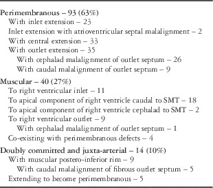
It proved an easy matter, on the basis of assessment of the borders of the communications providing the potential for shunting between the ventricles as viewed from the right ventricle, to place all the specimens into one of three groups (Table 1). By far the largest group, made up of 89 specimens, was distinguished by the presence of fibrous continuity between the leaflets of the aortic and tricuspid valves (Figure 2). With the exception of two hearts, in which we observed malalignment between the atrial and muscular ventricular septal structures (Figure 4), the fibrous continuity was found in the posterior quadrant of the defect as viewed from the right ventricle, although its extent varied markedly within the group. The two exceptional hearts were associated with straddling and overriding of the morphologically tricuspid valve. The muscular ventricular septum in these hearts was deviated away from the crux, inserting to the atrioventricular junction inferiorly within the right ventricle (Figure 4A). In these hearts, it is the plane of space representing the right ventricular margin of the channel floored by the deviated muscular ventricular septum that is the geometric interventricular communication, recognising that this space might not be entirely planar due to the potential curvature of the long axis of the muscular ventricular septum. The area of fibrous continuity between the aortic and tricuspid valves, however, was not part of this border, but was situated within the cavity of the left ventricle (Figure 4B). The malalignment between the muscular ventricular and the atrial septums, in fact, was the differentiating phenotypic feature of both hearts, although the channel produced between the ventricles was unequivocally an inlet defect (Figure 4).
Details of 147 hearts examined in the Birmingham Archive

In one subset of the defects, made up of 23 hearts, all characterised by aortic-to-tricuspid valvar continuity, but with the atrial and muscular ventricular septal structures aligned at the cardiac crux, the communication between the ventricles opened primarily into the inlet of the right ventricle (Figure 2A). In these hearts, the caudal margin of the defect was formed by an extensive area of fibrous continuity between the leaflets of the tricuspid and mitral valves, the continuity with the aortic valve being found at the roof of the defect (Figure 5A). The ventral margin of these defects was made up of the outlet septum, which supported a normal free-standing muscular subpulmonary infundibulum. The aortic valve was supported almost exclusively within the left ventricle, its margin of continuity with the tricuspid valve now forming the superior margin of the geometric interventricular communication (Figure 5A). In all of these hearts, the apex of the triangle of Koch was deviated inferiorly, and the line drawn from the apex of the triangle to the medial papillary muscle, which was extensive, ran inferiorly relative to the hole between the ventricles (Figure 2A). In some of these hearts, the holes permitting ventricular shunting were virtually closed by adherence of the septal leaflet of the tricuspid valve along the right ventricular border of the defect.
In a second subset of 33 hearts with fibrous continuity between the leaflets of the aortic and tricuspid valves, the area of valvar continuity was located postero-inferiorly relative to the defect when viewed from the right ventricle (Figure 2B). The long axis of these defects, which were positioned centrally relative to the inner curve of the right ventricle, extended towards the anterior margin of the right ventricle. When viewed from the aspect of the left ventricle (Figure 5B), the roof of the geometric interventricular communication was made up of the muscular outlet septum, continuous dorsally in some of the hearts with a remnant of the fibrous interventricular component of the membranous septum. The aortic valve was again supported almost exclusively above the cavity of the left ventricle. When seen from the right ventricle, the muscular outlet septum was continuous cranially with the sleeve of free-standing muscular subpulmonary infundibulum (Figure 2B). The apex of the triangle of Koch was positioned more superiorly in this group when compared to those in which the defect opened into the inlet of the right ventricle, but the line drawn to the medial papillary muscle, although shorter, continued to run inferiorly when viewed from the right ventricular aspect (Figure 2B). In several of the hearts, the defects were partially occluded by tissue tags derived from the leaflets of the tricuspid valve. In one of the hearts, a large aneurysmal sac extended into the cavity of the right ventricle, the initial defect itself being closed, although a small opening into the sac was visible on the left ventricular aspect.
In a third subset of 35 hearts with fibrous continuity between the leaflets of the aortic and tricuspid valves, there was overriding of the orifice of the aortic valve relative to the muscular ventricular septum, the aortic valvar leaflets being supported in part above the cavity of the right ventricle (Figure 2C). As an integral part of the valvar overriding, in the majority the muscular outlet septum was situated exclusively within the right ventricle, with its septal end attached to the antero-cephalad limb of the septomarginal trabeculation. This was the arrangement in 26 of the hearts. In these hearts, however, there was no obstruction of the subpulmonary outflow tract. Indeed, the subpulmonary infundibulum was well formed, extending distally from the leading edge of the muscular outlet septum, and supporting the leaflets of the pulmonary valve. In all the hearts fulfilling this phenotypic feature, the aortic valve remained supported in its greater part by the morphologically left ventricle, so the ventriculo-arterial connections remained concordant. When viewed from the left ventricle, there was an extensive area of fibrous continuity dorsally between the leaflets of the aortic and tricuspid valves (Figure 5C). It was the recognition of the right ventricular attachments of the overriding aortic valve that served to distinguish these defects with outlet extension from those extending so as to open centrally within the right ventricle. In the defects with valvar overriding, and the outlet septum deviated into the right ventricle, the right ventricular margin of the channel between the ventricles no longer represented the geometric interventricular communication. This was because, by virtue of the malalignment of the muscular outlet septum, the curved surface now representing the right ventricular border of the channel between the ventricles was located exclusively within the cavity of the right ventricle (Figure 2C). None of the hearts with these defects associated with aortic valvar overriding showed any evidence of spontaneous diminution in size. In the remaining 9 hearts opening between the ventricular outlets, the outlet septum was deviated into the left ventricle, with associated obstruction of the subaortic outflow tract (see below).
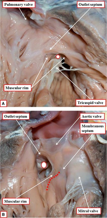
The feature of 40 hearts that could be catalogued within the second phenotypic grouping (Table 1) was the presence of exclusively muscular borders when viewed from the right ventricle. In all these hearts, it was possible to pass entirely round the right ventricular border whilst remaining on muscular tissue. The hearts again showed marked differences in the way the holes or channels opened into the right ventricular cavity. In 11 hearts, the defect opened into the right ventricle beneath the septal leaflet of the tricuspid valve (Figure 1A). The line extending from the apex of the triangle of Koch to the medial papillary muscle ran antero-cephalad relative to this hole (Figure 1A). In 18 hearts, the defects opened just anterior to the attachments of the septal leaflet of the tricuspid valve, but caudal relative to the body of the septomarginal trabeculation. The line from the apex of the triangle of Koch remained antero-cephalad relative to these defects. In 2 hearts, the muscular septum was deficient anterior to the septomarginal trabeculation. By virtue of the presence of the septoparietal trabeculations, multiple orifices were present between the ventricular cavities (Figure 1B). In 9 hearts, the muscular defects opened to the outlet of the right ventricle (Figure 1C). In these hearts, the caudal limb of the septomarginal trabeculation was fused with the ventriculo-infundibular fold, so that there was no continuity between the leaflets of the aortic and tricuspid valves. The roof of the defect was formed by the muscular outlet septum, which supported a short subpulmonary infundibulum. In all these hearts bar one, there was no obvious malalignment between the outlet septum and the floor of the defect, which opened into the right ventricle between the limbs of the septomarginal trabeculation. In the remaining heart, however, the muscular outlet septum was deviated in antero-cephalad fashion so that the aortic root arose in part from the right ventricle (Figure 6). In this heart, therefore, the curved surface representing the right ventricular border of the channel between the ventricles was again distinct from the relatively planar geometric interventricular communication. In four of the overall group of these hearts, the muscular defects co-existed with second defects characterised by fibrous continuity between the leaflets of the aortic and tricuspid valves. In all of these hearts, the line drawn from the apex of the triangle of Koch to the medial papillary muscle extended through the part of the muscular septum remaining between the defects.
The images show the solitary defect in the Birmingham archive with a muscular defect opening to the right ventricular outlet, but with antero-cephalad deviation of the muscular outlet septum into the right ventricle. Figure 6A shows the right ventricular aspect of the defect (white oval). There is no subpulmonary obstruction. Figure 6B, showing the left ventricular aspect of the defect, confirms the right ventricular location of the muscular outlet septum, and shows the muscular bar interposing between the leaflets of the aortic and tricuspid valve. The red dashed line shows the location of the atrioventricular conduction axis, which penetrates through an intact membranous septum.

We also found 14 hearts fulfilling a third phenotype (Table 1), namely fibrous continuity between the leaflets of the aortic and pulmonary valves (Figure 3). In 9 of these hearts, there was muscular continuity inferiorly and posteriorly between the caudal limb of the septomarginal trabeculation and the ventriculo-infundibular fold (Figure 3A), while in 5hearts the defect extended to become bordered by fibrous continuity between the leaflets of the aortic and tricuspid valves (Figure 3B). Variation was also noted in the cranial rim of the defect. In one heart, the rim was formed by fibrous continuity between the leaflet of the pulmonary valve and the base of the adjacent leaflet of the aortic valve. In another heart, this area of fibrous continuity was reinforced by a fibrous outlet septum, which hung down into the defect from the area of the conjoined arterial valves.
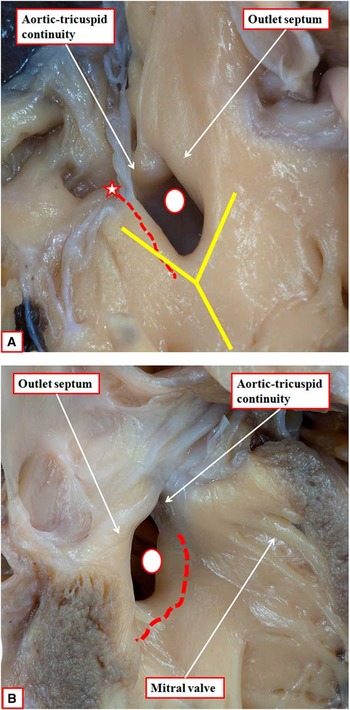
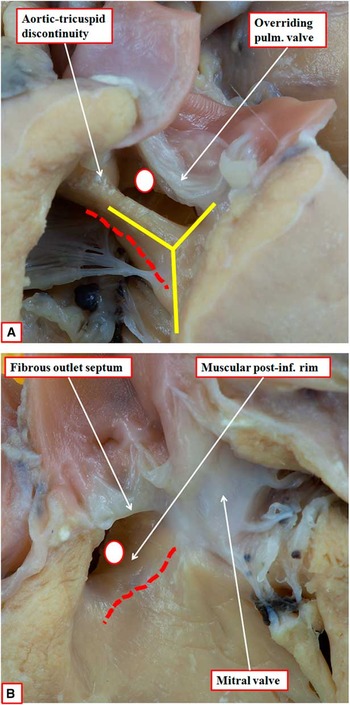
So as fully to categorise the differences between the various defects, it also proved necessary to account for any malalignment between the septal components. We have already described how the atrial and ventricular septal components were malaligned as part of the phenotype of the defects associated with overriding of the tricuspid valve (Figure 4). We have also described how malalignment of the muscular outlet septum into the right ventricle was part of the phenotype in the majority of those defects with aortic-to-tricuspid continuity opening to the outlet of the right ventricle, but in the absence of any subpulmonary obstruction. This variant, with antero-cephalad malalignment of the muscular outlet septum, was also found in one of the hearts with a muscular defect opening to the right ventricular outlet (Figure 6). Defects with aortic-to-tricuspid continuity opening between the ventricular outlets, however, along with those having aortic-to-pulmonary valvar continuity, were also observed with the muscular outlet septum, or its fibrous remnant, deviated postero-caudally into the left ventricle (Table 1). All of these hearts had associated aortic coarctation or interruption. In 9 of the hearts, it was the muscular outlet septum that was deviated into the left ventricle so as to obstruct the subaortic outflow tract (Figure 7). These hearts, therefore, fulfilled the criterion for classification as being perimembranous. In 5 of the hearts, in contrast, it was a fibrous outlet septum hanging down from an area of aortic-to-pulmonary valvar continuity which obstructed the entrance of the aortic outflow (Figure 8A). The hearts with a malaligned fibrous outlet septum all had a muscular postero-inferior margin to the defect (Figure 8B). Such hearts with a malaligned fibrous outlet septum must be anticipated to be not only doubly committed, but also perimembranous, although none were found in our archive in Birmingham.
The images show the right ventricular (A) and left ventricular (B) aspects of a perimembranous defect with caudal malalignment of the muscular outlet septum. The malaligned septum obstructs the subaortic outflow tract from the left ventricle. Abbreviations and symbols as before.

The images show the right ventricular (A) and left ventricular (B) aspects of a doubly committed ventricular septal defect with overriding of the pulmonary valve and malalignment of a fibrous outlet septum, the latter feature obstructing the left ventricular outflow tract. Note the muscular postero-inferior rim to the defect. Abbreviations and symbols as before.

Comment
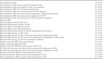
All the hearts we have examined thus far from the archive of Birmingham Children’s Hospital can be placed into one of the three phenotypic categories recognised and categorised in the European version of the International Paediatric and Congenital Cardiac Code. It was not possible, however, within the codes currently existing within the European code fully to categorise all the hearts examined. It proved necessary to create additional codes recognising the subtypes of caudal malalignment of the outlet septum, be it muscular or fibrous, so as to provide complete codification. These new codes have now been added to the European code (Table 2). In the European system, the different phenotypes, which provide the basis of categorisation, are described as being perimembranous, muscular, or doubly committed and juxta-arterial. The phenotypic feature underscoring the use of “perimembranous” is fibrous continuity between the leaflets of the aortic and tricuspid valves (Figure 2). In the majority of the hearts with this phenotypic feature, the area of fibrous continuity is part of the right ventricular margin of the defect. In a particular subset, however, specifically those characterised by malalignment between the atrial septum and the muscular ventricular septum, and with overriding of the tricuspid valvar orifice, the area of fibrous continuity is within the cavity of the left ventricle (Figure 4B).
Numbers from the Revised European version of the International Paediatric and Congenital Cardiac Code

Key: Abbreviations used: VSD – ventricular septal defect; AV – atrioventricular; OS – outlet septum; DC – doubly committed; RV – right ventricle;
*New code added to International Paediatric and Congenital Cardiac Code
In the hearts categorised as having muscular defects, the entirety of the margins of the holes or channels between the ventricles, as viewed from the right ventricle, is made up of myocardial tissue (Figure 1). The phenotypic feature of the third class of defect, described as being doubly committed and juxta-arterial, is that the roof of the defect is made up of fibrous continuity between the leaflets of the aortic and pulmonary valves (Figure 3). In the hearts examined in the Birmingham archive with this feature, it was also possible to recognise a fibrous outlet septum hanging down from the area of valvar continuity. These hearts could be further distinguished according to whether there was myocardial tissue interposed postero-caudally between the leaflets of the aortic and tricuspid valves, or whether there was additional aortic-to-tricuspid valvar continuity. In these latter hearts, the ventriculo-infundibular fold was not fused with the caudal limb of the septomarginal trabeculation. The latter defects, therefore, are not only doubly committed, but also extend to become perimembranous. Codes for both of these variants already exist within the current European version of the International Code (Table 2).Reference Franklin 2 Some of these hearts, however, also had postero-caudal malalignment of a fibrous outlet septum producing subaortic obstruction. This feature needed to be recognised as a phenotypic variant, and has led to a coding modification, as did the finding of postero-caudal malalignment of the muscular outlet septum.
In the third class of defect, with aortic-to-pulmonary fibrous continuity due to failure of formation of the muscular subpulmonary infundibulum, the defect was, of necessity, an outlet defect. All the holes of this type opened into the right ventricle between the limbs of the septomarginal trabeculation, or septal band. The defects falling into the other two phenotypic categories, in contrast, could open towards, or into, the inlet, the outlet, or the apical trabecular components of the morphologically right ventricle. Fully to describe the defects that are categorised as being perimembranous or muscular, therefore, it is necessary to account not only for their defining phenotypic feature, which depends on their borders, but also for their geographical location relative to the components of the morphologically right ventricle. This latter feature then provides important information as how best to approach those defects requiring surgical closure.
Equally important to the surgeon is knowledge of the relationship of the defects to the atrioventricular conduction axis. It is this second vital piece of clinical information that is provided by establishing the phenotype of the defect on the basis of its borders.Reference Soto, Becker and Moulaert 1 , Reference Milo, Ho and Wilkinson 7 With only one exception, when defects are characterised by aortic-to-tricuspid continuity, the conduction axis always runs postero-inferiorly when viewed from the right ventricle. In all these instances, the axis takes its origin from the atrioventricular node located at the apex of the triangle of Koch (Figure 2). The axis is deviated inferiorly when such typical perimembranous defects open to the inlet of the right ventricle, but still arises from an atrioventricular node located at the apex of the triangle of Koch, the triangle itself also being deviated inferiorly. This is a fundamentally different relationship when compared to muscular defects opening to the right ventricle (Figure 1A), and also when compared to the exceptional variant of defect characterised by aortic-to-tricuspid valvar continuity (Figure 4). This atypical variant also opens to the inlet of the morphologically right ventricle. Indeed, in the setting of the overriding of the tricuspid valvar orifice, the muscular ventricular septum runs the full width of the right atrioventricular junction. This accounts for its phenotypic feature, which is the malalignment between the atrial and muscular ventricular septums. The muscular septum then inserts inferiorly within the right atrioventricular junction. Because the atrioventricular conduction axis is itself carried on the crest of the malaligned muscular septum, it is no longer able to take its origin from the regular atrioventricular node.Reference Milo, Ho and Macartney 8 Instead, an anomalous atrioventricular node is formed at the site of union between the muscular septum and the atrioventricular junction (Figure 4A). Yet a fourth different disposition of the atrioventricular conduction axis is to be found in some hearts with channels permitting ventricular shunting opening to the inlet of the morphologically right ventricle.Reference Spicer, Anderson and Backer 6 This is found in hearts with atrioventricular septal defects, but with shunting confined at ventricular level. This is because, during ventricular systole, the bridging leaflets of the common atrioventricular valve coapt against the leading edge of the atrial septum.
All of this information highlights the deficiencies of systems that suggest there is need for description simply of an “inlet” ventricular septal defect. In the first place, description in this fashion fails to distinguish between the four potential phenotypic variants.Reference Spicer, Anderson and Backer 6 Specifically, such defects can be muscular, can be perimembranous opening to the inlet of the right ventricle, can be perimembranous with atrioventricular septal malalignment, or can be atrioventricular septal defects with shunting confined exclusively at ventricular level. In the second place, if the lesions are coded on the basis of the phenotypic variability (Table 1), there is then no need for a separate code for an “inlet” defect. In the third place, some of these hearts have been considered previously to show “atrioventricular canal type” defects, the suggestion being made that they exist because of absence of the “atrioventricular canal septum”.Reference Van Praagh, Geva and Kreutzer 3 We now know that there is no part of the muscular ventricular septum that is derived from the atrioventricular endocardial cushions.Reference Lamers, Wessels and Verbeek 9 It is possible, of course, to find hearts with a common atrioventricular junction and deficient atrioventricular septation in which shunting through the septal defect is confined at ventricular level. These are the true septal defects of “atrioventricular canal type”.Reference Spicer, Anderson and Backer 6 Their phenotypic feature is the presence of the common atrioventricular junction, and they make up one of the phenotypes emphasised above. The other defects that have previously been categorised in this fashion, namely large perimembranous defects opening to the inlet of the right ventricle, along with the defects associated with straddling and overriding of the tricuspid valve, lack this cardinal feature of atrioventricular canal defects, namely the common atrioventricular junction. It is suggested that the defects are of “atrioventricular canal type” simply because the muscular ventricular septum extends across the full width of an overriding atrioventricular valvar orifice.Reference Van Praagh, Geva and Kreutzer 3 This, however, is because of the malalignment between the atrial and ventricular septal structures, and not because of the presence of an atrioventricular canal. It is the septal malalignment that is of greatest significance, since this determines the anomalous course of the atrioventricular conduction axis in this specific phenotype.Reference Milo, Ho and Wilkinson 7
When categorising holes or channels between the ventricles, therefore, it is equally important to take note of the presence of septal malalignment. This is a feature not only of the defect associated with straddling and overriding of the tricuspid valve, but also the perimembranous defects that open to the outlet of the morphologically right ventricle, and the perimembranous and doubly committed defects associated with aortic coarctation or interruption. We also observed antero-cephalad malalignment of the outlet septum in one of the hearts with a muscular outlet defect. In these hearts, because of the malalignment of the muscular outlet septum, or its fibrous remnant, the malaligned septum becomes a right or left ventricular, rather than an interventricular, structure. The hearts we encountered with a muscular outlet septum malaligned rightward all exhibited a non-obstructed but completely muscular subpulmonary infundibulum. These hearts are also described as Eisenmenger defects. They form a major subset of the hearts, considered by some to represent conoventricular defects.Reference Van Praagh, Geva and Kreutzer 3 It is certainly the case that they show evidence of partial divorce between the subpulmonary conus and the body of the morphologically right ventricle. The hearts with perimembranous defects opening towards the apical trabecular component of the right ventricle also show divorce between the body of the right ventricle and the muscular outlet, or conal, septum. In these latter hearts, however, the aortic root remains supported exclusively by left ventricular structures, often with the remnant of the interventricular component of the membranous septum incorporated within their cranial borders. The Eisenmenger defect itself is characterised by cranial malalignment of the muscular outlet septum, but in the absence of subpulmonary obstruction. All the hearts of this type except one in the Birmingham archive had perimembranous defects (Figure 5C). The outstanding heart exhibited a muscular postero-inferior rim between the leaflets of the overriding aortic and the tricuspid valves (Figure 6). All of these defects are now catered for in the revised coding system, as are those with caudal malalignment of the outlet septum, be it muscular or fibrous (Table 2).
Those who focus on the conus when categorising holes between the ventricles also describe a set of defects considered to represent conal hyoplasia.Reference Van Praagh, Geva and Kreutzer 3 Our current study shows that, as with the “inlet defect”,Reference Spicer, Anderson and Backer 6 several phenotypic variants can be interpreted on the basis of incomplete formation of the components of the muscular subpulmonary infundibulum. In the normal heart, the infundibulum is formed almost exclusively by the ventriculo-infundibular fold, along with the free-standing sleeve of infundibular musculature that lifts the leaflets of the pulmonary valve away from the base of the ventricular mass.Reference Merrick, Yacoub and Ho 10 Both of these components are divorced from the apical muscular septum in hearts possessing outlet muscular defects. In these hearts, furthermore, it is possible to identify directly the different components of the normal infundibulum, namely the ventriculo-infundibular fold, the muscular outlet septum itself, and the hypoplastic infundibular sleeve that continues to lift the leaflets of the pulmonary valve away from the aortic root. It is the hearts with defects that are doubly committed and juxta-arterial, or directly subarterial, nonetheless, which show the extreme form of conal hypoplasia. In these hearts, there has been total failure of formation of the free-standing subpulmonary muscular sleeve. In many of these hearts, we did find it possible to identify a fibrous outlet septum, which hangs down from the area of arterial valvar fibrous continuity that is their defining phenotypic feature. It is then the postero-caudal deviation of this fibrous septum that obstructs the subaortic outflow tract.
Major criticisms have been directed against using the term “perimembranous” to describe the hearts defined on the basis of fibrous continuity between the leaflets of the aortic and tricuspid valves. It has been suggested that the defects would better be termed paramembranous.Reference Van Praagh, Geva and Kreutzer 3 Since they are directly adjacent to the atrioventricular component of the membranous septum, and often times possess a remnant of the interventricular component of the membranous septum as part of their border, there is much strength to this argument. For better or worse, nonetheless, the term “perimembranous” is now firmly embedded within the paediatric cardiac lexicon. The word was used in the first instance to emphasise that the defects exist because of deficiency of the muscular ventricular septum around the persisting atrioventricular component of the membranous septum, which then forms part of the perimeter of the hole.Reference Soto, Becker and Moulaert 1 This is the crucial feature since, when identified, and with the exception of those defects associated with straddling and overriding of the tricuspid valve, it permits the surgeon to predict with confidence the anticipated location of the atrioventricular conduction axis.Reference Soto, Becker and Moulaert 1 And it is precisely because the different parts of the muscular septum can be deficient around the margins of the hole that it can extend so as to open towards the inlet, the apical trabecular, or the outlet components of the right ventricle, the latter because of malalignment between the apical and outlet muscular septal components. It is noteworthy that, when Soto and colleagues proposed a modification of the system that introduced the term “perimembranous”,Reference Soto, Ceballo and Kirklin 11 they commented that the term itself was well understood by surgeons.
Conclusions
When categorising holes or channels between the ventricles, it is essential not only to distinguish between different phenotypes, but also to account for their geographical location relative to the components of the right ventricle. It is equally necessary to describe and code, when present, malalignment between the septal components. Our analysis thus far of the hearts held in the Birmingham archive shows that this is readily achievable using the numbers specified within the European version of the International Paediatric and Congenital Cardiac Code,Reference Franklin 2 and using the features previously defined as distinctive for these entities. New codes, nonetheless, have been necessary so as to account for the subtypes with caudal malalignment of either a muscular or fibrous outlet septum (Table 2). These numbers have been cross-mapped so as to provide a degree of correlation with those using alternative systems for categorisation.Reference Franklin, Jacobs and Krogmann 12 The alternative systems should also ideally account for both geographical location and phenotypic features. In order to reach agreement, however, it will be necessary to agree not only on the curved surface that is taken to represent the “ventricular septal defect”, but also the words that are used to describe it. Readers of our account will, by now, have realised that we have used “hole” to describe a communication between the ventricles that is relatively planar, whereas the communications existing in the setting of septal malalignment have been described as “channels”. This is because the curved surface closed by the surgeon when placing a patch to close the communication in the setting of septal malalignment is not always the same as the relatively planar geometric interventricular communication, the latter represented by the cranial continuation of the long axis of the muscular ventricular septum. The availability of three-dimensional techniques for imaging now show that it is possible to distinguish there virtual planes and real curved surfaces during clinical investigation.Reference Costello, Olivieri, Krieger, Thabit, Marshall, Yoo, Kim, Jonas and Nash 13 , Reference Anderson, Spicer, Henry, Rigsby, Hlavacek and Mohun 14 These geometric and linguistic aspects need also to be considered if we are to achieve the hoped-for consensus.
Acknowledgement
We are indebted to the staff of the Histopathology Department of Birmingham Children’s Hospital, who made it possible for us to gain access to the archive of congenitally malformed hearts.