Introduction
The coronavirus disease 2019 (COVID-19) pandemic, caused by severe acute respiratory syndrome coronavirus 2 (SARS-CoV-2) (Zhou et al., Reference Zhou, Yang, Wang, Hu, Zhang, Zhang, Si, Zhu, Li and Huang2020), has profoundly impacted the entire world and healthcare systems (Sahin et al., Reference Sahin, Akbulut and Yilmaz2020). According to the World Health Organization (Reference World Health2020), COVID-19 infections can be classified into five categories based on clinical severity and diagnostic findings: asymptomatic, mild, moderate, severe and critical. Asymptomatic individuals show no clinical symptoms or abnormalities on chest imaging but test positive for SARS-CoV-2 via RT-PCR. Mild cases present with symptoms such as fever <38◦C, fatigue, cough, sore throat and headache, yet exhibit no signs of pneumonia on imaging. Moderate infections involve similar clinical symptoms, but chest imaging reveals mild pneumonia. Severe cases are marked by respiratory distress, including a respiratory rate of 30 or more breaths per minute, oxygen saturation at or below 93% at rest, or a PaO2/FiO2 ratio of 300 mmHg or less; imaging may show rapid progression of lung lesions. Critical infections involve life-threatening complications such as respiratory failure requiring mechanical ventilation, shock or multi-organ failure, often requiring intensive care (World Health, Reference World Health2020). Membrane-bound angiotensin-converting enzyme 2 (ACE2), a key component of the renin-angiotensin system (RAS), serves as the human gateway that allows SARS-CoV-2 to enter host cells (Nazerian et al., Reference Nazerian, Vakili, Ebrahimi and Niknejad2021; Shang et al., Reference Shang, Wan, Luo, Ye, Geng, Auerbach and Li2020). In humans, ACE2 expression is highly prevalent in spermatogonia, Leydig cells and Sertoli cells within the testes of adult males (Pascolo et al., Reference Pascolo, Zito, Zupin, Luppi, Giolo, Martinelli, De Rocco, Crovella and Ricci2020; Wang and Xu, Reference Wang and Xu2020) and also widely expressed in the ovaries, uterus, vagina and placenta (Jing et al., Reference Jing, Run-Qian, Hao-Ran, Hao-Ran, Ya-Bin, Yang and Fei2020). Consequently, these tissues may represent potential target organs for viral infection (Cao et al., Reference Cao, Han, Feng, Lu, Wang, Sun, Zhao and Pan2024).
Throughout the history of vaccine development, the progression of COVID-19 vaccines has occurred at an extraordinary and unmatched speed (Krammer, Reference Krammer2020). COVID-19 vaccines are classified into four primary categories based on their underlying platforms: inactivated, protein subunit, viral vector and nucleic acid (Nagy and Alhatlani, Reference Nagy and Alhatlani2021). Research on the effects of COVID-19 on assisted reproductive technology (ART) outcomes has predominantly concentrated on its impact on female partners (Bentov et al., Reference Bentov, Beharier, Moav-Zafrir, Kabessa, Godin, Greenfield, Ketzinel-Gilad, Ash Broder, Holzer, Wolf, Oiknine-Djian, Barghouti, Goldman-Wohl, Yagel, Walfisch and Hersko Klement2021; Ding et al., Reference Ding, Wang, Zhang, Cui, Chen, Zhou, Yuan, Ma, Zhang and Rong2021; Jindal, Reference Jindal2023; Kolanska et al., Reference Kolanska, Hours, Jonquière, Mathieu d’Argent, Dabi, Dupont, Touboul, Antoine, Chabbert-Buffet and Daraï2021; Orvieto, Segev-Zahav, et al., Reference Orvieto, Noach-Hirsh, Segev-Zahav, Haas, Nahum and Aizer2021; Rageh et al., Reference Rageh, Farag, Behery, Badreldin and Ali2023). Notably, several studies have focused on examining the impact of COVID-19 infection on sperm parameters (Holtmann et al., Reference Holtmann, Edimiris, Andree, Doehmen, Baston-Buest, Adams, Kruessel and Bielfeld2020; Mittal et al., Reference Mittal, Mantravadi and Rao Gadela2023; Ruan et al., Reference Ruan, Hu, Liu, Liu, Jiang, Li, Li, Luan, Liu and Yu2021; Stigliani et al., Reference Stigliani, Massarotti, Bovis, Maccarini, Anserini and Scaruffi2023).
Overall, a deeper understanding of the effects of COVID-19 infection and vaccines, particularly inactivated vaccines, on the outcomes of assisted reproductive treatments such as intracytoplasmic sperm injection (ICSI), especially when both men and women are infected, is essential for providing more precise guidance to infertile couples and healthcare providers.
Materials and methods
This retrospective case–control study was conducted on infertile couples who attended the Royan Institute between August 2020 and March 2022. The Ethics Committee of Royan Institute approved the study protocol (approval code: IR.ACECR.ROYAN.REC.1401.029). Patients’ data were systematically collected using the Royan Research Institute database. The couples were divided into two groups: one consisting of individuals with a history of SARS-CoV-2 infection without prior vaccination and a control group comprising individuals with neither a history of infection nor vaccination. The positive PCR results were validated through the diagnostic laboratory at the Royan Institute. The infected group was further divided based on symptom severity into asymptomatic, mild, moderate and severe cases, with only individuals presenting mild symptoms such as fever, cough and taste/smell changes included in the analysis (Gandhi, Reference Gandhi2020), with no abnormal radiological findings (Feng et al., Reference Feng, Ling, Bai, Xie, Huang, Li, Xiong, Yang, Chen, Lu, Lu, Liu, Chen, Li, Li, Summah, Lin, Yan, Zhou, Lu and Qu2020). In the vaccination phase, couples who underwent two ICSI treatment cycles – one before and one after receiving the inactivated SARS-CoV-2 vaccine (Sinopharm) – were analysed. Clinical data related to the ICSI treatment cycle were collected and compared between groups. Patients’ information was obtained through telephone-administered questionnaires, which gathered data on infections and vaccinations from couples, as well as through a review of their medical records, which included details about the ICSI cycle.
This study evaluated and analysed several reproductive parameters, including ovarian parameters: the number of antral follicles at the start of the cycle, the number of oocyte–cumulus complexes retrieved by follicular puncture, the number of mature and immature oocytes obtained and the number of fertilized oocytes. Sperm parameters: Concentration, total sperm count, motility (including progressive motility) and sperm morphology. Embryo parameters: The number of embryos and their quality (grading) (Salemi et al., Reference Salemi, Yahyaei, Vesali and Ghaffari2021). Semen analysis was performed following the World Health Organization (WHO) laboratory guidelines, 6th edition (2021), including standardized protocols for semen collection and assessment of volume, concentration, motility and morphology, emphasizing clinical decision limits and methodological standardization. All assessments were conducted by trained laboratory personnel using validated equipment and under strict quality control measures. Pregnancy outcomes: On day 14 following embryo transfer, a positive beta-HCG resulted in the diagnosis of pregnancy. Two to three weeks following a positive pregnancy test, the presence of a gestational sac, as determined by an ultrasound evaluation, was considered a clinical pregnancy. The loss of a clinical pregnancy before 20 full weeks of gestation (18 weeks following fertilization) was referred to as a miscarriage. The delivery of at least one live fetus after 20 full weeks of gestation was referred to as a live birth. In addition, the time interval between recovery from infection and the initiation of the ART cycle, the time between the second dose of the vaccine and the start of the treatment cycle, and the type of vaccine administered were also recorded.
Inclusion criteria: couples who presented for ICSI treatment during the defined study period and after having a history of either SARS-CoV-2 infection or vaccination were included. The control group consisted of couples with no history of infection or vaccination. Exclusion criteria: Women with a history of more than three failed implantations, women requiring oocyte or embryo donation, couples undergoing preimplantation genetic diagnosis (PGD), women aged over 40 years, couples with underlying health conditions, women diagnosed with ovarian hyperstimulation syndrome (OHSS) and women with uterine anomalies.
Ovarian stimulation protocol and embryo transfer
Ovarian stimulation was carried out during the IVF cycles using a conventional GnRH antagonist protocol. From days two through three of the menstrual cycle, all patients were given 150–225 IU/day of Cinnal-fⓇ (follitropin alfa; CinnaGen, Iran). When at least two or three follicles were 12–14 mm in size, 0.25 mg of Cetrotide (cetrorelix acetate; Merck Serono, Eysins, Switzerland) was administered daily. After at least three follicles reached a size of 17–18 mm, 250 μg of Ovitrelle (choriogonadotropin alfa; Merck Serono Eysins, Switzerland) was administered. After 36 hours, the ovum was carried out. An embryo transfer catheter (Labotect Labor-Technik-Göttingen GmbH, Rosdorf, Germany) was used to transfer the embryo using a routine procedure. 2–3 days after oocyte retrieval, one to three embryos were put in the embryo transfer catheter, which was positioned 1–2 cm above the internal entrance of the cervical cavity. Up to the day of the beta-HCG test, luteal-phase support was given with 400 mg twice daily of vaginal progesterone (Aburaihan Pharmacy Company, Tehran, Iran). Fourteen days following embryo transfer, patients’ serum beta-HCG levels were measured. The exact progesterone dosage was maintained until 12 weeks of pregnancy if the beta-HCG test returned positive.
Statistical analysis
Statistical analyses were performed using SPSS (version 20). The Kolmogorov–Smirnov test was used to assess data normality. Continuous variables were analysed using independent t-tests or the Mann–Whitney U test, while categorical variables were assessed using Chi-square tests. A P-value less than 0.05 was considered statistically significant.
Results
Baseline characteristics
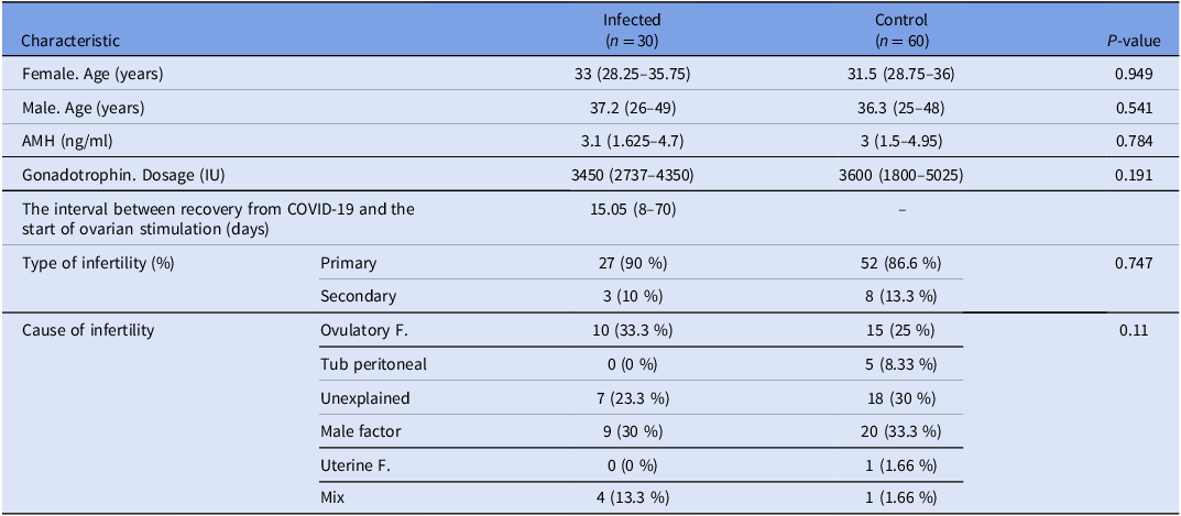
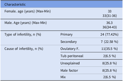
Following the application of exclusion criteria, 90 couples underwent ICSI in the COVID-19 infection phase, and 31 couples underwent ICSI in the vaccination phase. Patients in the COVID-19 infection phase were stratified into two groups based on their history of SARS-CoV-2 infection: the couples who both had a history of COVID-19 (n = 30) and a control group consisting of the couples who did not have any history of COVID-19 infection or vaccination (n = 60), as illustrated in Figure 1. Baseline patient characteristics in the infection phase are detailed in Table 1. There were no significant differences between the two groups of couples with a history of COVID-19 and the control group. Regarding female age, male age, anti-Müllerian hormone (AMH) and gonadotropin dosage (IU), the interval between COVID-19 and OPU (days), type, and cause of infertility. Table 2 provides the baseline characteristics of patients pre- and post-SARS-CoV-2 vaccination. The median of the interval time between vaccination and start of ovarian stimulation was 75 days (Min:30, Max:142). There were no significant differences in gonadotropin dosage (IU) in consecutive cycles (before and after vaccination) (P = 0.41).
Flowchart of the sampling process.
ICSI: intracytoplasmic sperm injection; OHSS: ovarian hyperstimulation syndrome; PGD: preimplantation genetic diagnosis; DOR: diminished ovarian reserve.
*Underlying diseases included: Chronic heart, lung, kidney, and liver disease, diabetes, obesity (BMI > 30 kg/m2), immunocompromising conditions or medications, and smoking.

Baseline characteristics of patients in the infected and control groups

Values are presented as the mean ± standard deviation, interquartile range (IQR), and number (percent).
* Statistical analyses were performed using the Mann–Whitney, chi-square tests, and Fisher’s exact test, with a significance threshold set at p < 0.05. AMH: anti-Müllerian hormone.
Baseline characteristics of couples who underwent SARS-CoV-2 vaccination (n = 31)
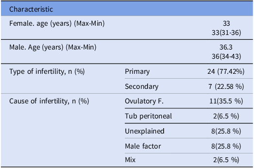
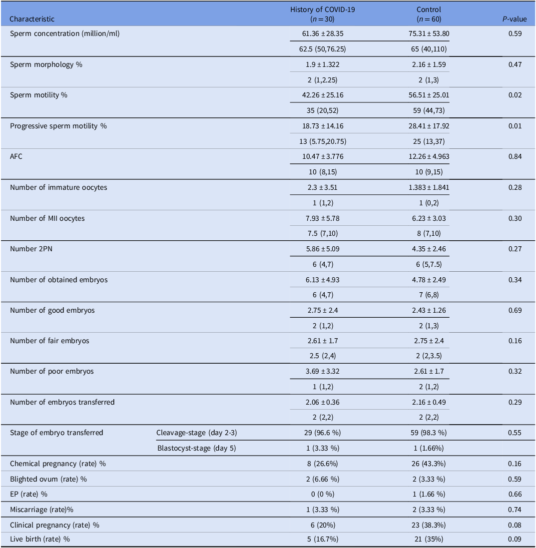
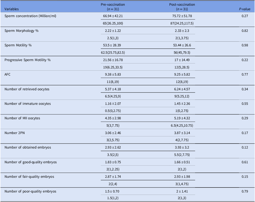
In this study, the variables including sperm concentration, sperm morphology, sperm motility, progressive sperm motility, antral follicle count (AFC) number, number of immature oocytes, number of metaphase II (MII) oocytes, number of two-pronuclei (2PN), number of good, fair and poor embryos, pregnancy outcomes, chemical pregnancy, blighted ovum, ectopic pregnancy (EP), clinical pregnancy, gestational sac, fetal heart rate (FHR), early miscarriage and birth rate were examined and compared between two groups: the control group (non-infected) and the COVID-19-infected group (Table 3). Additionally, all variables were assessed and compared in both groups before and after vaccination (Table 4).
Laboratory outcomes in couples with and without a history of SARS-CoV-2 infection

Values are presented as the mean ± standard deviation, interquartile range (IQR), and number (percent).
* It was obtained by an independent Mann–Whitney and chi-square tests, and the statistically significant level was 0.05. AFC, antral follicle counts; 2PN, two-pronuclei; EP, ectopic pregnancy; FHR, fetal heart rate.
Laboratory outcomes in vaccination group (pre- and post-SARS-CoV2 vaccination)

Values are presented as the mean ± Standard deviation, IQR (interquartile range), and number (percent).
*The results were obtained using the Mann-Whitney and the chi-square tests, with statistical significance defined as p < 0.05. AFC, antral follicle counts; MII oocytes, Metaphase 2 oocytes; 2PN, two-pronuclei.
Sperm parameters outcomes
The mean sperm concentration was lower in the COVID-19 history group (61.36 ± 28.35 million/ml) compared to the control group (75.31 ± 53.80 million/ml), though this difference was not statistically significant (p = 0.59). Similarly, sperm morphology showed a slight reduction in the COVID-19 group (1.9 ± 1.32%) compared to the control group (2.16 ± 1.59%), but this was not significant (p = 0.47). Notably, sperm motility and progressive motility were significantly lower in the COVID-19 group (42.26 ± 25.16%, p = 0.02; 18.73 ± 14.16%, p = 0.01, respectively) compared to the control group (56.51 ± 25.01%; 28.41 ± 17.92%). Comprehensive data on sperm parameters across infection groups are provided in Table 3.
The mean sperm concentration exhibited a modest increase from 66.94 ± 42.21 million/ml before vaccination to 75.72 ± 51.78 million/ml post-vaccination, though this difference was not statistically significant (p = 0.27). Similarly, sperm morphology showed minimal variation between the pre- and post-vaccination groups (2.22 ± 1.22% vs. 2.33 ± 2.3%; p = 0.82). Sperm motility remained virtually unchanged (53.5 ± 28.39% vs. 53.44 ± 26.6%; p = 0.98). Progressive motility demonstrated a non-significant decline following vaccination (21.56 ± 16.78% vs. 17 ± 14.49%; p = 0.22), as shown in Table 4.
Ovarian and embryological outcomes
Detailed ovarian stimulation and embryological outcomes are summarized in Table 3 for the infected and control groups. The AFC was slightly lower in the COVID-19 group (10.47 ± 3.77) compared to the control group (12.26 ± 4.96), but the difference was not statistically significant (p = 0.84). The mean number of immature oocytes was higher in the COVID-19 group (2.3 ± 3.51) compared to the control group (1.38 ± 1.84), though this difference was not significant (p = 0.28).
The number of MII oocytes and 2PN embryos was similar across groups, with no significant differences observed (MII: 7.93 ± 5.78 vs. 6.23 ± 3.03, p = 0.3; 2PN: 5.86 ± 5.09 vs. 4.35 ± 2.46, p = 0.27). Additionally, the total number of embryos, good-quality embryos and poor-quality embryos did not significantly differ between the two groups (p > 0.05). The AFC showed no notable change between the pre- and post-vaccination groups (9.38 ± 5.83 vs. 9.25 ± 5.82; p = 0.77). Similarly, the mean number of oocytes retrieved increased slightly post-vaccination (5.37 ± 4.18 vs. 6.24 ± 4.57), but this difference was not statistically significant (p = 0.34).
An increase was observed in the number of immature oocytes (1.16 ± 2.07 vs. 1.45 ± 2.36; p = 0.55) and MII oocytes (4.35 ± 2.98 vs. 5.19 ± 4.32; p = 0.29) in the post-vaccination group. The number of 2PN embryos also increased after vaccination (3.06 ± 2.46 vs. 3.87 ± 3.14), though this difference was not statistically significant (p = 0.17). The total number of embryos increased from (2.93 ± 2.62) pre-vaccination to (3.93 ± 3.2) post-vaccination, but this increase was not statistically significant (p = 0.12). The mean number of good-quality embryos showed no notable change (1.83 ± 0.75 vs. 1.66 ± 0.51; p = 0.61). The number of fair-quality embryos remained consistent between the groups (2.87 ± 1.74 vs. 2.93 ± 1.98; p = 0.15), and finally, the number of poor-quality embryos slightly improved post-vaccination, but this difference was not statistically significant (1.5 ± 0.7 vs. 2 ± 1.41; p = 0.79) as presented in Table 4.
Pregnancy and live birth outcomes
Chemical pregnancy rates were lower in the COVID-19 group (26.6%) than in the control group (43.3%), but this difference was not statistically significant (p = 0.16). Similarly, the rates of blighted ovum (6.66% vs. 3.33%; p = 0.59), EP (0% vs. 1.66%; p = 0.66) and miscarriage (3.33% in both groups; p = 0.74) were comparable between the two groups. Although the clinical pregnancy and live birth rates in the COVID-19 group were lower than those in the control group, these differences were not statistically significant (20% vs. 38.3%; p = 0.08 and 16.7% vs. 35%; p = 0.09). Table 3 presents a comparative overview of pregnancy-related outcomes, including clinical pregnancy and live birth rates.
Discussion
The duration of the ongoing COVID-19 pandemic remains uncertain, but periodic surges of SARS-CoV-2 are anticipated in the future (Rageh et al., Reference Rageh, Farag, Behery, Badreldin and Ali2023). This study is among the initial efforts to explore the effects of previous COVID-19 exposure on ovarian, sperm and embryonic variables and pregnancy outcomes. It also assesses the impact of the inactivated COVID-19 vaccine (Sinopharm) on ICSI cycle outcomes. SARS-CoV-2 may affect female fertility through multiple pathways, such as invading ovarian and granulosa cells, potentially leading to reduced ovarian function and oocyte quality. Furthermore, the virus might damage endometrial epithelial cells, interfering with the initial stages of embryo implantation (Vaz-Silva et al., Reference Vaz-Silva, Carneiro, Ferreira, Pinheiro, Silva, Silva-Filho, Witz, Reis, Santos and Reis2009).
Beyond ACE2, TMPRSS2 is present in spermatogonia, Leydig cells and Sertoli cells, all of which may serve as entry points for the virus (Wang and Xu, Reference Wang and Xu2020). In COVID-19 patients, Sertoli cell vacuolation, detachment from the basement membrane and reductions in Leydig and spermatogenic cells have been observed (Bian, Reference Bian2020; Yang et al., Reference Yang, Chen, Huang, Zhong, Su, Chen, Cao, Ma, He, Li, Li, Zhou, Fan, Luo, Chang, Arkun, Zhou and Nie2020). Sertoli and Leydig cells are essential for seminiferous tubule homeostasis and spermatogenesis (Griswold, Reference Griswold1998), suggesting that COVID-19 could hinder sperm production. Sperm parameter analysis plays a crucial role in assessing male fertility, with reduced sperm parameters often indicating male infertility. Some studies, however, have shown no significant changes in sperm morphology following SARS-CoV-2 infection. In this study, no substantial reduction in sperm morphology was observed. In contrast, other studies reported notable declines in sperm morphology after severe COVID-19, likely linked to immune responses and systemic inflammation, which may cause cellular and tissue damage (Das et al., Reference Das, Mondal, Samanta, Chakraborty and Sengupta2022; Yang et al., Reference Yang, Chen, Huang, Zhong, Su, Chen, Cao, Ma, He, Li, Li, Zhou, Fan, Luo, Chang, Arkun, Zhou and Nie2020). The lack of significant changes in our study might be due to the mild nature of the cases, as severe cases are more likely to cause changes in sperm morphology. A retrospective analysis by Villani et al. of over 22,000 ART cycles found that fertilization rates were significantly lower in patients with abnormal sperm parameters compared to those with normal sperm parameters. Both progressive and post-capacitation motility were significant predictors of fertilization, pregnancy and live birth rates (Villani et al., Reference Villani, Morini, Spaggiari, Falbo, Melli, La Sala, Romeo, Simoni, Aguzzoli and Santi2022). Holtmann et al. observed reduced sperm concentration and motility in men with moderate COVID-19, suggesting that fever associated with the virus may alter semen parameters, with recovery taking up to three months (Bendayan and Boitrelle, Reference Bendayan and Boitrelle2020). Our study recorded an average interval of 106.2 days between recovery and the initiation of ICSI cycles, with a significant reduction in sperm motility and progressive motility (p = 0.02; p = 0.01). These findings indicate that a prolonged recovery period might be necessary for complete normalization of sperm parameters. A similar study noted a decrease in progressive motility from 59% to 0% a month after COVID-19 infection (Çiçek et al., Reference Çiçek, Ayaz, Unal, Onal, Arici, Yucel and Polat2021), which aligns with our findings of substantial reductions in sperm motility. Further studies with larger sample sizes and varying recovery periods are necessary to better understand sperm quality recovery.
Studies reported that SARS-CoV-2 may negatively impact both ovarian tissue and granulosa cells, in addition to a detrimental effect on endometrial epithelial cells (Li et al., Reference Li, Yin, Fang, Li, Chen, Wang, Hao, Wu, Duan, Wang, Cheng, Zhou, Zafar, Xiong, Li, Yang and Qiao2020). Basigin (BSG), a crucial receptor for SARS-CoV-2, facilitates the virus’s entry into host cells. BSG is found not only in the uterus but also in ovarian stromal and granulosa cells, which it might influence follicular development, corpus luteum formation and embryo implantation (Chang et al., Reference Chang, Ni, Ma, Xu, Kadomatsu, Muramatsu and Yang2004). The entry of SARS-CoV-2 into cells, requiring ACE2 and TMPRSS2 receptor expression, warrants the examination of oocytes during ART to determine if the virus can infect them and embryos. RNA sequencing databases confirm ACE2 expression in human ovarian tissue, suggesting its vulnerability to COVID-19 (Wilkins and Al-Inizi, Reference Wilkins and Al-Inizi2021). Kong et al. found high ACE2 expression in cumulus-oocyte complexes (COCs) but low or negligible TMPRSS2 expression. This suggests that cumulus cells might facilitate the entry of SARS-CoV-2 into oocytes (Kong et al., Reference Kong, Yan, Yuan, Liu, Chen, Yang, Chen, Song, Yan and Yan2021). Moreover, increased ACE2 and TMPRSS2 expression during oocyte maturation could elevate the risk of viral infection and transmission (Boudry et al., Reference Boudry, Essahib, Mateizel, Van de Velde, De Geyter, Piérard, Waelput, Uvin, Tournaye and De Vos2022). SARS-CoV-2 requires ACE2 and TMPRSS2 to effectively enter host cells (Hoffmann et al., Reference Hoffmann, Kleine-Weber, Schroeder, Krüger, Herrler, Erichsen, Schiergens, Herrler, Wu and Nitsche2020). In one study, AMH levels were compared between 14 women with mild COVID-19 and a control group of 118 women undergoing IVF. No significant differences in AMH levels were found between the two groups (Kolanska et al., Reference Kolanska, Hours, Jonquière, Mathieu d’Argent, Dabi, Dupont, Touboul, Antoine, Chabbert-Buffet and Daraï2021). Previous studies have not reported any adverse effects of the SARS-CoV-2 virus on human oocytes. Similarly, the present investigation found no significant difference in the number of mature and immature oocytes between individuals with a history of SARS-CoV-2 infection and those in the control group. It is plausible that assessing ICSI outcomes during the active phase of COVID-19 infection could have provided more precise insights into the virus’s impact on oocyte numbers. As recommended by Orvieto et al., delaying IVF treatment by at least three months post-recovery may optimize outcomes by enabling the selection of healthy oocytes not exposed to the infection (Orvieto, Segev-Zahav, et al., Reference Orvieto, Segev-Zahav and Aizer2021).
Some studies have found no effects of SARS-CoV-2 on fertilization rates, embryo numbers or grading (Li et al., Reference Li, Ding, Luo, Li, Liu and Ding2023; Wang et al., Reference Wang, Yang, Ren, Hu, Li, Long, Xi, Zhu and Jin2021). However, Li et al. (Reference Li, Yin, Fang, Li, Chen, Wang, Hao, Wu, Duan, Wang, Cheng, Zhou, Zafar, Xiong, Li, Yang and Qiao2020) observed lower blastocyst formation rates 31–60 days after infection, possibly due to heightened pro-inflammatory cytokines and oxidative stress caused by SARS-CoV-2 (Li et al., Reference Li, Ding, Luo, Li, Liu and Ding2023). Wang et al. (Reference Wang, Yang, Ren, Hu, Li, Long, Xi, Zhu and Jin2021) found no differences in biochemical or clinical pregnancy rates between individuals with mild or asymptomatic COVID-19 and controls; however, they did not report live birth outcomes (Wang et al., Reference Wang, Yang, Ren, Hu, Li, Long, Xi, Zhu and Jin2021). The present study showed lower biochemical pregnancy rates in the infected group than in controls, though the difference was not statistically significant. This may be attributed to the potential impact of SARS-CoV-2 on early embryo implantation. Clinical pregnancy rates were higher in non-infected patients (38.3%) compared to those previously exposed to SARS-CoV-2 (20%), though this difference was not statistically significant. This could be associated with the impact of SARS-CoV-2 on embryonic development. A multicenter cohort study involving 585 infertile couples found that those with SARS-CoV-2 infection had significantly lower rates of top-quality embryos, blastocysts and blastocyst formation than uninfected couples (Tian et al., Reference Tian, Li, Li, Zhao, Luo, Zhang, Mao, Zhang, Li, Tang, Zhang, Li, Zhang and Zhao2023). Thromboembolic complications in COVID-19 patients may also contribute to these findings (Spratt and Buchsbaum, Reference Spratt and Buchsbaum2020). Similar to our findings, Banker et al. (Reference Banker, Arora, Banker, Shah, Gupta and Shah2022) reported no statistically significant differences in live birth rates between ART outcomes observed before the pandemic and those following COVID-19 infection. (Banker et al., Reference Banker, Arora, Banker, Shah, Gupta and Shah2022). Additionally, a study in 2021 noted placental damage in asymptomatic or mildly symptomatic pregnant women with SARS-CoV-2 infections, raising concerns about pregnancy outcomes (Jaiswal et al., Reference Jaiswal, Puri, Agarwal, Singh, Yadav, Tiwary, Tayal and Vats2021). Stress from external disasters, such as earthquakes and terrorist attacks, has also been shown to negatively impact pregnant women’s mental health and pregnancy outcomes (Hübner et al., Reference Hübner, Wolfgang, Theis, Steber, Wiedenmann, Wöckel, Diessner, Hein, Gründahl, Kämmerer, Kittel-Schneider and Bartmann2022), with previous COVID-19 infections potentially adding further risks to pregnant individuals.
Although numerous COVID-19 vaccines have been developed, there is limited clinical evidence on their effects on human fertility (Han et al., Reference Han, Lee, Kim, Choo, Park, Lee, Choi, Jun, Rhee and Kim2022). Most studies have focused on mRNA vaccines, so this research investigates the impact of the inactivated COVID-19 vaccine on ART outcomes. Accumulating evidence indicates that mRNA SARS-CoV-2 vaccines, particularly BNT162b2 (Pfizer-BioNTec) and mRNA-1273 (Moderna), do not adversely affect human reproductive physiology(Chen et al., Reference Chen, Zhu, Dai, Hao, Luan, Guo, Meng and Zhang2021; Chen et al., Reference Chen, Zhang, Lin, Gong and Hocher2023). In women, post-vaccination assessments of ovarian reserve – such as AMH levels (Mohr-Sasson et al., Reference Mohr-Sasson, Haas, Abuhasira, Sivan, Doitch Amdurski, Dadon, Blumenfeld, Derazne, Hemi, Orvieto, Afek and Rabinovici2021) and analysis of follicular fluid, revealed no measurable alterations in follicle function or quality, suggesting that follicular function remains intact (Bentov et al., Reference Bentov, Beharier, Moav-Zafrir, Kabessa, Godin, Greenfield, Ketzinel-Gilad, Ash Broder, Holzer, Wolf, Oiknine-Djian, Barghouti, Goldman-Wohl, Yagel, Walfisch and Hersko Klement2021). Likewise, male reproductive evaluations demonstrate stable semen parameters, including sperm volume, concentration and motility, with no histological signs of testicular impairment following mRNA vaccination (Gonzalez et al., Reference Gonzalez, Nassau, Khodamoradi, Ibrahim, Blachman-Braun, Ory and Ramasamy2021). These findings collectively support the safety of mRNA vaccines in preserving gamete integrity. In ART, clinical studies have shown no adverse effects on blastulation and fertilization rate or embryo quality (Avraham et al., Reference Avraham, Kedem, Zur, Youngster, Yaakov, Yerushalmi, Gat, Gidoni, Hochberg, Baum, Hourvitz and Maman2022; Jacobs et al., Reference Jacobs, Summers, Sparks and Mejia2022). For instance, Orvieto et al. (Reference Orvieto, Noach-Hirsh, Segev-Zahav, Haas, Nahum and Aizer2021) reported no significant differences in IVF performance between vaccinated and unvaccinated individuals (Orvieto, Noach-Hirsh, et al., Reference Orvieto, Segev-Zahav and Aizer2021). These insights offer a robust comparative framework for interpreting the reproductive implications of inactivated COVID-19 vaccines, as investigated in the present study. In alignment with findings from mRNA COVID-19 vaccine studies, our investigation of the inactivated SARS-CoV-2 vaccine revealed no detrimental effects on ovarian function, sperm quality or embryonic development. However, due to limitations in statistical power, pregnancy outcomes could not be conclusively evaluated.
Previous studies indicate that inactivated COVID-19 vaccines do not affect serum AMH levels. For instance, Huang et al. reported no statistically significant differences in AMH concentrations among 474 women when comparing values before and after vaccination (Huang et al., Reference Huang, Guan, Tian, Xia, Xu, Wu, Huang, Chen, Fang, Xiong, Nie, Wang, Li, Zhao and Wu2023). Unfortunately, we did not report AMH levels pre- and post-vaccination due to data limitations. The average interval between vaccination and semen analysis in this study was 75.8 days, aligning with the natural spermatogenesis cycle (Gonzalez et al., Reference Gonzalez, Nassau, Khodamoradi, Ibrahim, Blachman-Braun, Ory and Ramasamy2021; Lifshitz et al., Reference Lifshitz, Haas, Lebovitz, Raviv, Orvieto and Aizer2022). Mild side effects reported in the vaccinated group were consistent with existing literature, and no cases of azoospermia were observed. Our findings suggest that the inactivated COVID-19 vaccine did not adversely affect sperm parameters, oocytes or embryo quality in subsequent ICSI cycles. No adverse effects of inactivated SARS-CoV-2 vaccines on ICSI outcomes have been reported. The moderate systemic inflammation induced by vaccines, with minimal effects on folliculogenesis and spermatogenesis, could explain the results (Orvieto, Segev-Zahav, et al., Reference Orvieto, Segev-Zahav and Aizer2021). Huang et al. compared vaccinated and unvaccinated individuals and found similar outcomes in oocyte retrieval, embryo quality and clinical pregnancy rates, though they did not assess sperm parameters(J. Huang et al., Reference Huang, Xia, Lin, Liu, Zhao, Xin, Ai, Cao, Zhang, Tian and Wu2022).
This study is the first to evaluate the effects of inactivated SARS-CoV-2 vaccines on sperm and ovarian parameters. Most existing research has focused on male and female reproductive health separately. A retrospective study on the inactivated SARS-CoV-2 vaccine’s impact on male fertility concluded that the vaccine does not impair male fertility (Dong et al., Reference Dong, Li, Li, Zhu, Wei, He, Cheng, Yang and Chen2023). Another study found no adverse effects on sperm parameters or embryo quality during IVF treatments (Xia et al., Reference Xia, Zhao, Hu, Fang and Wu2022). Research has also shown that inactivated COVID-19 vaccines do not impact female ovarian reserve (Huang et al., Reference Huang, Guan, Tian, Xia, Xu, Wu, Huang, Chen, Fang, Xiong, Nie, Wang, Li, Zhao and Wu2023), and no detrimental effects were observed on embryo ploidy during IVF treatments (Huang et al., Reference Huang, Xia, Tian, Fan, Xu, Ai, Wu, Chen, Xing, Huang, Zuo, Chen, Li, Zhang, Liu, Lin and Wu2022). While these studies provide reassuring insights into the safety of inactivated vaccines, there has been little comprehensive research assessing the effects of inactivated SARS-CoV-2 vaccines on both male and female reproductive health simultaneously. Further investigations are needed to gain a holistic understanding of the vaccine’s effects on reproductive health across genders. A key strength of our study lies in its comprehensive evaluation of ovarian, sperm and embryo parameters before and after inactivated SARS-CoV-2 vaccination during ICSI cycles.
Conclusion
Based on the available evidence, the study concludes that COVID-19 infection, even in mild cases, may negatively impact male reproductive parameters, particularly sperm and progressive motility. These changes suggest the need for careful planning of ICSI cycles post-recovery. No significant adverse effects were observed on ovarian function, oocyte quality or embryonic development.
Under the methodological constraints of the present study, the inactivated Sinopharm vaccine demonstrated no statistically significant adverse effects on sperm parameters, ovarian function or embryonic development during ICSI cycles. These findings support its safety and utility as a protective measure against severe COVID-19, particularly in the context of ART treatments. Couples undergoing ART can confidently proceed with vaccination without concerns of adverse effects on fertility outcomes.
Availability of data and materials
The datasets used or analysed during the current study are available from the corresponding authors on reasonable request.
Acknowledgements
We express our heartfelt gratitude to all participants and contributors to this study for their invaluable support and cooperation.
Author contributions
F.GH. designed the research. F.SA., A.M., B.E. and A.A. contributed to patient selection, data collection, interpretation of data and manuscript writing/editing. F.SA. and F.GH. wrote the manuscript. Z.Z. performed the analysis of the data. All authors read and approved the final manuscript.
Funding
The study didn’t have any funding support.
Competing interests
All authors have nothing to disclose.
Ethics approval and consent to participate
The Institutional Review Boards and the Ethics Committees of Royan Institute approved this study (Ethics approval number: IR.ACECR.ROYAN.REC.1401.029). All procedures conducted in this study involving human participants adhered to the ethical standards of the 1964 Helsinki Declaration and its later amendments. The eligible patients signed written informed consent forms before participation in the study.
Consent for publication
Not applicable.
Declaration of generative AI (Artificial Intelligence) and AI-assisted technologies in the writing process
During the preparation of this article, the authors utilized Grammarly (an AI writing tool) to enhance the language, improving grammar and revising sentence structure. After using this service, the authors reviewed and edited the content as necessary and take full responsibility for the final publication.