Atrial septal defect is one of the most commonly recognised CHDs that may present in adulthood.Reference Hoffman and Kaplan1 Over the years, percutaneous device closure using femoral venous access has been accepted as an effective treatment for patients with secundum atrial septal defect.Reference Du, Hijazi, Kleinman, Silverman and Larntz2 In patients with bilateral femoral venous occlusion or interrupted inferior caval vein, transfemoral approach becomes impossible. Alternative percutaneous approaches include trans-hepatic and trans-jugular access.Reference Narin, Pamukcu, Baykan, Argun, Ozyurt and Uzum3,Reference Singh, Neuzil and Skoka4 However, trans-hepatic access carries a significant risk of haemorrhage when using large introducer sheaths necessary for large devices, and trans-jugular access imposes significant technical difficulties concerning orientation of device during deployment particularly with large defects.
Hybrid closure of atrial septal defect using a small sub-mammary incision is a reasonable alternative for patients with interrupted inferior caval vein.Reference Sheth, Jain, Joshi and Dalvi5
Case report
The patient is a 31-year-old male who presented with palpitation and exercise intolerance and was found to have a large secundum atrial septal defect with a maximum diameter of 28 mm. During a planned cardiac catheterisation for atrial septal defect closure, the patient was also found to have interrupted inferior caval vein, precluding percutaneous closure by femoral access. Trans-oesophageal echocardiography showed a large defect measuring 28 mm × 23 mm, with deficient anterior rim. Planning for per-atrial closure was undertaken and explained to the patient; the patient’s consent was obtained to perform the procedure.
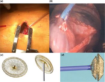
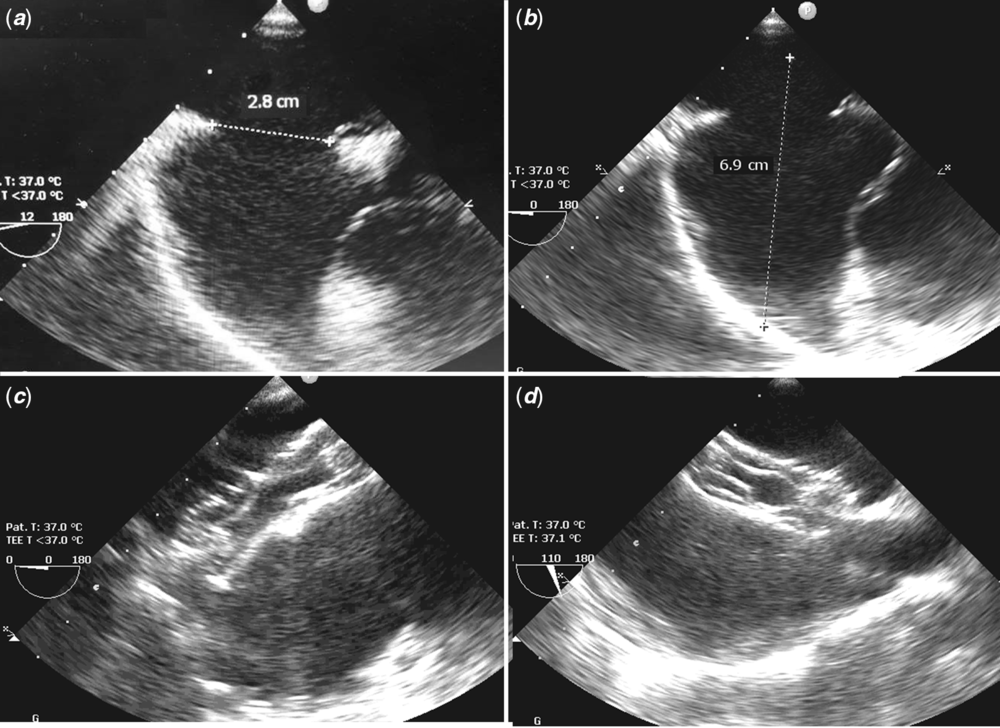
The procedure was done in the operating theatre, with trans-oesophageal guidance. Under general anaesthesia, and single left lung ventilation, a right sub-mammary, 3-cm long transverse incision was made along the fourth intercostal space (Fig 1). The pericardium was opened, and exposure of the right atrial wall was established. Double purse-string sutures were placed. A small stab puncture in the anterolateral wall of the right atrium, 2 cm above the atrioventricular junction, was done to access the right atrium. A 33-mm Occlutech Figula Flex II device (Occlutech, La Cours Gata 2, Helsingborg, Sweden) was preloaded into a 30-cm long, 12-French sheath (Cook Medical, Bloomington, IN, United States of America). The sheath-device assembly was introduced through the puncture hole in the anterolateral wall of the right atrium, while securing haemostasis by the purse-string sutures. The approximate length of the sheath to be introduced was pre-determined by measuring the distance between the mid-left atrial cavity and the free wall of the right atrium (Fig 2). With trans-oesophageal guidance, the assembly was introduced in postero-leftward direction until the tip of the sheath was seen into the left atrial cavity. The left atrial disc was deployed in the usual manner, and the assembly was pulled back till the device was in parallel with the interatrial septum, then the right atrial disc was deployed. After confirming correct position of the device by trans-oesophageal imaging, the device was released, and the sheath was pulled out. The atrial puncture site was secured by tightening the purse-string suture. A small drain was placed, and the chest wall was closed without complication.
(a) A small right, inframammary incision to obtain access to the right atrial wall. (b) Image through endoscope camera showing the delivery sheath (blue) inserted through the right atrial free wall for closure of the atrial septal defect. (c and d) Photographs of the Occlutech Figula Flex II device, free, attached by cable, and partially introduced within the delivery sheath.

Intraoperative trans-oesophageal echocardiographic images during hybrid closure of a large atrial septal defect in a patient with interrupted inferior vena cava: (a) measurement of the atrial septal defect, (b) measurement between the atrial free wall to the mid-cavity of the left atrium to estimate the length of the sheath to be introduced, (c and d) following deployment of a 33-mm Occlutech septal occluder, with device in good position.

The patient was discharged from the hospital 48 hours after the procedure. Follow-up was done after 1 week and 1 month. The patient was in sinus rhythm, and the device was in good position with no residual shunting. The patient expressed significant improvement of his exercise tolerance.
Discussion
Interrupted inferior caval vein is a rare anomaly that is present in 0.6% of patients with CHD.Reference Anderson, Adams and Burke6 In the setting of atrial septal defect, this anomaly precludes percutaneous closure via femoral vein approach.
The available options for our patient were to undergo surgical closure of atrial septal defect with the use of cardiopulmonary bypass, or device closure using trans-jugular, trans-hepatic, or per-atrial approach.
Surgical closure of atrial septal defect is accomplished with success rates like that of percutaneous device closure. However, complications of cardiopulmonary bypass, longer hospital stay, and physical and psychological distress due to the sternotomy scar of surgical repair make it a less favourable choice for patients.Reference Du, Hijazi, Kleinman, Silverman and Larntz2,Reference Huffmyer and Groves7
As the defect was large, measuring 28 × 23 mm, a 12-French sheath would be required to insert the device. A trans-hepatic approach using a large sheath carries significant risks, such as haemorrhage, hepatic vein thrombosis, and liver and biliary system injury.Reference Singh, Neuzil and Skoka4,Reference Hoevels, Lunderquist and Owman8 Internal jugular vein approach was believed to result in technical difficulties during deployment of a large device, as the orientation of the device during deployment will be perpendicular to the atrial septum. In addition, a large sheath may result in vascular neck injury. Taking all these reasons into consideration, a hybrid approach through a small sub-mammary incision without the use of cardiopulmonary bypass was chosen.
For large atrial septal defects, self-centring devices have been the only available and widely spread options for transcatheter closure. Commonly used devices are Amplatzer septal occluders (Abbot, Abbot Park, IL, United States of America), Lifetech septal occluders (Lifetech Scientific, Nanshan District, Shenzhen, China), Occlutech septal occluders, among few others. The technique for deployment of these devices is generally similar. At our institution, we commonly use Occlutech devices as in this patient, but we believe any other self-centring device could have been used with success.
The rationale behind the use of the hybrid approach over surgical approach was the many advantages it offers: a shorter hospital stay, shorter time for recovery, the pleasant cosmetic result and, most importantly, the avoidance of cardiopulmonary bypass.Reference Sheth, Jain, Joshi and Dalvi5,Reference Hongxin, Wenbin and Lijun9
Conclusion
In patients with large atrial septal defect and interrupted inferior caval vein, transcatheter device closure can successfully be accomplished using a minimally invasive per-atrial hybrid approach.
Acknowledgements
None.
Financial support
This research received no specific grant from any funding agency, commercial or not-for-profit sectors.
Conflict of interest
None.
Ethical Standards
This report does not involve human or animal experimentation.