A small number of phenotypically specific, congenitally malformed hearts account for the majority of congenital cardiac diseases seen in childhood. With sound training and experience, the paediatric echocardiographer gains familiarity with these more common lesions. More challenging are the very rare, but often life-threatening, conditions that might be encountered only a handful of times throughout an individual’s career. Owing to their rare nature, these lesions are very often missed or misdiagnosed, even by those working in busy centres dealing with congenital cardiac disease. It is important, therefore, that paediatric echocardiographers be able to identify, and to image adequately, these rare conditions.
Those rare lesions that can result in significant haemodynamic impairment, but which fortunately are amenable to complete anatomic repair, include the aortopulmonary window, the aorto-ventricular tunnel, and aneurysm of the aortic sinuses of Valsalva. All of these lesions may be confused with a number of more common congenital cardiac malformations, or may occur in association with other lesions, which may mask the characteristic findings. In this review, we highlight the key anatomic details that permit the echocardiographer to make unequivocal diagnoses and plan appropriate surgical treatment.
Aortopulmonary window
In the current era, patients with aortopulmonary windows are usually diagnosed echocardiographically. Despite the windows often being of considerable size, and producing significant haemodynamic changes, they are commonly misdiagnosed or overlooked, even in experienced hands.Reference Kiran, Singh, Shah, John and Maheshwari1 Misdiagnosis may result from an overemphasis on the use of colour Doppler interrogation to identify those lesions associated with shunting of blood. In addition, up to half of the patients with aortopulmonary windows also have other congenital cardiac lesions.Reference van Son, Puga and Danielson2 The tendency to focus on the associated lesion may lead to one’s overlooking the anatomic features of the window. When using systematic principles of segmental imaging, nonetheless, the windows should be identified in all cases.
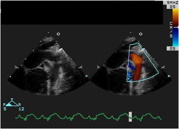
A number of inferential findings can alert the echocardiographer to the presence of an aortopulmonary window. The physiology is typically that of a large left-to-right shunt.Reference Alboliras, Chin, Barber, Helton and Pigott3 As such, one typically notes enlargement of the left heart chambers, an increased pulmonary venous return, and increased flow across the mitral and aortic valves. When the window is an isolated lesion, there are characteristic physiologic features that are evident by cross-sectional, pulsed Doppler, and colour Doppler interrogation of the great arterial trunks. Owing to the large quantity of flow from the aorta to the pulmonary trunk in diastole, the thoracic aorta has prominent pulsatility. This feature can be appreciated by suprasternal imaging of the aortic arch, as well as with subcostal costal imaging, the latter typically demonstrating a very pulsatile aorta at the level of the diaphragm. Cross-sectional imaging may also show enlarged right and left pulmonary arteries, since the ratio of pulmonary to systemic flow across the window usually exceeds 3 to 1.Reference Rice, Seward, Hagler, Mair and Tajik4 Doppler interrogation also provides many important clues to assist in diagnosis. Owing to the extensive shunt, there is considerable reversal of diastolic flow in the descending aorta (Fig 1).Reference Horimi, Hasegawa, Shiraishi, Endo and Yanagisawa5 This same feature may be present in the setting of a large patent arterial duct, and therefore it is also important to establish the reversal of flow in the transverse aortic arch and ascending aorta. Aortic insufficiency, nonetheless, can also produce such reversal of flow. Careful assessment of the function of the aortic valve, therefore, is always warranted. Aorto-ventricular tunnels, and aneurysms of the aortic sinuses of Valsalva, to be discussed below, can similarly produce the marked reversal of flow in the ascending aorta.
Suprasternal sagittal image of the thoracic aorta shows a significant quantity of retrograde flow in the transverse aortic arch in the setting of an aortopulmonary window.

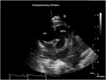
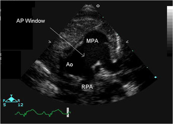
These inferential findings might suggest an aortopulmonary window, and therefore the diagnosis rests upon careful cross-sectional imaging of the intrapericardial arterial trunks. The defect is usually large, and pressure in the pulmonary arteries is typically systemic, and therefore the shunt will typically not be turbulent, and hence can be challenging to identify with colour Doppler interrogation.Reference Kiran, Singh, Shah, John and Maheshwari1 The key to this echocardiographic diagnosis, therefore, is recognition of the deficiency between the adjacent walls of the arterial trunks. The window is most commonly located near the origin of the right pulmonary artery (Fig 2). Hence, parasternal imaging typically provides the most valuable plane with which to confirm the diagnosis. Numerous additional significant lesions can occur in association with the windows. Owing to this, methodical segmental diagnosis is essential. Important variants, such as interruption of the aortic arch, or anomalous origin of one of the pulmonary arteries (Fig 3), will impact on the planned surgical treatment.
Parasternal short-axis image shows the typical location of aortopulmonary windows.

This parasternal short-axis image shows an aortopulmonary window in which the right pulmonary artery arises from the aorta.

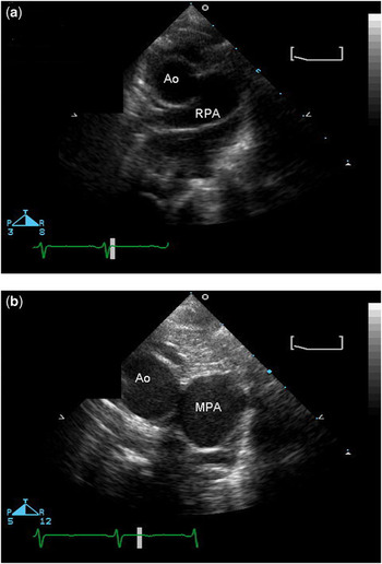
As already emphasised, the key diagnostic feature is the deficiency in the adjacent walls of the intrapericardial arterial trunks. This must be distinguished from the artefactual dropout that may occur when the ultrasonic beam in parallel with the tissue plane usually interposed between the arterial trunks (Figs 4a and b). Specular reflections are maximised when interfaces between tissues lie perpendicular to the ultrasonic beam. Hence, the artefact is overcome by repositioning the ultrasound to a more favourable point of vantage. Another example of this phenomenon is the impaired ability to visualise thin linear structures, such as the tendinous cords of the mitral valve, when they are parallel to the ultrasonic beam, as in the five-chamber view. When these structures are perpendicular to the beam, as in the long-axis view, they are easily visualised. As such, subcostal imaging often provides an excellent alternative plane of imaging with which to identify the aortopulmonary window.
The parasternal short-axis view (a) shows an artefactual dropout, which may be mistaken for an aortopulmonary window. Better delineation (b) shows the tissue plane interposing between the walls of the aorta and pulmonary trunk, confirming the absence of a window.

Imaging is now supplemented by the use of three-dimensional echocardiography, although this technique is seldom used currently for purposes of surveillance, practitioners not usually using the three-dimensional technique to identify those lesions readily recognised using conventional echocardiography. The three-dimensional format, nonetheless, can provide more robust spatial orientation, and assist the surgeon in planning operative repair.Reference Singh, Mehmood, Romp, Nanda and Mallavarapu6
Aorto-ventricular tunnels
Most tunnels originate from the left ventricle, but even that lesion is extremely rare, estimated to occur in approximately one of each 300,000 live births. Even experienced echocardiographers are likely to see fewer than five such cases in their careers. The haemodynamic features are such that the lesion is usually readily apparent. It may be misdiagnosed at first not only because of its rarity, but also because of the overlap with other entities, such as severe aortic valvar insufficiency.
As with the aortopulmonary window, there are a number of physiologic features that are readily identified using conventional cross-sectional and Doppler techniques. The tunnel essentially provides unrestricted diastolic flow from the ascending aorta to the left ventricular cavity.Reference Jureidini, de, Nouri and Kanter7 As with the window, therefore, the thoracic aorta shows prominent pulsatility. Reversal of diastolic flow can be appreciated at the level of the diaphragm when using subcostal planes of imaging. The left ventricle is dilated as a compensatory mechanism, and secondary mitral insufficiency may occur.
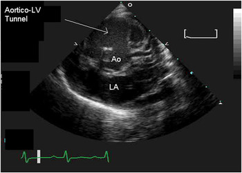
The key feature distinguishing the tunnel from aortic insufficiency or ruptured aneurysm of an aortic sinus of Valsalva relates to the pathway of flow of blood from the aorta to the left ventricle. Most frequently, the aortic opening for the tunnel is within the right coronary aortic sinus of Valsalva (Fig 5),Reference Cooley, Harris and Rodin8 the tunnel then typically communicating with the left ventricle beneath the zone of apposition between the leaflets of the right and left coronary aortic sinuses.Reference Perez-Martinez, Quero, Castro, Moreno, Brito and Merino9 Less commonly, the tunnel may have its aortic origin in relation to the left coronary aortic sinus, in which case the pathway to the left ventricle tends to be longer.
The parasternal short-axis view shows an aorto-left ventricular tunnel bypassing the right coronary aortic leaflet.

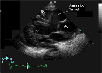
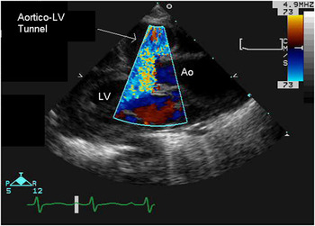
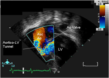
It is the parasternal long-axis plane that tends to provide the clearest anatomic picture of the tunnel (Fig 6). In this view, the right coronary aortic leaflet can be seen to be separated from its anticipated hinge point,Reference Wu, Huang, Chen, Lin and Roan10 producing the potential to mistake the tunnel for a sizeable perimembranous ventricular septal defect. Colour Doppler interrogation, however, shows the significant quantity of diastolic flow into the left ventricle (Fig 7), with lack of shunting into the right ventricle. Cross-sectional imaging also shows the membranous septum to be intact. The diameter of the tunnel often exceeds the diameter of the aortic outflow tract (Fig 8). In the parasternal short-axis view, the tunnel is seen to bypass the hinge of the aortic valvar leaflet. Apical or subcostal imaging can also show important features of the tunnel. In an apical imaging plane, with the transducer angled anteriorly towards the left ventricular outflow tract, the tunnel is seen to be parallel to the native outflow tract. Colour Doppler interrogation from this plane shows two parallel passages of blood flow in systole, with significant diastolic flow from the aorta to the left ventricle. As with the aortopulmonary window, three-dimensional echocardiography can provide supplementary images to assist in reconstruction of the lesion and planning of surgery.
The parasternal long-axis view shows the relationship of the aorto-left ventricular tunnel to the leaflet of the aortic valve. Note the intact membranous septum.

The parasternal long-axis view shows significant retrograde flow through a tunnel connecting the aorta and the left ventricle.

The apical image shows the reversal of flow through the tunnel running from the ascending aorta to the left ventricle. The flow bypasses the detached hinge of the aortic valvar leaflet.

Tunnels extending to the right heart are less common entities. Such tunnels still originate just above either the right or left aortic coronary sinuses, but communicate with either the right ventricle or right atrium. When running to the atrium, the tunnel generally terminates in the atrial roof, sometimes near the junction with the mouth of the superior caval vein. When the tunnel originates in relation to the left aortic sinus of Valsalva, the tunnel courses posterior to the ascending aorta before emptying into the right atrium.Reference Rosenberg, Williams and Trusler11 When the tunnel arises from the right aortic sinus of Valsalva, it takes an anterior course relative to the ascending aorta before joining the right atrium.
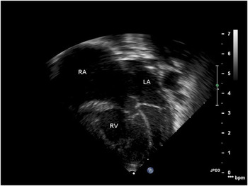
Some of the physiologic features of the atrial tunnels overlap with those connecting the aorta with the left ventricle. Thus, imaging of the aortic arch, either from suprasternal or subcostal planes, shows a very pulsatile aorta with diastolic reversal of flow into the root of the aorta.Reference Kursaklioglu, Iyisoy, Celik, Kose, Amasyali and Isik12 When the tunnel connects with the right atrium, the left-to-right shunting tends to result in dilation of all the cardiac chambers, though dilation of the right atrium is most prominent (Fig 9). In addition, shunting tends to occur throughout the cardiac cycle, since aortic pressure remains higher than right atrial pressure in both diastole and systole.
The apical four-chamber view shows marked dilation of the right atrium in a child with an aorto-right atrial tunnel.

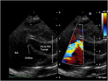
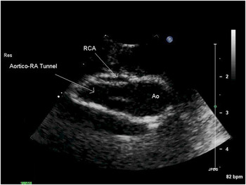
The anatomy of the atrial tunnel is best defined in the parasternal short-axis view. In many cases, the entirety of the tunnel can be shown in a single plane, with the tunnel often tapering near its junction with the right atrium (Fig 10). Colour Doppler interrogation may show flow acceleration and turbulence in this area. As the differential diagnosis includes coronary-cameral fistulas, it is important to define coronary arterial anatomy, as is also the case for those tunnels joining the left ventricle. A large coronary-cameral fistula to the right atrium can yield very similar haemodynamic findings, however, to those produced by the aorto-right atrial tunnels. In the setting of the tunnel, nonetheless, the coronary arteries maintain their anticipated course and dimensions, and the fistula can be distinguished from the tunnel by both cross-sectional and colour Doppler imaging (Fig 11).
The parasternal short-axis image showing Doppler colour interrogation reveals acceleration of flow at the orifice of an aorto-right atrial tunnel joining the roof of the right atrium.

The parasternal short-axis view shows a separate origin of the right coronary artery in the setting of an aorto-right atrial tunnel.

Tunnels connecting the aorta with the right ventricle are even rarer than those joining the right atrium.Reference Bharati, Lev and Cassels13, Reference Vargas, Molina, Martinez, Ranzini and Vazquez14 Many of the features of the right ventricular tunnels are similar to those already described above. The right ventricular tunnel is typically found in relation to the right aortic coronary sinus, and joins the subpulmonary infundibulum. The lesion can be identified in both the parasternal long-axis and short-axis imaging planes.
Aneurysms of the aortic sinuses of Valsalva
Aneurysms involving the aortic sinuses of the Valsalva aneurysm are the most common of the lesions discussed in this review. Unlike the lesions described thus far, however, the aneurysms may either be congenital or acquired, and may not result in any physiologic perturbations or symptoms. The aneurysms are prone to sudden rupture, and it is this event that may produce sudden haemodynamic changes. Owing to these features, the aneurysms are usually not detected in childhood. Median age at presentation has been reported to be in the fourth or fifth decade of life. The aneurysms may occur in isolation, or in association with congenital cardiac malformations,Reference Nakamura, Suzuki and Satomi15 typically in the setting of perimembranous ventricular septal defects.Reference Lewis and Agathangelou16
Diagnosis can be confirmed in nearly all cases by echocardiography, although the subtle anatomical features mean that, on occasion, the diagnosis is only made with certainty by surgical inspection. As the name suggests, the defect represents an aneurysm, localised or diffuse, of the sinus of Valsalva, which typically protrudes or ruptures into the right ventricle or right atrium. By definition, the aneurysm is distal to the semilunar origin of the aortic valvar leaflets, permitting the distinction of ruptured aneurysms from the aorto-atrial or ventricular tunnels. The tunnels typically are elongated, whereas a ruptured aneurysm tends to have a short course.
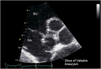
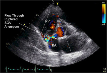
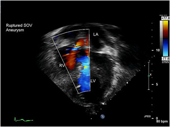
The haemodynamic features of the aneurysms depend on whether or not there has been a rupture. If a rupture has occurred, the same features are seen as for all the other entities described in this review, notably a dilated and pulsatile thoracic aorta with a significant diastolic reversal of flow. The cross-sectional echocardiographic features obviously include the aneurysmal dilation of the involved aortic valvar sinus, which may assume the appearance of a windsock (Fig 12).Reference Hands, Lloyd and Hung17 When the rupture is to the right ventricle, there may also be an end-diastolic opening of the pulmonary valve, indicating a progressive rise of right ventricular diastolic pressure. Features seen on Doppler interrogation include continuous unidirectional flow through the lesion at high velocity (Fig 13). The diagnosis is usually confirmed on colour flow imaging, which reveals a unidirectional and continuous mosaic jet from the aorta to the right atrium or ventricle (Fig 14). These features may be seen more clearly on transoesophageal echocardiography.
The parasternal short-axis view shows an aneurysm of the non-coronary aortic sinus of Valsalva.

The parasternal short-axis view shows a ruptured aneurysm of an aortic sinus of Valsalva, with flow into the right atrium.

The apical image shows systolic flow into the right ventricle secondary to rupture of an aortic sinus of Valsalva.

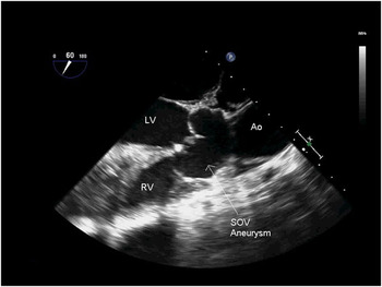
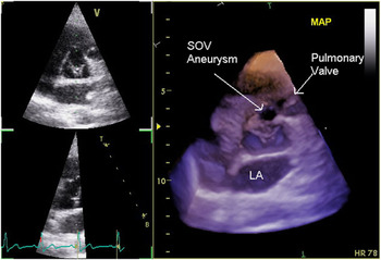
When the aneurysm occurs in association with a ventricular septal defect, it may produce features that can be confused with prolapse of an aortic valvar leaflet. In the latter setting, typically involving the right or left aortic coronary sinus, the leaflet becomes elongated and distorted, often producing significant aortic insufficiency. As discussed, when the sinus of Valsalva is aneurysmal, then of necessity the lesion is distal to the hinge of the aortic valvar leaflet (Fig 15), although such distinction can be challenging for echocardiographic confirmation in isolation. Several additional echocardiographic features, nonetheless, help to distinguish the aneurysm from a prolapsed valvar leaflet. The aneurysm extends from the sinutubular junction to the hinge of the involved valvar leaflet, and is often elongated, with a length exceeding 1 centimetre. If ruptured, there can be a significant quantity of diastolic flow from the aorta to the right ventricle. Prolapse of a valvar leaflet can also result in significant aortic valvar insufficiency, and therefore the diastolic jet in this setting emanates from the central portion of the involved leaflet, and is often directed towards the aortic leaflet of the mitral valve as the right coronary aortic leaflet is pulled into the ventricular septal defect. Three-dimensional echocardiography can more clearly delineate this anatomy in some cases (Fig 16).
Transoesophageal imaging shows an aneurysmal aortic sinus of Valsalva ruptured to the right ventricle in a patient with a doubly committed and juxtaarterial ventricular septal defect.

A three-dimensional echocardiographic image of aneurysmal aortic sinus of Valsalva.

Transoesophageal echocardiography supplements the transthoracic approach. Imaging in the long-axis plane provides an excellent definition of the relationship of the aneurysm to the right ventricle, while short-axis imaging can show the relationship of the aneurysm to the aortic valvar leaflets and the coronary arteries.