Introduction
Trichinellosis is an important zoonotic disease caused by the infectious nematodes of the genus Trichinella (Fu et al., Reference Fu, Liu, Yao, Li, Li, Wanga, Wub, Zhang, Cai, Blaga and Boireau2009; Gottstein et al., Reference Gottstein, Pozio and Nöckler2009; Pozio et al., Reference Pozio, Hoberg, La Rosa and Zarlenga2009; Krivokapich et al., Reference Krivokapich, Pozio, Gatti, Gonzales Prous, Ribicich, Marucci, La Rosa and Confalonieri2012). The parasite belongs to the family Trichinellidae, phylum Nematoda, and there are currently nine encapsulated species and genotypes, namely Trichinella britovi, T. murrelli, T. nativa, T. spiralis, T. nelsoni, T. patagoniensis, Trichinella T6, Trichinella T8 and Trichinella T9, with three additional non-encapsulated species: T. pseudospiralis, T. zimbabwensis and T. papuae (Pozio & Zarlenga, Reference Pozio and Zarlenga2013).
Trichinella spp. have a direct life cycle characterized by completing both intermediary and definitive stages in a single host (Pozio, Reference Pozio2007; Gottstein et al., Reference Gottstein, Pozio and Nöckler2009; Pozio et al., Reference Pozio, Hoberg, La Rosa and Zarlenga2009). Unlike other nematodes, Trichinella spp. are also characterized by an infective first larval stage (L1) in contrast to the typical infective third-stage larvae (L3) found in most nematode genera (Gajadhar et al., Reference Gajadhar, Pozio, Gamble, Nöckler, Maddox-Hyttel, Forbes, Vallee, Rossi, Marinculic and Boireau2009; Pozio et al., Reference Pozio, Hoberg, La Rosa and Zarlenga2009). Vertebrates, including humans, are infected through the ingestion of raw or undercooked meat infected with Trichinella larvae in the muscle tissue (Gottstein et al., Reference Gottstein, Pozio and Nöckler2009). Newborn larvae (NBL) are transported passively to the striated muscles (Dupouy-Camet, Reference Dupouy-Camet2000; Gottstein et al., Reference Gottstein, Pozio and Nöckler2009) via the host lymphatic and blood vessels (Gottstein et al., Reference Gottstein, Pozio and Nöckler2009). Based on the findings by Gottstein et al. (Reference Gottstein, Pozio and Nöckler2009), Trichinella larvae may remain viable in the nurse muscle for many years after encysting. This however, may depend on the Trichinella species/genotype and the host's immune response.
Cases of trichinellosis have been reported worldwide with the exception of Antarctica (Pozio et al., Reference Pozio, Hoberg, La Rosa and Zarlenga2009; Mukaratirwa et al., Reference Mukaratirwa, La Grange and Pfukenyi2013). Trichinella zimbabwensis, T. britovi, T. nelsoni and genotype T8 have been reported in sub-Saharan Africa and occur mainly in carnivorous and omnivorous sylvatics with T. zimbabwensis being the most prevalent species (Mukaratirwa et al., Reference Mukaratirwa, La Grange and Pfukenyi2013). According to Blaga et al. (Reference Blaga, Gherman, Cozma, Zocevic, Pozio and Boireau2009), 10,000 cases of human trichinellosis have been reported globally, with an annual mortality rate as low as 0.2% (Pozio, Reference Pozio2007). Generally, animals remain asymptomatic and, in the absence of clinical disease manifestation, infection with Trichinella spp. is referred to as Trichinella infection rather than trichinellosis (Gottstein et al., Reference Gottstein, Pozio and Nöckler2009).
Trichinella zimbabwensis, T. nelsoni and Trichinella T8 have been reported in wildlife from South Africa during epidemiological investigations in the Greater Kruger National Park (GKNP) and adjacent areas of the Limpopo and Mpumalanga provinces (Marucci et al., Reference Marucci, La Grange, La Rosa and Pozio2009; La Grange et al., Reference La Grange, Marucci and Pozio2010; Mukaratirwa et al., Reference Mukaratirwa, La Grange and Pfukenyi2013; La Grange et al., Reference La Grange, Reininghaus and Mukaratirwa2014). The main reservoirs for Trichinella spp. in the Kruger National Park are carnivorous wildlife with scavenging and cannibalistic behaviour (Mukaratirwa et al., Reference Mukaratirwa, La Grange and Pfukenyi2013). The spotted hyaena (Crocuta crocuta) and the lion (Panthera leo) appear to be the major reservoirs for Trichinella infections as they currently have the highest documented prevalence in South Africa (Marucci et al., Reference Marucci, La Grange, La Rosa and Pozio2009; Mukaratirwa et al., Reference Mukaratirwa, La Grange and Pfukenyi2013). Mixed infections of T. nelsoni and Trichinella T8 have been reported in both a lion and a leopard (Marucci et al., Reference Marucci, La Grange, La Rosa and Pozio2009; La Grange et al., Reference La Grange, Reininghaus and Mukaratirwa2014) and T. zimbabwensis has also been reported in a lion (La Grange et al., Reference La Grange, Marucci and Pozio2010). To date, no report of human infections or cases involving domestic animals have been documented in South Africa, despite this country having the highest documented prevalence of Trichinella infections in wildlife in sub-Saharan Africa (Mukaratirwa et al., Reference Mukaratirwa, La Grange and Pfukenyi2013).
There is paucity of information on Trichinella infections in humans, domestic and wild animals in most of sub-Saharan Africa (Dupouy-Camet, Reference Dupouy-Camet2000; Pozio, Reference Pozio2007) including South Africa. Hence, the aim of this study was to close the gap by determining the prevalence of Trichinella infection in convenience samples collected from wildlife species from the GKNP and identifying the Trichinella spp. larvae isolates using molecular techniques.
Materials and methods
Sample collection and processing
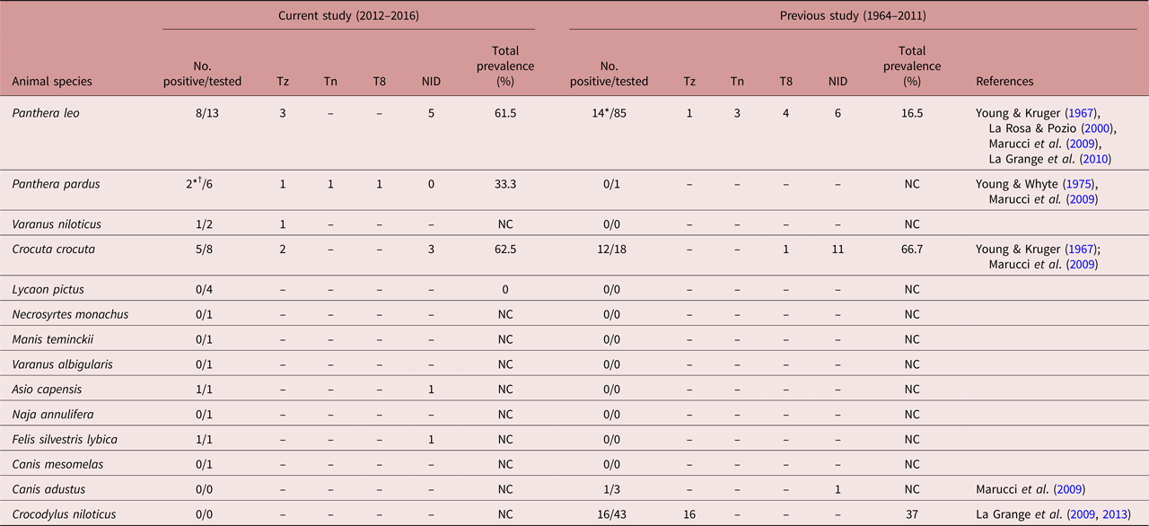
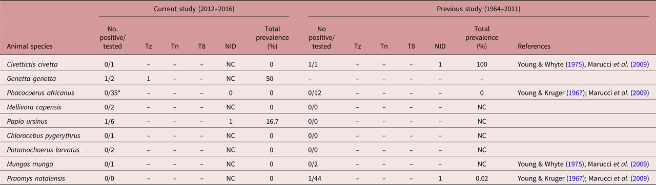
Muscle samples were conveniently collected from carcasses of wildlife, either culled from nature reserves, killed by hunters or animals that died of natural causes or as a result of vehicular accidents and/or poisoning in the GKNP, private nature reserves and towns neighbouring the GKNP in Limpopo and Mpumalanga provinces. Ninety samples representing 15 mammal, three reptile and two bird species were collected and tested during the period 2012–2016 (tables 1 and 2). Samples were digested as described by Nöckler & Kapel (Reference Nöckler, Kapel, Dupouy-Camet and Murrell2007) as a preliminary screening for the presence of Trichinella larvae in the muscle sample. From the positive samples, Trichinella larvae were collected in small vials containing 70% ethanol and later used for DNA extraction. Due to logistic constraints, some of the muscle samples were either frozen and kept for extended periods of time prior to testing or, in other instances, sample collection was delayed, resulting in larvae being degraded and not suitable for molecular analysis.
Wild carnivores from the greater Kruger National Park, South Africa, screened for Trichinella spp. larvae and the prevalence of Trichinella spp. in each species for the period 2012–2016 and previous studies (1964–2011).
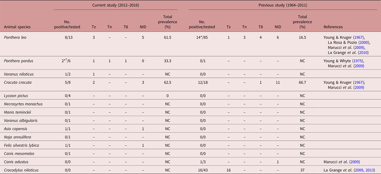
Tz, Trichinella zimbabwensis; Tn, Trichinella nelsoni; T8, Trichinella genotype T8; NID, not identified; NC, not calculated due to sample size < 4.
*One animal represents a mixed infection of Trichinella nelsoni and Trichinella T8.
†Four animals reported by La Grange et al. (Reference La Grange, Reininghaus and Mukaratirwa2014) included in present work.
Wild omnivores from the greater Kruger National Park, South Africa, screened for Trichinella spp. larvae and the prevalence of Trichinella spp. in each species for the period 2012–2016 and previous studies (1964–2011).
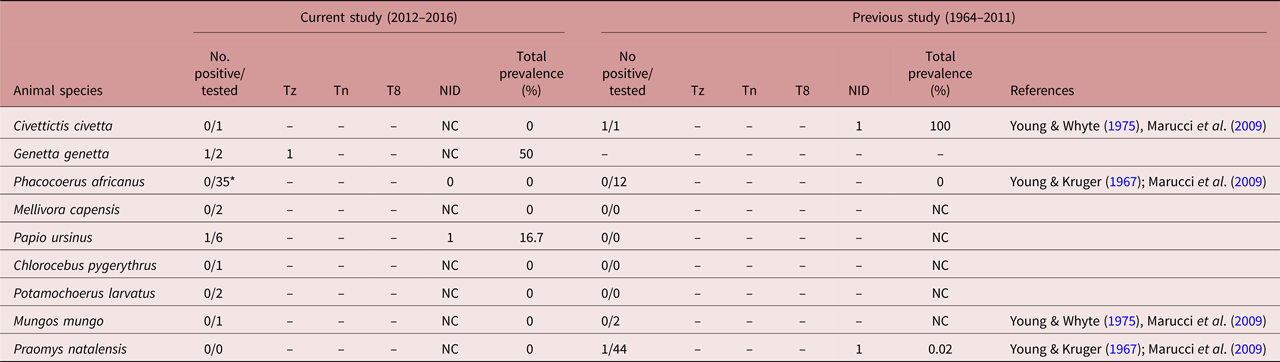
Tz, Trichinella zimbabwensis; Tn, Trichinella nelsoni; T8, Trichinella genotype T8; NC, not calculated due to sample size < 4; NID, not identified.
*17 Warthogs reported by La Grange et al. (Reference La Grange, Reininghaus and Mukaratirwa2014) included in present work.
DNA extraction from Trichinella larvae
Genomic DNA was extracted from at least five larvae of each positive sample whenever possible, using the genomic DNA™ tissue mini-prep kit (Zymo Research Corporation, Irvine, California, USA) according to the manufacturer's instructions.
Polymerase chain reaction and sequencing
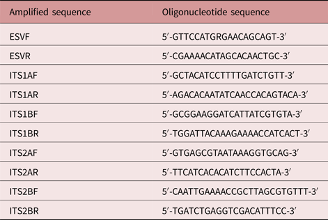
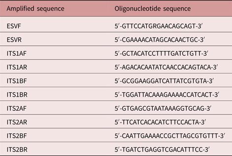
DNA was subjected to multiplex polymerase chain reaction (PCR) using primers ESVIF + ESVIR, ITS1AF + ITS1AR, ITS1BF + ITS1BR, ITS2AF + ITS2AR and ITS2BF + ITS2BR as described by Zarlenga et al. (Reference Zarlenga, Chute, Martin and Kapel1999) (table 3). The amplifications were performed in reactions of 50 μl volume containing 20 μl TopTaq master mix, 2 μl of each primer (forward and reverse) and 10 μl of DNA. Thermal cycling was carried out at 94°C for 3 min; followed by 40 cycles of 94°C for 1 min, 60°C for 1 min and 72°C for 2 min; followed by a final extension at 72°C for 7 min. Amplicons were separated by electrophoresis in 2% agarose gels, and visualized by staining with ethidium bromide. A laboratory-maintained reference strain of T. zimbabwensis was used as control. PCR amplification products of the expansion segment V (ESV) region were sent for sequencing by the Sanger dideoxy method at Inqaba Biotechnical Industries (Pty) Ltd, South Africa.
Nucleotide sequences amplified during multiplex PCR with respective forward and reverse oligonucleotide sequences.
Data and molecular analysis
The number of wildlife species with positive isolates from digestion of muscle were tabulated and compared with the data reported in the period 1964–2011.
Sequence alignments were analysed using the maximum parsimony method in PAUP 4.0b10 (Swofford, Reference Swofford2002) and Bayesian inference as implemented in MrBayes 3.0b4 (Huelsenbeck & Ronquist, Reference Huelsenbeck and Ronquist2001). For parsimony analyses, starting trees were obtained by stepwise addition. The addition sequence was random, with one tree held at each step and with ten replicates. Node support was estimated using 1000 bootstrap replicates. Bayesian analyses were run using four Markov chains, sampling every 100 generations, for 500,000 generations, or until the standard deviation of the split frequencies was less than 0.01. The chains were heated with the temperature scaling factor T = 0.02. We discarded the first 2000 trees as burn-in, in each case having checked in a preliminary run that this was more than sufficient to achieve stationarity. Bayesian inference trees were presented, with node support indicated as Bayesian posterior probabilities and maximum parsimony bootstrap values.
Analyses included T. zimbabwensis, T. nativa, T. papuae, T. spiralis, T. britovi, T. pseudospiralis, genotype 12 and genotype T8 as in-groups and Paratrichosoma sp. and Trichuris arvicolae as outgroups. Individual pairwise genetic p-distances between the sequences were determined using MEGA6 (Tamura et al., Reference Tamura, Stecher, Peterson, Filipski and Kumar2013).
Results
Prevalence of Trichinella spp. from digestion
Extrapolated data from previously published reports (1964–2011) were combined with the findings of this study (2012–2016) on the screening of Trichinella larvae and identification of Trichinella spp. in wildlife carnivores (table 1) and omnivores (table 2) from South Africa. Results show that T. zimbabwensis was the most prevalent species recorded to date. The species has been reported in five wildlife species (lion, hyaena, leopard (Panthera pardus), genet (Genetta genetta) and Nile monitor lizard (Varanus niloticus)) (tables 1 and 2). Trichinella nelsoni was reported in a leopard and Trichinella T8 was reported once in a leopard as a mixed infection with T. nelsoni (table 1). A similar mixed infection was also reported previously in a lion (Marucci et al., Reference Marucci, La Grange, La Rosa and Pozio2009) (table 1).
Ninety muscle samples from 20 wildlife species (15 mammals, 3 reptiles and 2 birds) were screened. Twenty samples from seven wildlife species were positive for Trichinella spp. through digestion (six mammals and one reptile) (tables 1 and 2) and Trichinella prevalence was higher in carnivores (18.9%, 18/90) than in omnivores (2.2%, 2/90). In the period from 1964–2011, 45 samples from nine wildlife species were positive for Trichinella spp. through digestion (seven mammals and one reptile) (table 1) with a prevalence of 21.4% (45/210). The prevalence of Trichinella was 20.5% (43/210) in carnivores and 0.9% (2/210) in omnivores.
Multiplex PCR analysis and phylogenetic analysis
Electrophoresis of multiplex PCR amplicons of putative Trichinella isolates produced two general types of amplification pattern. One of these contained two main bands, with sizes of approximately 270 and 350 bp, and was shared by isolates from hyaena, lion, monitor lizard and a T. zimbabwensis laboratory reference strain. This is generally consistent with the identification of these isolates as T. zimbabwensis. A contrasting amplification pattern, comprising two smaller main bands of approximately 150 and 250 nucleotides, was shared by a genet, lion and leopard. These isolates remain unidentified. It is interesting that a similar main banding pattern (bands of 127 and 253 nucleotides) was exhibited by Trichinella isolate T3 (Zarlenga et al., Reference Zarlenga, Chute, Martin and Kapel1999). Isolates from the African wildcat (Felis silvestris lybica), Chacma baboon (Papio ursinus) and Marsh owl (Asio capensis) did not produce amplification patterns.
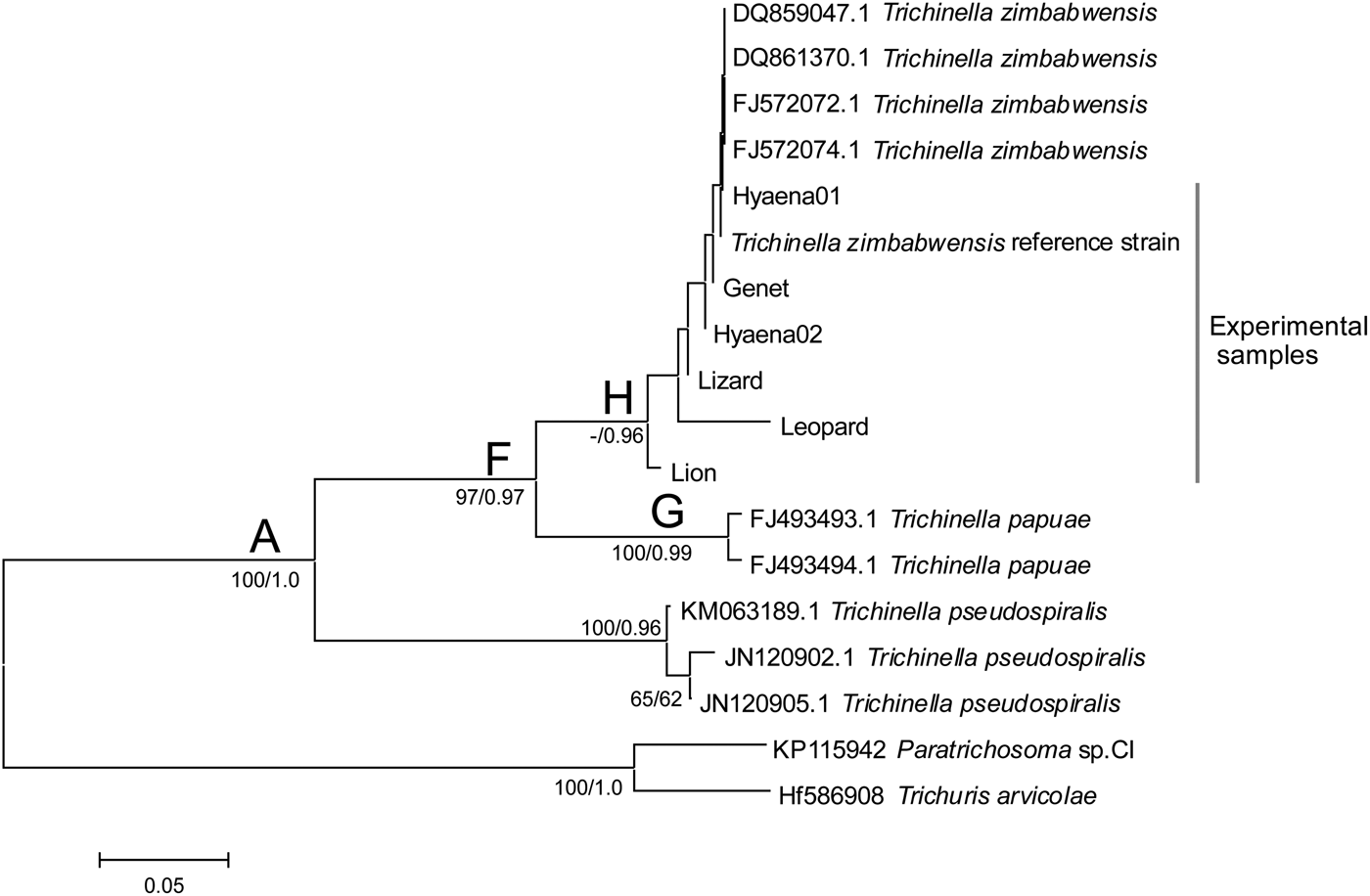
Alignment of ESV DNA sequences was created, based on 105 nucleotides (fig. 1) as the sequences contained areas of microsatellite repeats, and did not all yield good-quality sequence for the entire length. The alignment resolved phylogenetic relationships with support values as shown in fig. 1.
Bayesian inference tree based on 105 nucleotides of the ESV DNA region depicting relationships between experimental samples and sequences downloaded from GenBank (National Center for Biotechnology Information). Nodal support from maximum parsimony and Bayesian analyses is shown in that order.

Trichinella isolates formed a monophyletic clade (A) with reference to the outgroups (fig. 1). There was strong support for clade (F), which included T. papuae and T. zimbabwensis from GenBank and experimental isolates from our study. The T. papuae clade (G) was strongly supported and sister to T. zimbabwensis (fig. 1), a well-supported clade (H) comprising GenBank T. zimbabwensis samples and experimental Trichinella isolates from two hyaenas, lion, leopard, monitor lizard, genet and T. zimbabwensis reference isolate. Based on the phylogenetic species concept (Cracraft, Reference Cracraft1983), these experimental isolates are T. zimbabwensis. Further, genetic distances separating these isolates from the T. zimbabwensis reference sample are small (0.00–0.02), and consistent with those separating other GenBank samples of T. zimbabwensis from the reference strain (also 0.00–0.02). This is consistent with the identification of the experimental isolates as T. zimbabwensis based on the genetic species concept (Baker & Bradley, Reference Baker and Bradley2006). In contrast, and as would be expected, genetic distances between T. zimbabwensis isolates and other species of Trichinella are considerably higher, consistent with a greater level of taxonomic separation from T. papuae (0.11–0.16), T. pseudospiralis (0.26–0.31), T. nativa (0.36–0.39), T. britovi (0.38–0.41) and T. spiralis (0.43–0.47).
A third hyaena isolate was present in a strongly supported unresolved clade (D) containing GenBank samples of the encapsulated species T. spiralis, T. britovi and T. nativa (figure not shown), although it could not be identified to species level.
Distribution and prevalence of Trichinella spp. in wildlife species
Trichinella spp. larvae were isolated from eight lions during the course of this study (8/13, prevalence 61.5%). Of these, three were T. zimbabwensis (37.5%) and five isolates were unidentified to species level (table 1). Previous studies (1964–2011) reported isolates from 14 lions (14/85, prevalence 16.5%). Of these, one was T. zimbabwensis, three were T. nelsoni, four were Trichinella T8 and six were unidentified to species level (table 1).
Of eight hyaenas tested in this study, five were found to harbour Trichinella spp. larvae (5/8, prevalence 62.5%) (table 1). Based on ESV sequence analysis, two were T. zimbabwensis (40%) (fig. 1), and a third fell under the encapsulated clade, although it was not possible to identify this isolate to species level. The other two isolates were also unidentified to species level. Twelve isolates from hyaena have been reported from 18 screened during the period from 1964 to 2011 (12/18, prevalence 66.7%). Of these, one was Trichinella T8 and the remaining 11 were unidentified.
Two Trichinella spp. isolates were recovered from six screened leopards (2/6, prevalence 33.3%). One leopard had a mixed infection of T. nelsoni and T8, the results of which were previously published (see La Grange et al., Reference La Grange, Reininghaus and Mukaratirwa2014). The remaining isolate from this study was T. zimbabwensis. This is the first report of a natural infection with T. zimbabwensis in this host. From the previous studies (1964–2011) only one leopard was screened and was found to be negative (table 1).
Two Nile monitor lizards were screened in our study and one was positive for T. zimbabwensis (1/2, prevalence 50%). Isolates from a single African wildcat in this study and from a side-striped jackal (Canis adustus) in the period (1964–2011) were unidentified.
Of the two small spotted genets tested, only one (1/2, prevalence 50%) tested positive. The isolate was identified as T. zimbabwensis and this is the first report of a natural infection with T. zimbabwensis in this host.
Six Chacma baboons were screened during the course of this study; one was positive (1/6, prevalence 16.7%) and a single larva recovered. The sample was in an advanced state of autolysis and the identification to species level was unresolved.
Samples from one African wildcat and one Marsh owl were positive from screening. Only a few larvae were detected and, similar to that of baboon, the samples were in an advanced state of autolysis and no species identification was possible.
Discussion
Results from this study are consistent with the commonly accepted postulate (Mukaratirwa et al., Reference Mukaratirwa, La Grange and Pfukenyi2013) that the most important route for Trichinella transmission in wild animals appears to be via predation, cannibalism and scavenging. In a review by Mukaratirwa et al. (Reference Mukaratirwa, La Grange and Pfukenyi2013), Trichinella spp. prevalence was reported to be high in wild carnivores, which is consistent with results of the present study, which revealed a high prevalence in carnivores with predatory and scavenging behaviour compared to omnivorous animals.
The ability of T. zimbabwensis to infect mammalian hosts has been demonstrated in several experimental studies (Mukaratirwa & Foggin, Reference Mukaratirwa and Foggin1999; Pozio et al., Reference Pozio, Marucci and Casulli2004; Mukaratirwa et al., Reference Mukaratirwa, Dzoma, Matenga, Ruziwa, Sacchi and Pozio2008) and proven to occur in nature (La Grange et al., Reference La Grange, Marucci and Pozio2010). This species has been documented in Nile crocodiles of Zimbabwe, Mozambique, Ethiopia and South Africa, in Nile monitor lizards of Zimbabwe and in a lion from the Kruger National Park, South Africa (Mukaratirwa & Foggin, Reference Mukaratirwa and Foggin1999; Pozio et al., Reference Pozio, Foggin, Maruccia, La Rosa, Sacchi, Corona, Rossi and Mukaratirwa2002; La Grange et al., Reference La Grange, Marucci and Pozio2009, Reference La Grange, Marucci and Pozio2010, Reference La Grange, Govender and Mukaratirwa2013). Our results represent the second report of natural infection with T. zimbabwensis in a lion and this confirms the previous report by La Grange et al. (Reference La Grange, Marucci and Pozio2010) that the lion is an exceptional host for all three Trichinella taxa (Trichinella T8, T. nelsoni and T. zimbabwensis) circulating in South Africa (La Grange et al., Reference La Grange, Marucci and Pozio2010).
Results from this study confirm that hyaenas are equally important hosts for at least two Trichinella taxa known to circulate in South Africa. Mukaratirwa et al. (Reference Mukaratirwa, La Grange and Pfukenyi2013) suggested the existence of a maintenance cycle for T. nelsoni and Trichinella T8 between lions and hyaenas. Results from this study suggest a similar maintenance cycle for T. zimbabwensis between these two carnivorous species. One isolate formed a well-supported association with a clade of encapsulated species and was unidentified to species level. Most likely this isolate was T. nelsoni or Trichinella T8, since these are the only two encapsulated species to have been reported in lions and hyaenas in South Africa to date (Marucci et al., Reference Marucci, La Grange, La Rosa and Pozio2009; Mukaratirwa et al., Reference Mukaratirwa, La Grange and Pfukenyi2013).
An important finding from this study is that of T. zimbabwensis infection in the small spotted genet. Lariviere & Calzada (Reference Lariviere and Calzada2001) reported the diet of the small spotted genet to be euryphagous. The diet of this opportunist omnivore consists of small mammals, amphibians, reptiles, fruits and birds (Lariviere & Calzada, Reference Lariviere and Calzada2001) and the genet may have acquired infection from feeding on infected small mammals, such as rodents or reptiles.
Mukaratirwa et al. (Reference Mukaratirwa, La Grange and Pfukenyi2013) postulated that carnivorous reptiles from the families Crocodylidae and Varanidae are likely to be the main reservoir hosts for T. zimbabwensis. Natural T. zimbabwensis infection has been reported in Nile crocodiles of South Africa (La Grange et al., Reference La Grange, Marucci and Pozio2009, Reference La Grange, Govender and Mukaratirwa2013) and this study reports for the first time a natural infection of T. zimbabwensis in a Nile monitor lizard from South Africa. Results from reports to date show a high prevalence of Trichinella spp. in carnivores with cannibalistic and scavenging behaviour (Mukaratirwa et al., Reference Mukaratirwa, La Grange and Pfukenyi2013). This study also reports the occurrence of T. zimbabwensis in a leopard for the first time.
Trichinella sp. larvae were detected for the first time in a baboon, although with a significantly low number of larvae. Species identification was not possible due to the small number of larvae and degradation of larval DNA. For the first time, a Trichinella spp.-like infection was reported in a bird from South Africa. Trichinella pseudospiralis is the only Trichinella taxon known to infect birds and it has been reported in birds and mammals from Asia, North America, Europe and Tasmania (Pozio & Murrell, Reference Pozio and Murrell2006). This parasite species has not been reported in Africa and, despite preliminary data from this study suggesting the existence of this parasite in South Africa, the absence of conclusive molecular evidence precludes a definitive report.
Previous reports of natural infections involving T. zimbabwensis may suggest a propensity of this parasite species towards infecting reptiles, but results from this study clearly show that T. zimbabwensis infects a variety of ecto- and endothermic host species indiscriminately. Additionally, previous reports of a lion (La Grange et al., Reference La Grange, Marucci and Pozio2010) and the current report of spotted hyena, leopard, lion and a small spotted genet naturally infected with T. zimbabwensis confirm previous suggestions of the significant epidemiological role of mammals in the parasite epidemiology (La Grange et al., Reference La Grange, Marucci and Pozio2010; Mukaratirwa et al., Reference Mukaratirwa, La Grange and Pfukenyi2013).
The infection in a small spotted genet may also suggest the existence of a large biomass of this parasite maintained in a number of smaller rodent and/or reptile species. Similarly, the infection in a leopard suggests that smaller carnivores, such as the genet, are frequently infected in nature and supports the previous hypothesis. Leopards predate small carnivorous mammals (Hayward et al., Reference Hayward, Henschel, O'Brien, Hofmeyr, Balme and Kerley2006) and their potential to serve as sources of infection to leopards has previously been alluded to (La Grange et al., Reference La Grange, Reininghaus and Mukaratirwa2014). This is a cause of concern from a veterinary public health perspective, since the potential risk of transmission of the parasite from the natural sylvatic cycle to domestic animals can be through rodent infestations.
The presence of Trichinella spp. in at least four different omnivorous species in GKNP certainly suggests that they act as maintenance hosts, although results from this study together with reports by Young & Whyte (Reference Young and Whyte1975) and Young & Kruger (Reference Young and Kruger1967) represent the only four cases of Trichinella infections reported in omnivores in the GKNP and adjacent areas.
Epidemiological investigations on Trichinella species have been carried out mostly on wild animals from the GKNP and surrounding areas in South Africa. More surveys aimed at elucidating the prevalence and species richness of this parasite genus in the rest of South Africa are required. Such surveys will certainly prove invaluable in adding to the body of knowledge on Trichinella. However, more importantly, they will prove crucial in determining the risk of human infection, which will inevitably increase alongside the expansion of the game industry, population growth and the search for alternative food sources to ensure food security for the country's inhabitants.
Acknowledgements
Our sincere appreciation to the Mpumalanga Department of Agriculture, Rural Development, Land and Environmental Affairs for logistical and financial support provided through the Chief Directorate of Veterinary Services; Dr J.P. Raath and staff of Wildlifevets.com and Wildlife Pharmaceuticals for financial, logistical and technical support.
Financial support
Financial support was provided by the National Research Foundation of South Africa from the Incentive for Rated Researchers scheme for S.M. for the laboratory work, and by the Chief Directorate of Veterinary Services, Mpumalanga Department of Agriculture, Rural Development, Land and Environmental Affairs and Wildlife Pharmaceuticals for collection of samples.
Conflict of interest
None.