


Allergy to peanuts (Arachis hypogaea) is a burgeoning health problem. Approximately 1% of the population suffers from peanut allergy (Ref. Reference Sicherer, Munoz-Furlong and Sampson1), but there is evidence that the prevalence is increasing (Ref. Reference Grundy2). For many peanut-allergic individuals, exposure to minute quantities of peanut can lead to severe reactions including anaphylaxis, which can sometimes prove fatal. Currently, there is no cure for this condition and strategies for the prevention of severe reactions are limited to avoidance of exposure to peanuts and administration of adrenaline as an emergency treatment after inadvertent contact. There is a clear need for a safe and effective specific therapy for patients with peanut allergy.
Molecular characterisation of allergenic components of peanuts and elucidation of immune mechanisms that determine clinical responses are required for the development of specific therapies and improved management for peanut allergy. This review begins by summarising the clinical features of peanut allergy and the mucosal immune response to food allergens, and then discusses the molecular characterisation of peanut and crossreactive allergens, and how this knowledge is being utilised for the development of potential therapeutic modalities for the treatment and prevention of peanut allergy.
Clinical features of peanut allergy
Peanut allergy generally develops early in life and is commonly associated with other atopic disorders such as asthma, eczema and rhinitis. Sensitisation is thought to occur through the consumption of foods such as peanut butter or, in some cases, the use of topical preparations containing peanut oil (Ref. Reference Lack5). Exposure can also occur in utero (Ref. Reference Kaufman6) or through breast milk (Ref. Reference Vadas7), although the importance of this route for sensitisation has been contentious (Ref. Reference Lack5). It is becoming apparent that the increasing consumption of peanuts together with other, unidentified factors is leading to an increase in prevalence of peanut allergy, particularly in children (Refs Reference Sicherer, Munoz-Furlong and Sampson1, Reference Grundy2). This is likely to flow through to a higher prevalence in adults.
Allergic symptoms following the ingestion of peanuts occur from within minutes to a few hours, and manifestations range from oral pruritus, nausea, vomiting, urticaria and angioedema to bronchospasm (Ref. Reference Sampson8). In severe cases, anaphylaxis with angioedema, respiratory compromise and hypotension can prove fatal without the prompt administration of adrenaline. Peanuts account for the majority of food-related anaphylaxis in children, adolescents and adults (Refs Reference Sampson, Mendelson and Rosen3, Reference Bock, Munoz-Furlong and Sampson4). A characteristic of peanut allergy is its tendency to persist through to adulthood, with only 21.5% of peanut-allergic individuals experiencing resolution of this type of food allergy with increasing age (Refs Reference Bock and Atkins9, Reference Skolnick10). Thus, lifelong vigilance is essential for the majority of sufferers of peanut allergy.
The mucosal immune response to peanut allergens
Exposure of the immune system to a food allergen such as peanut generally occurs at the mucosal surface of the gut. Peanut allergens are taken up by specialised epithelial cells called M cells, and transferred to antigen-presenting cells such as dendritic cells where they are processed into peptide fragments and presented on the cell surface in the context of class II major histocompatibility complex (MHC) molecules (Refs Reference Prioult and Nagler-Anderson11, Reference Strobel and Mowat12) (Fig. 1). These peptides are presented to naive T helper (Th) cells via interaction of the MHC–peptide complex with the T-cell receptor, resulting in Th-cell priming and activation, which triggers the humoral and cellular events associated with allergic inflammation.
The major cellular interactions during the mucosal allergic immune response. (a) Sensitisation. In the gastric mucosa, peanut allergens are taken up by specialised epithelial cells called M cells and transferred to antigen-presenting cells such as dendritic cells, where they are processed into peptide fragments. Complexes of peanut-allergen peptide and major histocompatibility complex (MHC II) are presented to naive T cells, which differentiate into T helper 2 (Th2) cells. Activated Th2 cells recognise peanut-allergen-peptide–MHC complexes on the surface of B cells, releasing cytokines interleukin 4 (IL-4) and IL-13, which promote immunoglobulin E (IgE) antibody production by B cells. Secreted IgE antibodies bind to FcɛRI receptors on effector cells such as mast cells, which become sensitised. (b) Challenge. Upon secondary encounter with the same peanut allergen, the allergen crosslinks cell-bound IgE, activating mast cells to release inflammatory mediators. Additional production of IL-4 and IL-13 by mast cells and basophils results in further Th2-cell differentiation and IgE synthesis (not shown). Th2 cells and mast cells also produce TNF-α, IL-5 and chemokines which drives the recruitment of eosinophils to the site of inflammation (not shown). This cascade of events ultimately leads to the induction of symptoms commonly associated with peanut allergy.

In atopic individuals, the activation of Th cells results in the secretion of cytokines that stimulate B cells to synthesise IgE antibodies specific to the allergen. Th cells are polarised into two subgroups, defined by the dominant pattern of cytokine secretion (Ref. Reference Mosmann13). Th1-type cells mainly secrete interleukin 2 (IL-2), interferon γ (IFN-γ) and tumour necrosis factor α (TNF-α). By contrast, Th2-type cells secrete IL-4, IL-5, IL-9 and IL-13. Th2 cells play a pivotal role in the allergic response as it is their activation and secretion of IL-4 and IL-13 that drives allergen-stimulated B-cell differentiation into IgE-secreting plasma cells.
IgE antibodies are bound by high-affinity surface IgE receptors (FcɛRI) present on effector cells such as mast cells and basophils. In peanut-allergic individuals, subsequent exposure to peanut allergens induces inflammatory reactions largely governed by mast cells, basophils and eosinophils. Adjacent IgE antibodies bound by FcɛRI on the surface of mast cells and basophils are crosslinked by peanut allergens, resulting in the release of inflammatory mediators such as histamine, prostaglandins, leukotrienes, heparin and platelet-activating factor. Additional cytokine production occurs during this phase, most notably IL-4 and IL-13 by mast cells and basophils, further augmenting Th2-cell differentiation and IgE synthesis. Th2 cells and mast cells also produce TNF-α, IL-5 and chemokines, which results in the recruitment of eosinophils to the site of inflammation. Eosinophils are activated by IL-4, IL-5 and IL-13, inducing the release of inflammatory mediators such as major basic protein, eosinophil peroxidase, eosinophil cationic protein and eosinophil-derived neurotoxin (Ref. Reference Gleich14). These mechanisms ultimately lead to the manifestation of clinical symptoms such as vomiting, diarrhoea, urticaria, angioedema, asthma and anaphylaxis that are commonly associated with peanut allergy.
Current treatment options for peanut allergy
At present, there is no cure for peanut allergy. Management is only by strict avoidance of the offending food, and administration of adrenaline as an emergency treatment. Avoidance can be difficult as peanuts are widely used as additives in different foods, and some foods are inadequately labelled. Furthermore, contamination of foods with peanut proteins can occur inadvertently during the manufacturing process, posing the threat of ‘hidden allergens’ within these foods. Foods cooked using crude or cold-pressed peanut oil, which have been shown to contain peanut allergens (Refs Reference Hourihane15, Reference Hoffmann and Collins-Williams16), might also elicit an allergic reaction in sensitive individuals. Given the risks associated with peanut allergy, it is not surprising that accidental exposures are quite common for many patients, further emphasising the need to maintain vigilance.
Allergen-specific immunotherapy, commonly used as a therapeutic strategy for environmental allergies such as those to house dust mite and grass pollen, is not currently available for peanut allergy. This form of therapy usually involves subcutaneous injections of gradually increasing doses of allergen extract during an induction or updosing phase. Subsequently, a maintenance phase is typically given with stable doses at fixed intervals for 3 to 5 years in order to achieve clinical ‘tolerance’ upon subsequent exposure to the same allergen. Successful allergen-specific immunotherapy results in the modulation of several T-cell and B-cell responses. There is often an increase in the ratio of Th1 cytokines to Th2 cytokines, induction of T-cell anergy, the generation of allergen-specific T-regulatory cells and an increase in production of regulatory cytokines (Refs Reference Secrist17, Reference Gardner18, Reference Bellinghausen19, Reference Jutel20). B cells have also been shown to produce allergen-specific IgG antibodies that effectively compete with IgE antibodies for binding to allergens, thus blocking the downstream events associated with allergic inflammation (Ref. Reference Flicker and Valenta21).
There are a few reports where peanut-specific allergen immunotherapy using crude peanut extracts has been explored. In one case study, successful desensitisation was performed for peanut allergy (Ref. Reference Patriarca22) but this involved a patient with only relatively mild gastrointestinal manifestations and no data are available on duration of efficacy. In another study, the potency of crude peanut extract was highlighted when an attempt was made to desensitise peanut-allergic patients by traditional injection rush (accelerated) immunotherapy using a crude peanut extract (Ref. Reference Oppenheimer23). The rate of systemic reactions was 13.3% and the study was prematurely terminated after one participant suffered a fatal anaphylactic reaction through an administration error of active extract to a control patient (Ref. Reference Oppenheimer23).
Conventional immunotherapy for peanut allergy using crude peanut extracts is therefore not recommended currently because of the unacceptably high risk of anaphylaxis. A safe hypoallergenic formulation is needed to allow development of novel treatments for peanut allergy and perhaps for use as preventative agents in high-risk infants. For this, a detailed knowledge of immunoreactive components of peanut is required.
Allergenic components of peanut
To date, eight peanut allergens have been recognised officially, as summarised in Table 1. The three main peanut allergens – Ara h 1, Ara h 2 and Ara h 3 – are well characterised, with cloning of the allergens and derived sequence data. Ara h 1 and Ara h 2 are reported to be highly allergenic, with most studies showing a high frequency of peanut-allergic individuals having serum-specific IgE to these allergens (Refs Reference Burks24, Reference Burks25, Reference Burks26). The designation of Ara h 3 is not yet clear: one study demonstrated serum IgE reactivity to this protein in 8/18 peanut allergic subjects (44%) (Ref. Reference Rabjohn27), whereas another identified specific IgE against Ara h 3 in 95% of a group of 16 peanut-allergic children in Italy (Ref. Reference Restani28). Ara h 4–7 are less reactive with IgE in patient sera (Ref. Reference Kleber-Janke29) and are yet to be fully characterised. Ara h 8 is distinct from the other peanut allergens as it is found to be a major allergen in individuals in central Europe who exhibit co-allergy to peanut and birch pollen (Ref. Reference Mittag30).
Molecular characteristics of peanut allergens

Abbreviations: C, conformational; L, linear.
Most of the identified peanut allergens are members of seed storage protein families, with the exception of Ara h 5 and Ara h 8. Ara h 5 belongs to the profilin family, a group of actin-binding proteins responsible for cytoskeleton formation in plant cells (Ref. Reference Breiteneder and Ebner31) and known to be a major cause of pollen-associated food allergy. Ara h 8 is a member of the pathogenesis-related protein family PR-10, which is similarly involved in pollen-associated food allergy (Refs Reference Mittag30, Reference Breiteneder and Ebner31).
Biochemical properties of peanut allergens
Food allergens and their corresponding IgE-binding epitopes are thought to possess physicochemical properties that confer resistance to digestive enzymes and thermal processing. This increases their allergenicity by enhancing their ability to reach the intestinal mucosa. Ara h 1 is capable of forming stable dimers, trimers and larger complexes upon heating without affecting IgE reactivity (Ref. Reference Koppelman32). Treatment of Ara h 1 with gastrointestinal enzymes such as pepsin, trypsin and chymotrypsin produces large proteolytic fragments that retain binding affinity for serum IgE from peanut-allergic individuals (Ref. Reference Maleki33). The resistance of Ara h 1 to degradation following heating and treatment with digestive enzymes may be related to its stable, homotrimeric structure (Ref. Reference Maleki33). This monomer–monomer interaction reduces access to the catalytic sites within the protein, allowing Ara h 1 to survive intact during food processing or passage along the digestive tract, thus contributing to its potency as an allergen.
Similarly, Ara h 2 and Ara h 6 retain allergenicity following proteolytic digestion as well as heat treatment (Refs Reference Suhr34, Reference Lehmann35). Structural studies revealed that following digestion, both allergens yielded immunologically active core structures that were able to induce release of inflammatory mediators even in the context of decreased serum IgE binding (Ref. Reference Lehmann35). Ara h 2 shares sequence homology with trypsin inhibitors and can act as a weak trypsin inhibitor with increased activity following roasting, thus protecting itself as well as Ara h 1 from trypsin digestion (Ref. Reference Maleki36). This function of Ara h 2 may contribute to the overall allergenic properties of peanut proteins through increased resistance to digestive enzymes. By contrast, Ara h 8 has low stability to heat and gastric digestion (Ref. Reference Mittag30).
The Maillard reaction is also a contributing factor to the allergenicity of peanuts (Refs Reference Chung and Champagne37, Reference Maleki38, Reference Chung39). This is a nonenzymatic reaction between a protein and a reducing sugar that occurs during thermal processing and cooking (Ref. Reference Namiki40). The amino groups of proteins become glycosylated to form Amadori products, which degrade into dicarbonyl intermediates. These intermediary compounds react with amino groups of proteins to form stable end products known as advanced glycation end products. Chung and Champagne (Ref. Reference Chung and Champagne37) demonstrated that advanced glycation end products, formed by heating a previously nonallergenic peanut lectin protein in the presence of sugars, can effectively compete with untreated peanut allergens for serum IgE antibodies, suggesting that the Maillard reaction can convert a nonallergenic protein into a potentially allergenic protein. Similarly, Maleki and colleagues observed that roasted peanut proteins inhibited IgE binding to raw peanut proteins more effectively (90-fold higher) than raw peanut proteins (Ref. Reference Maleki38). Both studies concluded that the presence of advanced glycation end products in heat-treated peanuts contributed to their overall heightened allergenicity.
Epitope mapping
Identification of suitable peanut allergen preparations for specific immunotherapy requires detailed characterisation of sites that interact with antibodies/B cells and T cells. Antibodies and B cells interact with native allergens in their conformational state, whereas T cells interact with linear peptides presented in the context of MHC molecules on antigen-presenting cell surfaces. Conformational epitopes recognised by antibodies comprise discontinuous short linear sequences that are closely associated in the correctly folded molecule, but some antibody epitopes are longer linear sequences. It has been suggested that children with milk allergy whose predominant IgE reactivity is against conformational epitopes are more likely to develop tolerance to milk than those who react against linear epitopes (Ref. Reference Vila41), but whether this relationship occurs for peanut allergens is not known.
B-cell epitopes
To date, most studies on peanut IgE reactivity have focused on the more readily analysed linear epitopes. The linear IgE-binding epitopes of Ara h 1, Ara h 2 and Ara h 3 were mapped using synthetic peptides (Table 1). Typically these epitopes ranged from six to ten amino acids in length, and in all cases the epitopes could be rendered nonreactive to IgE by alanine substitution of a single amino acid residue (Refs Reference Rabjohn27, Reference Burks42, Reference Stanley43). Structural studies that generated the tertiary structure of Ara h 1, using the highly homologous phaseolin protein in homology-based modelling, allowed the positional identification of the linear IgE-binding epitopes of Ara h 1 within its native conformation. The majority of amino acid residues identified as critical for binding to IgE were distributed on the surface of the molecule in epitope clusters (Refs Reference Bannon44, Reference Shin45). The presentation of clustered epitopes to mast cells and basophils may result in a more efficient release of mediators, which may explain the severe clinical symptoms associated with peanut allergy. As mentioned previously, Ara h 1 is capable of higher-order aggregation, forming a stable trimeric complex through interactions between hydrophobic amino acid residues (Refs Reference Maleki33, Reference Bannon44). Structural analysis of the Ara h 1 monomer identified hydrophobic regions at each end of the molecule acting as contact points for trimer formation, with most of the IgE-binding epitopes clustered around these contact points (Fig. 2).
Molecular model of the Ara h 1 trimer. A space-filled, homology-based model of the Ara h 1 trimer shows that the majority of IgE-binding epitopes (red) are located in areas close to monomer–monomer contact. Reprinted from Ref. Reference Maleki33 (© 2000 The American Association of Immunologists, Inc.), with permission from The Journal of Immunology and Gary Bannon (Monsanto, St Louis, MO, USA).

Diversity of IgE recognition of linear epitopes of peanut allergens has been shown to correlate with the severity of clinical symptoms. Using 20-mer peptides corresponding to the entire primary sequences of Ara h 1, Ara h 2 and Ara h 3, it was found that peanut-specific serum IgE from subjects with a history of severe systemic allergic reactions displayed a higher degree of epitope diversity in comparison to those who experienced only cutaneous reactions (Ref. Reference Shreffler46). Analysis of individual patients revealed no distinct pattern of linear-epitope recognition, confirming the diversity of the IgE response to peanut allergens between peanut-allergic individuals.
T-cell epitopes
By contrast to B-cell epitopes, data on the T-cell epitopes of peanut allergens are extremely limited. To date, dominant sites of T-cell reactivity have been reported for only one peanut allergen, Ara h 2, and the core T-cell epitopes within these sites have not yet been identified. Glaspole et al. identified two highly immunogenic T-cell-reactive regions: Ara h 2 (19–47) and Ara h 2 (73–119). Peptides spanning these two regions induced strong T-cell proliferation associated with a Th2-type cytokine response (Ref. Reference Glaspole47). Knowledge of the dominant T-cell epitopes of allergens is critical information for the development of a T-cell-targeted vaccine for peanut-specific allergen immunotherapy.
Peanut allergen crossreactivity
Crossreactivity between peanut and tree nuts
Clinical studies indicate that peanut-allergic subjects, particularly adults, usually exhibit multiple sensitivities to both peanut and tree nuts. In specialist allergy clinics, the prevalence of co-allergy to peanut and tree nuts was reported as 35–40% (Refs Reference Ewan48, Reference Sicherer, Burks and Sampson49), although in our experience up to 80% of adult peanut-allergic patients are clinically allergic to one or more tree nuts (R. O'Hehir, unpublished). Sicherer and colleagues examined the serology of peanut-allergic children and found significant correlations between the level of peanut-specific IgE and tree-nut-specific IgE antibodies. In particular, peanut-specific IgE levels correlated with IgE levels for hazelnut, Brazil nut and almond (Ref. Reference Sicherer, Burks and Sampson49). Given this, several studies have investigated the presence of crossreactive allergens in peanut and various tree nuts. IgE crossreactivity was demonstrated between peanut and the tree-nuts pistachio, macadamia, almond, Brazil nut and hazelnut (Refs Reference Parra50, Reference Sutherland51, Reference de Leon52). In the majority of these cases the identity of the crossreacting allergen(s) was not established, with the exception of Ara h 2, which shares IgE-binding epitopes with almond and Brazil nut allergens (Ref. Reference de Leon53).
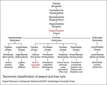
Although peanut is a legume and is taxonomically distantly related to tree nuts (Fig. 3), they are both defined as ‘edible’ seeds and perform similar functions in plant development. Indeed, several known peanut and tree-nut allergens belong to the same protein families. The major peanut allergen, Ara h 1, is a member of the vicilin family of seed storage proteins (Ref. Reference Burks24) together with allergens from walnut, cashew and hazelnut (Refs Reference Teuber54, Reference Wang55, Reference Pastorello56). Also, 2S albumin seed storage proteins, implicated as allergens in almond, Brazil nut, hazelnut and walnut (Refs Reference Pastorello56, Reference Poltronieri57, Reference Pastorello58, Reference Teuber59), are related to conglutin seed storage proteins, which include allergens from almond (Ref. Reference Poltronieri57) and peanut – namely Ara h 2, Ara h 6 and Ara h 7 (Refs Reference Burks25, Reference Kleber-Janke29). Legumins or 11S globulins are seed storage proteins shown to be allergenic in peanut (Ara h 3), cashew, hazelnut and walnut (Refs Reference Rabjohn27, Reference Pastorello56, Reference Wang60, Reference Beyer61, Reference Teuber62). Therefore, it is highly likely that a number of peanut allergens are responsible for the observed IgE crossreactivity between peanut and certain tree nuts.
Taxonomic classification of peanut and tree nuts. Many people suffering from peanut allergy are also allergic to one or more tree nuts. Although peanut and a number of tree nuts belong to different plant families, they arise from the same class, namely Magnoliopsida, and therefore may contain homologous proteins that play a role in allergenic crossreactivity. Tree nuts that have been shown to share IgE-binding epitopes with peanuts include almond, Brazil nut, hazelnut, macadamia and pistachio. The classification shown is based on information at the website of the US Department of Agriculture (http://plants.usda.gov/).
Clinical and biological relevance of IgE crossreactivity
Whether or not in vitro allergen crossreactivity translates into clinical hypersensitivity is controversial. In addition to peanut, the legume family includes soybean, pea, lima bean and green bean; however, although these foods are closely related taxonomically, clinical hypersensitivity to more than one legume is rare (Refs Reference Bernhisel-Broadbent and Sampson63, Reference Bernhisel-Broadbent, Taylor and Sampson64). van der Veen and colleagues observed that a third of patients sensitised to grass pollen (including timothy and orchard grass as confirmed by serum specific IgE test) had significant serum levels of peanut-specific IgE antibodies but no clinical symptoms (Ref. Reference van der Veen65). This phenomenon was attributed to crossreactive carbohydrate determinants (CCDs) – N-linked carbohydrate groups of glycoprotein allergens that induce the production of crossreactive IgE antibodies to food and grass pollen allergens (Refs Reference Batanero66, Reference Petersen67). Subjects with true peanut allergy had minimal levels of IgE antibodies specific for CCDs, in contrast to those with false-positive skin prick tests to peanut (Ref. Reference van der Veen65).
IgE antibodies to CCDs also appear to have negligible biological activity. In the same study by van der Veen and colleagues, higher concentrations of peanut extract (∼1000-fold) were required for basophil histamine release in patients with high levels of anti-CCD IgE antibodies with no peanut allergy compared with peanut-allergic patients, further validating the assertion that crossreactive IgE antibodies directed at CCDs are not indicative of clinical sensitivity. However, biological activity of peanut-specific IgE antibodies that crossreact with tree-nut allergens was demonstrated by basophil-activation assays (Ref. Reference de Leon68). Human basophils passively resensitised with peanut-specific serum IgE antibodies from peanut- and tree-nut-allergic subjects become activated following incubation with almond and Brazil-nut extracts, as indicated by detection of CD63 surface expression by flow cytometry (Ref. Reference de Leon68). Although the threshold doses of peanut, almond and Brazil-nut extracts required for basophil activation were not compared, it is highly likely that crosslinking of peanut-specific IgE antibodies present on effector cells by tree-nut allergens is clinically relevant given the high incidence of cosensitisation to peanut and tree nuts in peanut-allergic subjects.
Clinical implications: immunotherapeutic options
There is a critical need for a safe and effective treatment for peanut allergy given the severity and life-threatening nature of this condition. Currently, several immunotherapeutic methods are being developed as proposed treatments for peanut allergy, including the use of allergen derivatives as vaccines (Fig. 4). Conventional subcutaneous immunotherapy using crude allergen extracts in order to modify the immune response and obtain clinical tolerance to the allergen is not feasible for peanut allergy because of the high risk of severe systemic side effects including anaphylaxis and death (Ref. Reference Oppenheimer23), as discussed earlier in this review.
Potential specific immunotherapeutic strategies for the treatment of peanut allergy. The identification and characterisation of peanut allergens allows for the development of vaccines that could potentially be used for the treatment of peanut allergy. (a) Peanut allergens can be identified using serum IgE from peanut-allergic individuals and subsequently cloned, sequenced and expressed as recombinant proteins. (b) Following the identification of the IgE-binding sites on the peanut allergens, mutations can be introduced into the peanut allergen gene to render these sites nonreactive to IgE. Expression of the mutated peanut allergen construct will result in the production of hypoallergenic variants that can be used for immunotherapy. (c) Alternatively, overlapping peptides spanning the entire protein sequence of peanut allergens can be used in T-cell assays to identify peptides encoding the dominant T-cell epitopes that can also be utilised in a vaccine. Ultimately the aim of using hypoallergenic peanut-allergen variants and/or T-cell-epitope peptide vaccines is to induce tolerance to peanut allergens in susceptible individuals without the IgE-mediated side effects.

Modified peanut allergens with reduced IgE reactivity
Given the high-affinity IgE binding of peanut allergens, there is a need to develop suitable allergen preparations that do not crosslink IgE antibodies bound to effector cells. One approach is the engineering of recombinant allergens with conserved T-cell reactivity and decreased IgE binding. Knowledge of the IgE-binding epitopes of peanut allergens has allowed preparation of hypoallergenic variants with decreased IgE binding (Refs Reference Burks, King and Bannon69, Reference King70).
PCR mutagenesis targeting linear IgE-binding epitopes was utilised to produce a hypoallergenic Ara h 2 mutant; however, only 75% efficacy was achieved, with serum IgE from 12/16 patients demonstrating a considerable decrease in reactivity to the Ara h 2 mutant in comparison with the wild-type allergen (Ref. Reference Burks, King and Bannon69). Similarly, another hypoallergenic Ara h 2 mutant was produced but this variant also retained IgE reactivity in a small cohort of peanut-allergic subjects and induced significant mediator release from basophils passively sensitised by peanut-allergic sera (Ref. Reference King70).
Site-directed mutagenesis was utilised in the development of a hypoallergenic variant of Ara h 3. A 40 kDa acidic subunit of Ara h 3 containing the four identified Ara h 3 linear IgE-binding epitopes was expressed using a bacterial system, with the critical residues for IgE binding targeted for point mutations by substitution with an alanine residue (Ref. Reference Rabjohn71). The resulting modified protein was still IgE-reactive although binding was substantially decreased in comparison with the wild-type protein (Ref. Reference Rabjohn71). A hypoallergenic form of Ara h 1 with T-cell reactivity has also been produced (Ref. Reference Bannon72) but the characterisation of this modified allergen has been limited.
The retention of IgE reactivity with the Ara h 2 and Ara h 3 variants following mutation of the linear IgE-binding epitopes suggests that conformational B-cell epitopes should also be disrupted to avoid IgE-mediated adverse reactions in patients. In view of the multiple sensitivities to peanut allergens in most patients (Ref. Reference Lewis73), it is likely that a cocktail of hypoallergenic mutants would be required in a vaccine.
T-cell-epitope peptide vaccines
An alternative approach to specific immunotherapy uses short T-cell-epitope peptides that lack IgE binding. This strategy shows promise in the treatment of certain allergies and is particularly attractive because the peptides are too short to crosslink effector-cell-bound IgE antibodies. In preclinical studies, the administration of peptides based on dominant T-cell epitopes of allergens from cat and house dust mites induced specific T-cell tolerance in mice (Refs Reference Briner74, Reference Hoyne75). This approach has been encouraging in clinical trials using allergen peptides for cat allergy (Refs Reference Oldfield, Larche and Kay76, Reference Alexander77) and bee venom allergy (Ref. Reference Muller78), and experimental data with a potent allergen of natural rubber latex have shown some promise (Ref. Reference Drew79). Treatment with multiple doses of cat allergen (Fel d 1) peptides inhibited early- and late-phase allergic reactions to the whole allergen, with an associated decrease in Th1 and Th2 responses and an increase in IL-10 production by peripheral blood mononuclear cells (Ref. Reference Oldfield, Larche and Kay76). Furthermore, this treatment improved nasal symptom scores in subjects with allergic rhinitis (Ref. Reference Alexander77). In the case of allergy to bee venom, immunotherapy using T-cell-epitope peptides of the major bee venom allergen, phospholipase A2 (PLA2), protected patients from challenge with PLA2 (Ref. Reference Muller78). Concurrent decreases in PLA2-allergen-induced T-cell proliferation and cytokine secretion were detected, consistent with T-cell anergy.
A similar therapeutic approach for peanut allergy requires full characterisation of the dominant T-cell epitopes of the major peanut allergens. While dominant T-cell epitopes of Ara h 2 have been reported (Ref. Reference Glaspole47), there is no information available on Ara h 1 T-cell epitopes. In addition, if Ara h 3 is also confirmed as a major peanut allergen, T-cell epitopes of Ara h 3 will also need definition to ensure that the necessary dominant T-cell-reactive sites are included in a potential vaccine.
Nonspecific immunotherapeutic strategies
Non-allergen-specific strategies have also been considered for the treatment of peanut allergy. Anti-human-IgE therapy showed promise in studies of asthma and allergic rhinitis. One preparation used in such therapy is TNX-901, a humanised IgG1 monoclonal antibody specific for an epitope in the CH3 region of IgE – the region that binds to the high-affinity FcɛRI receptor on mast cells and basophils (Ref. Reference Leung80). In one study on peanut-allergic patients, TNX-901 was administered subcutaneously every 4 weeks for 16 weeks and subsequent peanut food challenges demonstrated a significant increase in the threshold dose of peanut flour eliciting symptoms (the new threshold dose was equivalent to approximately six to eight peanuts) (Ref. Reference Leung80). Although this treatment was not curative, the risk of a severe or fatal reaction after accidental ingestion was diminished. The nonspecific nature of this form of treatment may be advantageous in patients with food allergies where a strict diet is difficult to manage and specific immunotherapy is not available, as is the case with peanut allergy. Conceptually, it has appeal for patients who are extremely sensitive to more than one allergen from any given source. However, high patient commitment and compliance would be needed and the cost of such therapy is considerable. Currently, an alternative humanised anti-IgE antibody is being tested for peanut allergy for future therapeutic use (Ref. Reference Teuber and Beyer81). Other nonspecific therapies reported to suppress peanut-induced anaphylaxis in murine studies include the administration of IL-12 (Ref. Reference Lee82) and treatment with a Chinese herbal formula, FAHF-1 (Ref. Reference Li83), but efficacy and safety in humans have not been shown.
Humanised anti-IgE antibody treatment might be useful as an adjunct during the induction phase of conventional injection allergen-specific immunotherapy. Intuitively, the high potency of crude peanut allergens suggests that hypoallergenic extracts or T-cell-epitope peptide preparations would still be preferable for safety because of the difficulty in delineating the optimal dose of anti-IgE therapy that would give a satisfactory level of protection from adverse reactions for every peanut-allergic individual. Nevertheless, the possibilities of sublingual routes (see next section) with their apparently decreased adverse events remain to be fully explored.
Research in progress and outstanding research questions
Sublingual immunotherapy – an alternative route?
Currently, much of the research on peanut allergy is being driven by the need to provide an effective immunotherapy for this life-threatening food allergy. As discussed above, several immunotherapeutic interventions have been proposed for peanut allergy, with the ultimate goal of inducing tolerance to peanuts. In general, the route of administration for conventional immunotherapy is subcutaneous; however, local routes, most notably sublingual, are being investigated as an alternative. Sublingual immunotherapy (SLIT) has raised substantial interest because it is noninvasive and is reported to be efficacious with minimal adverse events. In this immunotherapy regimen, allergen extract is placed under the tongue for 1–2 min and then either swallowed or spat out (Ref. Reference Moingeon84). While SLIT has been utilised successfully for environmental allergens (Refs Reference Dahl85, Reference Bousquet86), it is conceptually difficult to imagine that it can be used for peanut-allergic individuals, given that accidental ingestion of peanut extract can lead to life-threatening anaphylactic reactions. However, SLIT using standardised hazelnut extract to desensitise patients who were allergic to hazelnut showed promising results. Following treatment, there was a significant increase in the threshold dose of hazelnut that elicited objective symptoms, together with an increase in the level of hazelnut-specific IgG4 (Ref. Reference Enrique87). A low rate of side effects was reported in the study (three reactions out of 1466 doses), which included subjects who previously experienced both local and systemic reactions after hazelnut ingestion (Ref. Reference Enrique87). Although complete tolerance to hazelnut was not achieved, the higher threshold dose after treatment would nevertheless provide a level of protection in cases of accidental ingestion. It is yet to be seen if similar results can be achieved with SLIT using peanut extract, although the primary concern would be the risk of extreme side effects or the development of apparent initial benefit followed by tachyphylaxis (resistance to benefit) with the risk of a subsequent severe reaction.
An alternative strategy to avoid IgE-mediated reactions is to use hypoallergenic preparations for SLIT. However, whether IgE-mediated uptake of the allergen at the oral mucosa is important for tolerance induction remains to be resolved (Ref. Reference Moingeon84). The need for IgE-reactive allergen preparations for successful SLIT could pose difficulties in the treatment of peanut allergy, especially in extremely sensitive patients. A high level of caution would be needed during clinical investigation with perhaps an emphasis on the desirability of good animal models for proof of concept studies.
Burks and colleagues are currently investigating oral immunotherapy in children with peanut allergy (http://www.foodallergyproject.org/Duke-Burks.pdf). The challenge will be to determine the duration of treatment required (3 years, 5 years or lifelong) and a safe dosing interval. Again, compliance concerns with chronic therapy and risks of tachyphylaxis will need careful consideration with the use of crude extracts, because a lapse of regimen could result in a heightened risk of a catastrophic allergic reaction.
Identification of crossreactive peanut and tree-nut allergens
With the observation that cosensitisation to peanuts and tree nuts might be due to allergenic crossreactivity, there is a need to identify the crossreactive components. Such information will improve the diagnosis and management of these allergies. The seed storage proteins, which are conserved throughout different plant families, are strong candidates for mediating the observed crossreactivity. Membership of the same protein family, however, does not necessarily translate into allergenic crossreactivity. For example, no crossreactivity was demonstrated between Ara h 1 and another member of the vicilin family in walnut, Jug r 2 (Ref. Reference Teuber54). The cashew allergen Ana o 1, another vicilin protein, also showed no common IgE-binding epitopes despite sharing 45% amino acid sequence similarity with Ara h 1 (Ref. Reference Wang55). It is likely that 50–70% amino acid sequence homology is required for immunological B-cell crossreactivity to occur between proteins (Ref. Reference Aalberse88). Alternatively, homology between two proteins limited to a small stretch of amino acids might result in crossreactivity if there are similarities in the tertiary structure (Ref. Reference Aalberse, Akkerdaas and van Ree89). Further characterisation of potential crossreactive peanut and tree-nut allergens at the level of secondary and tertiary molecular structure would provide valuable insight into the molecular characteristics required for immunologically relevant crossreactivity to occur. Evaluation using functional assays such as basophil-activation assays will provide convincing evidence for clinically relevant crossreactivity.
Transgenic plants for the production of hypoallergenic peanuts
A novel strategy for decreasing the incidence of peanut allergy and, most significantly, avoiding the risk of exposure to peanut allergens, is the engineering of transgenic plants to produce hypoallergenic peanuts. This may be achieved by introducing antisense RNA copies of the allergen gene into the plant to suppress allergen gene expression (Ref. Reference Moseley90). Using this approach, expression of a 16 kDa allergen was decreased in rice seeds (Ref. Reference Tada91) and expression of the major allergen Gly m Bd 30 K was silenced in soybean seeds (Ref. Reference Herman92). Importantly, suppression of the allergen in transgenic soybeans did not interfere with the normal plant life cycle, with no obvious differences in growth, development, reproduction and seed maturation compared with wild-type soybeans (Ref. Reference Herman92).
An alternative strategy, post-transcriptional gene silencing (PTGS) by RNA degradation, is being trialled for the production of hypoallergenic peanuts (Ref. Reference Dodo, Konan and Viquez93). For this, an extra copy of an endogenous gene is introduced into plants, leading to the degradation of RNA encoded by both the transgene and homologous endogenous gene (Ref. Reference Vaucheret, Beclin and Fagard94). Preliminary studies showed that transformation of peanut embryos with a fragment of Ara h 2 mRNA results in the integration of the Ara h 2 transgene into the peanut genome followed by stable expression throughout plant development (Ref. Reference Dodo, Konan and Viquez93). It is hypothesised that the truncated Ara h 2 mRNA will be synthesised in the peanut seeds and subsequently trigger the specific degradation of endogenous Ara h 2 mRNA (Ref. Reference Dodo, Konan and Viquez93). The success of this strategy is yet to be seen as no results have been reported on the production of peanuts lacking the Ara h 2 protein, if it is indeed possible. For clinical effect, it is likely that peanuts also lacking Ara h 1 and Ara h 3 would need to be engineered as minimum requirements for decreased allergenicity in the peanut-allergic population. However, the deletion of at least three genes from the peanut genome might have deleterious effects on plant growth and development and thus hamper the production of a hypoallergenic peanut.
Concluding remarks
An optimal strategy for the treatment and prevention of peanut allergy is currently lacking. Potential specific and nonspecific strategies have been considered and their combined use may be warranted. A T-cell-targeted approach offers prospects for effective and safe specific treatment of peanut allergy but requires further elucidation of dominant T- and B-cell epitopes of peanut allergens. Optimal strategies for downregulating adverse immune responses to peanut allergens then require careful evaluation in preclinical studies using either short linear peptides based on the T-cell epitopes or T-cell-reactive hypoallergenic mutants. Hypoallergenic mutants have been proposed for specific treatment of allergic disease arising from other allergens, but in the case of the potent peanut allergens, careful consideration must be made of the possibility of severe side effects. The IgE reactivity of any peanut mutant would need to be completely abolished before clinical use. To date, this has not been achieved and a more complete understanding of IgE-reactive sites of peanut allergens is required. While linear IgE epitopes of some peanut allergens have been identified, there is scant information regarding the conformational IgE epitopes of peanut allergens. This is important given that spatial clustering of conformational IgE-binding epitopes on the allergen surface might be crucial for allergenic activity and might influence the intensity of the allergic response (Ref. Reference Flicker95). This is particularly relevant for peanut allergens given the high incidence of adverse allergic reactions with potentially fatal consequences. Therefore, further molecular characterisation of peanut allergens, coupled with improved understanding of the immune mechanisms underlying the allergic response to peanut, are essential components for the development of immunotherapeutic strategies.
Acknowledgements and funding
Financial support from the National Health and Medical Research Council of Australia, the Alfred Research Trusts and the Ilhan Food Allergy Foundation is gratefully acknowledged. The authors thank the reviewers for their helpful comments.


