Introduction
The genus Trypethelium includes tropical, crustose and pyrenocarpous lichen-forming fungi classified in the family Trypetheliaceae. Currently there are c. 50 accepted species in the genus worldwide (Kirk et al. Reference Kirk, Cannon, Minter and Stalpers2008). The genus was recently shown to be polyphyletic (Nelsen et al. Reference Nelsen, Lücking, Aptroot, Andrew, Cáceres, Rivas Plata, Gueidan, da Silva Canêz, Knight and Ludwig2014) but the generic delimitations in the family were known for some time to be artificial, since they were based on only a few characters which were incongruent with evolutionary relationships (Harris Reference Harris1989, Reference Harris1995; Del Prado et al. Reference Del Prado, Schmitt, Kautz, Palice, Lücking and Lumbsch2006; Aptroot et al. Reference Aptroot, Lücking, Sipman, Umaña and Chaves2008; Nelsen et al. Reference Nelsen, Lücking, Grube, Mbatchou, Muggia, Plata and Lumbsch2009, Reference Nelsen, Lücking, Mbatchou, Andrew, Spielmann and Lumbsch2011). However, there is a monophyletic group of Trypethelium species related to the type species, T. eluteriae Spreng., and which represents Trypethelium s. str., that is a sister group to the Marcelaria purpurina group (Nelsen et al. Reference Nelsen, Lücking, Aptroot, Andrew, Cáceres, Rivas Plata, Gueidan, da Silva Canêz, Knight and Ludwig2014). Currently, the diversity of Trypetheliaceae in South-East Asia, including Thailand, is poorly known (Makhija & Patwardhan Reference Makhija and Patwardhan1993; Wolseley et al. Reference Wolseley, Aguirre-Hudson and McCarthy2002; Aptroot et al. Reference Aptroot, Saipunkaew, Sipman, Sparrius and Wolseley2007). In fact, only one species of Trypethelium s. str., T. eluteriae, is currently known from Thailand. This species has numerous ascomata aggregated within pseudostromata that contain a yellow to orange, KOH+ red to purple pigment, apical ostioles, branched and anastomosing paraphyses, and the asci contain hyaline, transversely septate ascospores (Makhija & Patwardhan Reference Makhija and Patwardhan1993; Aptroot Reference Aptroot2009). While studying the diversity of Trypetheliaceae in Thailand, we soon realized that material identified as T. eluteriae was not homogeneous and that there might be a greater diversity present than previously recognized. In order to examine this further, we employed a polyphasic approach (Montes et al. Reference Montes, Belloch, Galiana, Garcia, Andres, Ferrer, Torres-Rodriguez and Guinea1999; Hong et al. Reference Hong, Go, Shin, Frisvad and Samson2005, Reference Hong, Maccaroni, Figuli, Pryor and Belisario2006; Knutsen et al. Reference Knutsen, Robert, Poot, Epping, Figge, Holst-Jensen, Skaar and Smith2007; Groenewald et al. Reference Groenewald, Daniel, Robert, Poot and Smith2008; Cai et al. Reference Cai, Hyde, Taylor, Weir, Waller, Abang, Zhang, Yang, Phoulivong and Liu2009; Samson et al. Reference Samson, Houbraken, Varga and Frisvad2009; Favaro et al. Reference Favaro, de Lima, de Melo, Aguilar-Vildoso and Araujo2011) to characterize this diversity. This approach included the study of the morphology, anatomy, and chemistry of the lichenized specimens, and the generation of DNA sequence data from mycobiont cultures of the nuclear ITS region and the mitochondrial SSU rDNA. The aims of this study were to 1) identify how many species of Trypethelium s. str. occur in Thailand, and 2) characterize the distinct lineages with morphological and chemical traits.
Materials and Methods
Taxonomic sampling and mycobiont isolation
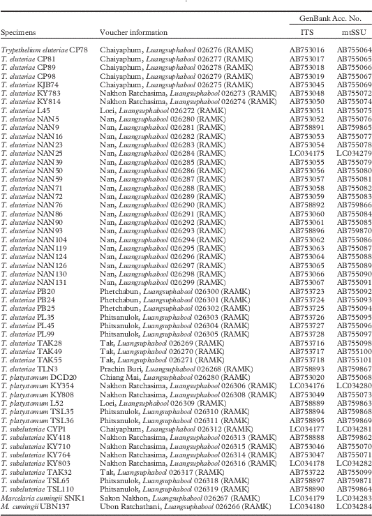
Lichen thalli were collected from various locations in Thailand (Table 1) and were deposited in RAMK. Fifty-four mycobionts were isolated from collected material by ascospore germination on Malt-Yeast-Extract medium as described previously (Sangvichien et al. Reference Sangvichien, Hawksworth and Whalley2011). The cultures were incubated at room temperature (32–35 °C) for 9 weeks and then used for DNA extraction. The cultures are maintained in the lichen research unit at Ramkhamhaeng University (Bangkok).
Voucher information, geographical origin, herbarium accession numbers and GenBank accession numbers for the species and specimens used in this study

DNA extraction, amplification and sequencing
Total genomic DNA was extracted from mycobiont cultures using the CTAB precipitation and chloroform extraction protocol (Cubero & Crespo Reference Cubero and Crespo2002). The ITS and mtSSU rDNA loci of each culture were amplified using the primer pairs ITS1F (Gardes & Bruns Reference Gardes and Bruns1993) with ITS4 (White et al. Reference White, Bruns, Lee and Taylor1990), and mrSSU1 (Zoller et al. Reference Zoller, Scheidegger and Sperisen1999) with MSU7 (Zhou & Stanosz Reference Zhou and Stanosz2001), respectively. Amplifications were performed in a 50 µl reaction mixture containing 5 µl 10× Pfu Buffer with MgSO4, 2 mM of dNTP mix, 20 µM of each primer, 1·25 U of Pfu DNA Polymerase (Thermo Fisher Scientific Inc.), and 5 µl of DNA solution. The amplification reactions were carried out in a thermal cycler TP600 (Takara Shuzo Co., Tokyo) and performed using the following program: initial denaturation for 1 min at 94 °C and 38 cycles of 94 °C for 1 min, 51 °C for 1 min (ITS1F/ITS4) and 53 °C for 45 s (mrSSU1/MSU7), followed by an extension at 72 °C for 1 min, and a final extension at 72 °C for 5 min. The products were cleaned with QIAquick PCR Purification Kit (QIAGEN, Hilden, Germany) according to the manufacturer’s instructions. We sequenced complementary strands using BigDye v3.1 (Applied Biosystems, Foster City, CA, USA) and the same primers used for amplifications. Sequenced PCR products were run on an ABI 3730 automated sequencer (Applied Biosystems) at the Pritzker Laboratory for Molecular Systematics and Evolution at the Field Museum, Chicago, USA.
Phylogenetic analysis
ITS and mtSSU sequences listed in Table 1 were aligned separately using Clustal W (Thompson et al. Reference Thompson, Higgins and Gibson1994) and the alignment was subsequently improved manually using MEGA v.6 software (Tamura et al. Reference Tamura, Stecher, Peterson, Filipski and Kumar2013). Dothidea insculpta (AF027764, FJ190602), D. sambuci (AY930109, AY544739) and Marcelaria cumingii were selected as outgroup, with M. cumingii as a representative of the M. purpurina group, which has been shown to be the sister group to Trypethelium s. str. (Nelsen et al. Reference Nelsen, Lücking, Aptroot, Andrew, Cáceres, Rivas Plata, Gueidan, da Silva Canêz, Knight and Ludwig2014). The best-fit model of nucleotide substitution was chosen using jModelTest v.2.1.4 (Darriba et al. Reference Darriba, Taboada, Doallo and Posada2012) with the Akaike Information Criterion (AIC). Phylogenetic trees were constructed using maximum likelihood (ML) and Bayesian inference (BI). The ML analysis was performed using the program RAxML v.8.0.9 (Stamatakis Reference Stamatakis2006) with the GTRGAMMA model and bootstrap values were calculated using 1000 pseudoreplicates (Stamatakis et al. Reference Stamatakis, Hoover and Rougemont2008). The BI tree and posterior probabilities were calculated using MrBayes v.3.2.1 (Ronquist & Huelsenbeck Reference Ronquist and Huelsenbeck2003) with the same nucleotide substitution model as in the ML analysis. Two independent runs were performed with four Markov chains with 10 000 000 generations, stoprule option and aborting the analyses were set at the average standard deviation of split frequencies of 0·01. Every 100th tree was saved into a file. The phylogenetic trees were viewed using FigTree v.1.3.1 (http://tree.bio.ed.ac.uk/software/figtree/).
Chemical studies
10% KOH was used for spot tests and TLC was performed using standardized methods with solvent systems A and C (Culberson Reference Culberson1972; Lumbsch Reference Lumbsch2002).
Results and Discussion
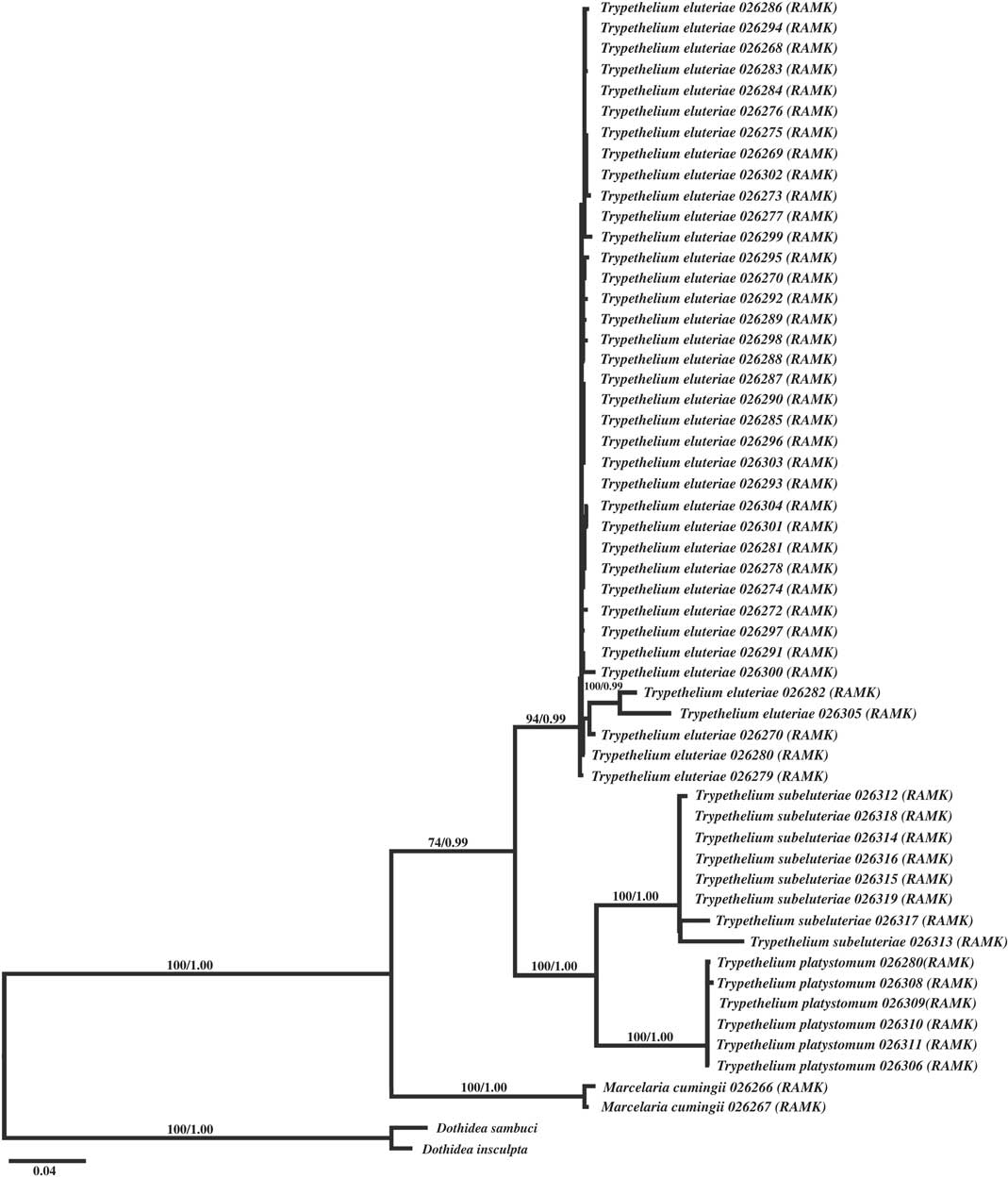
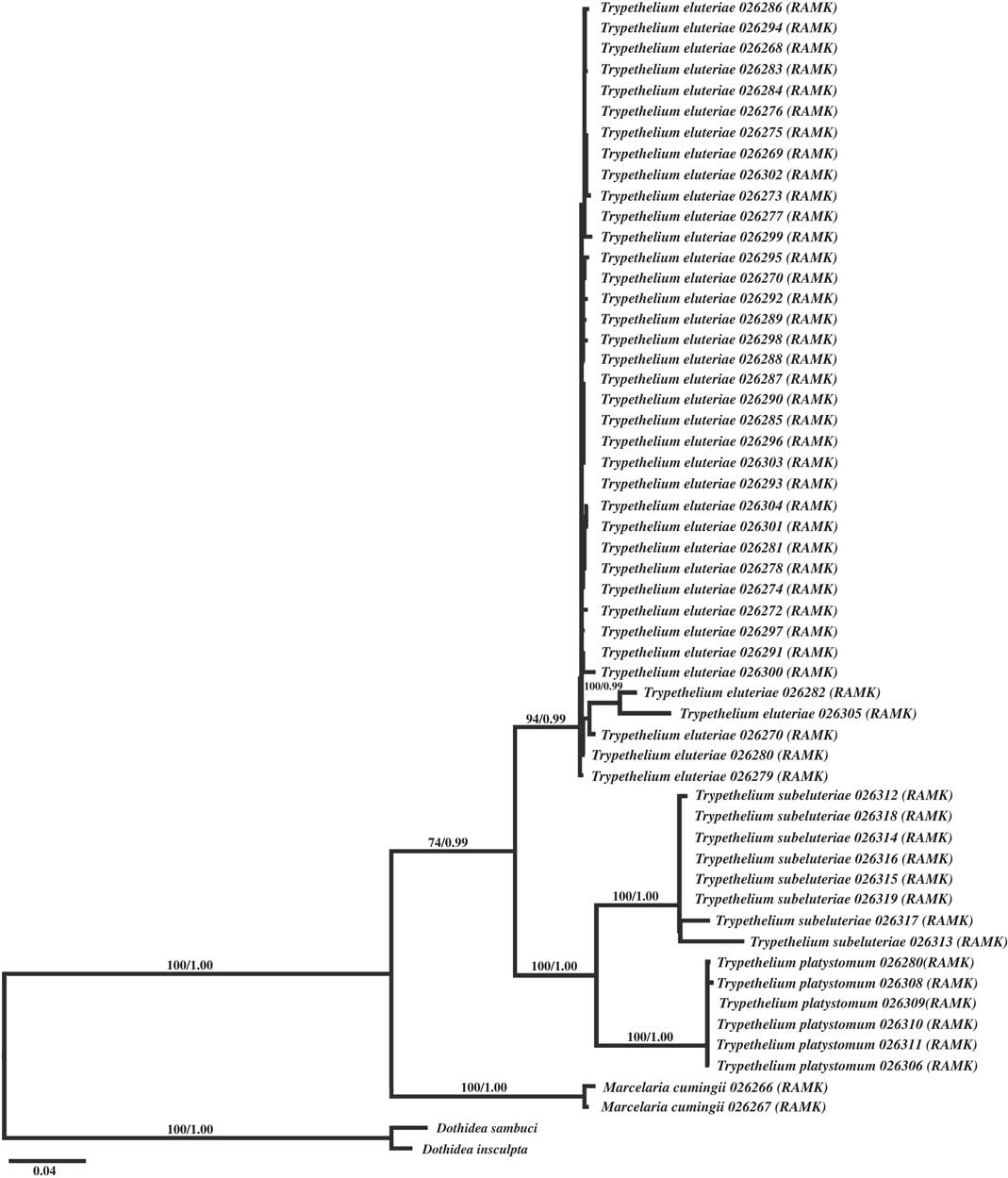
We generated 108 new DNA sequences for this study (54 each for ITS and mtSSU rDNA). The matrix of the ITS data set had 592 unambiguously aligned nucleotide position characters and 341 were variable sites, while the mtSSU data set included 668 unambiguously aligned nucleotide position characters and 210 were variable. A total of 1372 positions were constant in the combined data set. The TIM2+G model was chosen as the best-fit model of nucleotide substitution for the analysis, and the closest model implemented in RAxML and MrBayes (GTR+G) was employed. The two single locus ML trees did not show any incongruence and hence concatenated analyses were performed. The Bayesian and ML trees generated from the combined data set had an identical topology. Here we show the full tree of the results (Fig. 1). The Trypethelium eluteriae s. lat. samples cluster in three distinct and well-supported clades. The majority of samples (38) clustered in a well-supported group (94%/0·99) that morphologically agrees with T. eluteriae s. str. and forms a sister group to a clade including the two other well-supported clades (both 100%/1·00), one including six specimens identified as T. platystomum, and the other clade identified as T. subeluteriae, with eight samples.
Maximum likelihood tree for the Trypethelium eluteriae group based on partial ITS and mtSSU rDNA sequences. ML bootstrap values ≥70% and posterior probabilities ≥0·95 for the Bayesian analysis are given above the branches.
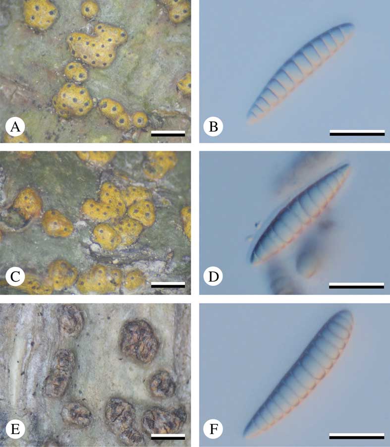
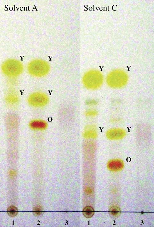
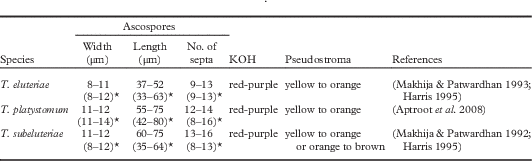
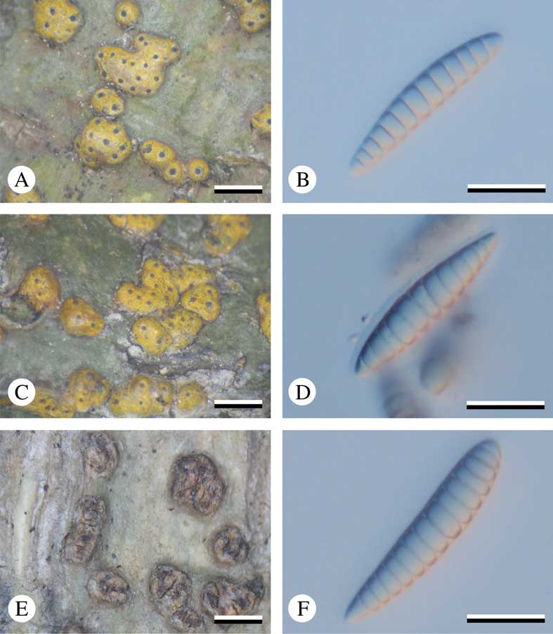
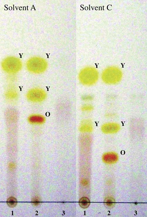
In contrast to some taxonomic treatments that applied a broad circumscription of T. eluteriae (Aptroot Reference Aptroot2009), we found that the T. eluteriae group consists of more than one distinct lineage and that some of the previously synonymized names in fact represent distinct species. The reason for the ambiguity in species delimitation in the group is that some phenotypic characters previously used to distinguish species overlap (Table 2; Fig. 2) and before molecular data were available it was difficult to assess whether this was due to intraspecific variation or not. In particular, the ascospore size and septation, albeit different, shows considerable overlap. In order to search for additional phenotypic characters to distinguish the species in this complex without molecular data, we re-examined the secondary chemistry of the clades and found differences in the chemical profiles. Trypethelium subeluteriae produced three main substances, parietin, emodin and secalonic acid A, whereas T. eluteriae contains only parietin and emodin; T. platystomum did not produce these three substances, but did produce other unidentified anthraquinones (Fig. 3). In the taxonomy of lichenized fungi, secondary metabolites play an important role (Hawksworth Reference Hawksworth1976; Lumbsch Reference Lumbsch1998a , Reference Lumbsch b ) and our results indicate that this character set is potentially also useful in delimiting species in lichenized pyrenomycetes. The distinction of the three species is also supported by the fact that no shared haplotypes were found among species, even when growing sympatrically, such as in Nakhon Ratchasima (data not shown).
T. eluteriae group. A & B, T. eluteriae; A, thallus; B, ascospores. C & D, T. subeluteriae; C, thallus; D, ascospores. E & F, T. platystomum; E, thallus; F, ascospores. Scales: A, C & E=1 mm; B, D & F=20 µm. In colour online.
TLC plates showing the anthraquinone pigments found in the T. eluteriae group. The two major yellow major pigments (Y), parietin (above) and emodin (below), together with the major orange pigment (O) secalonic acid A, are revealed. Key to species: 1=T. eluteriae; 2=T. subeluteriae; 3=T. platystomum. In colour online.
Diagnostic characters of Trypethelium eluteriae, T. platystomum and T. subeluteriae

*Found in this study.
Our data shows that the T. eluteriae group is represented by three species in Thailand and that secondary chemistry is useful in these lichens to characterize taxa phenotypically, in addition to traditionally used ascospore characters.
We would like to thank Kansri Boonpragob, Kajonsak Vongshewarat and Natwida Dangphui (Bangkok) for their suggestions and Warinthorn Chavasiri and Suekanya Jarupinthusophon (Bangkok) for their help in identifying lichen substances. This study was funded by the Royal Thai Government through Ramkhamhaeng University, CU Graduate School Thesis Grant and a scholarship from the Human Resource Development in Science Project (Science Achievement Scholarship of Thailand).