Case report
A female infant was delivered via scheduled caesarean section at 38 weeks of gestation, with a birth weight of 3,006 g. Prenatal echocardiography identified right atrial isomerism, a common atrium, single-ventricle physiology, supracardiac total anomalous pulmonary venous connection, and pulmonary atresia. Severe vertical vein stenosis was also suspected.
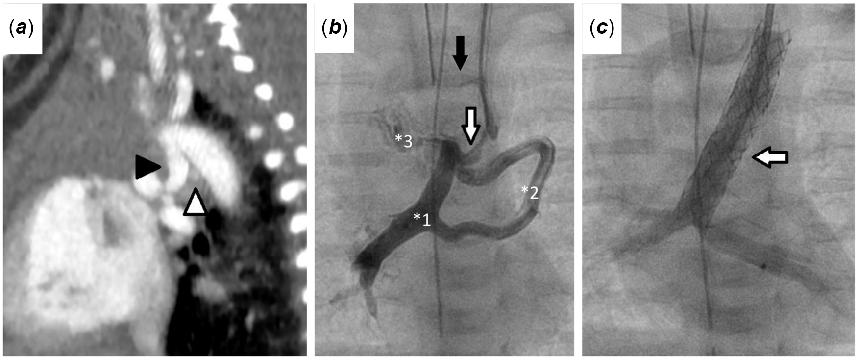
Immediately after birth, she developed respiratory failure resulting from severe pulmonary venous obstruction and required intubation. Despite positive pressure ventilation with pure oxygen, her oxygen saturation remained at 70%. Postnatal echocardiography confirmed the prenatal findings. The four pulmonary veins drained into a common chamber dorsal to the common atrium before emptying into the innominate vein via an ascending vertical vein. The vertical vein exhibited a midline curvature with severe stenosis in the bend. Chest X-ray revealed pulmonary congestion, whereas contrast-enhanced CT showed that the vertical vein ascended tortuously dorsal to the ductus arteriosus, with critical stenosis at the junction (Figure 1a).
( a ) Sagittal CT view demonstrating the crossing site of the vertical vein (white arrow) and ductus arteriosus (black arrow). ( b ) Anteroposterior (AP) cine venogram reveals a common chamber of the pulmonary veins (*1), a critically obstructed vertical vein with a thread-like lumen (white arrow), and the innominate vein (black arrow). It also shows the collateral drainage vein (*2) leading to the mediastinal capillary veins (*3) originating from the common chamber. ( c ) AP cine venogram post-stent implantation showing restored flow through the vertical vein and well-aligned stents (arrow).

At two hours of life, she was transferred to the catheterisation lab. A 5-Fr Prelude™ sheath introducer (Merit Medical, UT, USA) was inserted into the left internal jugular and right femoral veins. The femoral venous introducer was placed as an additional vascular access for the vertical vein and as an emergency route for drug administration. A 0.025-inch Radifocus® guidewire (TERUMO, Tokyo, Japan) and a 4 Fr Judkins right coronary catheter facilitate the delivery of a Sniper® 2 microcatheter (TERUMO) from the left internal jugular vein through the innominate vein into the common chamber. The mean pressures in the common chamber and innominate vein were 17 mmHg and 4 mmHg, respectively. Angiography demonstrated a vertical vein origin diameter of 3.8 mm, with near total occlusion at the midline bend (Figure 1b).
The guidewire was exchanged for a Spindle® XS (ASAHI INTECC, Aichi, Japan), and pre-dilation of the stenosis was performed using a 3-mm Coyote™ balloon (Boston Scientific, MA, USA). The sheath introducer in the left internal jugular vein was advanced into the common chamber over the Spindle® XS guidewire. Three Palmaz Genesis® (5 x15 mm) stents (Cordis, FL, USA) were deployed along the vertical vein: one at the junction with the common chamber, another at the midline curvature, and a third at the connection to the innominate vein. The stents were partially overlapped, lining the entire vertical vein. Post-dilation with a 6 × 20 mm Yoroi® balloon (KANEKA MEDIX, Osaka, Japan) achieved a smoother venous contour (Figure 1c). Following stenting, mean pressures in the common chamber and innominate vein improved to 8 mmHg and 7 mmHg, respectively.
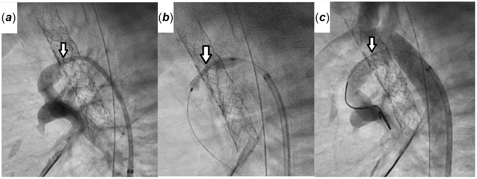
Despite successfully relieving venous obstruction, oxygen saturation dropped to 50% under pure oxygen. Suspecting compression of the ductus arteriosus by the stents, ductal angiography via the single ventricle confirmed severe stenosis. A 4-Fr sheath introducer (Terumo) was then inserted into the right femoral artery. Ductus arteriosus stenting was performed using a 4-Fr Judkins catheter, Sniper® 2 microcatheter, and a 0.025-inch Radifocus® guidewire. The guidewire was exchanged for a Spindle® XS, and a Parent Plus® 30 (MEDIKIT, Tokyo, Japan) was advanced into the ductus arteriosus. A 3.5 mm × 12 mm Synergy™ stent (Boston Scientific) was deployed at the stenotic segment (Figure 2). Vertical vein stent integrity was preserved post-stenting, and oxygen saturation rapidly improved to 80% on ambient air. The stent did not cover the pulmonary end of the ductus arteriosus, and lipo-prostaglandin E1 was continued to maintain ductus arteriosus patency. Anticoagulation therapy with heparin was initiated and later transitioned to aspirin and warfarin.
( a ) Lateral cine angiogram depicting severe ductus arteriosus (DA) obstruction (arrow) and the pulmonary artery. ( b ) Lateral cine views illustrating balloon dilatation of the DA (arrow). ( c ) Lateral cine angiogram following DA stent implantation, demonstrating restored flow through the DA (arrow).

The patient was extubated on day eight post-intervention and discharged from the intensive care unit three days later. Post-procedure recovery was uneventful, with steady weight gain.
At two months of age, balloon re-dilatation was required for stenosis in the vertical vein stents, using a 7 mm × 20 mm Yoroi® balloon across the entire stented segment. Recurrent restenosis necessitated a third catheterisation, during which an Express™ Vascular LD (7 mm × 17 mm stent, Boston Scientific) stent was deployed at 12 atm within the prior stents. Following intervention, pulmonary venous wedge pressure—a surrogate for pulmonary arterial pressure—and common chamber pressures were 17 mmHg and 12 mmHg, respectively. Pulmonary vascular resistance was calculated at 0.89 Wood unit/m2. Right and left pulmonary artery diameters were 5.4 mm and 5.9 mm, with a calculated pulmonary arterial index of 209. These parameters indicated the patient was a suitable candidate for Glenn surgery.
At four months of age, she underwent total anomalous pulmonary venous connection repair and bidirectional Glenn surgery. Although the surgeons attempted to remove both stents, this was not feasible because of strong adhesion to the vessel wall. The vertical vein was divided at the side of the common chamber; however, the innominate vein side was deliberately left untouched because of limited surgical accessibility. The ductus arteriosus was clipped at the aortic side, whereas the pulmonary artery side remained untouched, as surgical intervention was deemed unfeasible. She was extubated on postoperative day two, and her recovery was uneventful.
Discussion
Total anomalous pulmonary venous connection with drainage obstruction typically necessitates urgent surgical intervention. It is an independent risk factor for early and long-term outcomes, particularly in cases of single-ventricle physiology. Reference Kirshbom, Myung and Gaynor1,Reference Hoashi, Kagisaki and Oda2 Several reports have described drainage vein stenting as a temporary measure to facilitate a more stable preoperative condition, allowing for delayed yet optimised open-heart surgery. Reference Kyser, Bengur and Siwik3–Reference Meadows, Marshall, Lock, Scheurer, Laussen and Bacha6 Similarly, ductus arteriosus stenting is well-established in patients dependent on ductal patency for pulmonary or systemic circulation, with numerous reports documenting favourable outcomes. Reference Tseng, Truong and Peck7
In this case, vertical vein stenting inadvertently compressed the ductus arteriosus, necessitating subsequent ductal stenting to restore patency. To our knowledge, no prior reports have documented this specific complication. Total anomalous pulmonary venous connection associated with right atrial isomerism presents a unique anatomic configuration, Reference Cheung, Cheng, Chau, Chiu, Yung and Leung8 and preoperative imaging in our patient revealed a meandering and stenotic vertical vein crossing near the ductus arteriosus. This finding suggested that vertical vein stenting could induce secondary ductal compression and obstruction. Recognising this anatomic relationship facilitated early recognition of the complication when oxygenation failed to improve post-stenting. The decision to stent the ductus arteriosus without extending the stent along its entire length was intentional, aiming to minimise interference between the adjacent stents. We elected not to stent the entire length of the ductus arteriosus for two reasons. First, full stenting could have resulted in straightening of the ductus arteriosus, potentially interfering with the vertical vein. Second, we sought to maintain the ability to modulate pulmonary blood flow by adjusting prostaglandin therapy.
In-stent restenosis resulting from neointimal hyperplasia is commonly observed following vertical vein stenting. Reference Kitano, Yazaki, Kagisaki and Kurosaki9–Reference Itoi10 When using bare-metal stents, a stent diameter of 6 mm or larger is frequently reported. Indeed, in the present case, despite post-dilation to 6 mm, two re-interventions were required prior to the Glenn procedure. Several reports describe the use of 4–5 mm drug-eluting stents, and we have experience with such cases at our institution. However, our outcomes with 4–5 mm drug-eluting stents have not been favourable. Therefore, in this case, we opted for post-dilation with 6 mm for the vertical vein stents. Additionally, the patient’s vertical vein was markedly tortuous, and we anticipated that stenting only the stenotic segment would result in irregular post-stent anatomy, increasing the risk of early restenosis. Thus, despite concerns regarding potential compression of the ductus arteriosus, we chose to place a stent along the entire length of the vertical vein.
If there is anatomic overlap between the vertical vein and ductus arteriosus, stent implantation in the vertical vein may lead to ductal stenosis or occlusion. However, early recognition and proactive management can mitigate this complication, ensuring timely intervention and improved patient outcomes.
Acknowledgements
The authors thank Editage (www.editage.jp) for English language editing.
Financial support
This research received no specific grant from any funding agency, commercial, or not-for-profit sectors.
Competing interests
The authors declare no conflict of interest.
Ethical standard
Ethical approval was not required for this case report as per Kanagawa Children’s Medical Center guidelines. Written informed consent for publication was obtained from the patient’s parents.