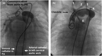
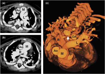
Crossed pulmonary arteries are a very rare form of pulmonary arterial malposition, usually found in association with abnormalities of the ventricular outflow tracts or common arterial trunk. The left pulmonary artery originates from the pulmonary trunk to the right and usually above the origin of the right pulmonary artery.Reference Jue, Lockman and Edwards1 Pulmonary angiography in a four month old female (Fig. 1a) demonstrated this relationship and showed a small amount of radiographic contrast passing into the aorta through a large communication (arrow) between the aorta and pulmonary trunk. Her aortogram (Fig. 1b) revealed a left sided cervical aortic arch, giving off an aberrant right subclavian artery as its final branch (5) after the left subclavian (4), left vertebral (3), left carotid (2) and right carotid (1) arteries. In this picture, a guidewire passes from the pulmonary trunk through the communication into the first branch of the cervical aortic arch. Axial slices of a contrast computed tomographic study, moving from top (Fig. 2a) to bottom (Fig. 2b), show origin of left pulmonary artery (L) from the pulmonary trunk (M) in a plane superior to that of the right pulmonary artery (R). Viewed from above, three dimensional reconstruction (Fig. 2c) demonstrates fusion of contrast between the aorta (A) and pulmonary trunk (M) through a large communication, with the crossing pulmonary arteries lying posteriorly. The retroesophageal aberrant subclavian artery (arrow) is also seen arising from the descending aorta, and passing anterior to the vertebral column. Knowledge of these abnormalities of the pulmonary arteries will help minimise technical difficulties in surgical repair and further catheter interventions.
(a) Pulmonary angiogram. (b) Aortogram.

Axial computed tomogram (a, b) and 3-dimensional reconstruction of computed tomogram (c).