Introduction
In visuo-constructive tasks, demented patients may show the tendency to copying drawings near to or superimposed on the original model. This phenomenon is labeled closing-in (CI) and has been first described by Mayer-Gross (1935). CI is common in normally developing children (Ambron, McIntosh, & Della Sala, Reference Ambron, McIntosh and Della Sala2010; Gainotti, Reference Gainotti1972) and is more often observed in progressive mental deterioration than after focal brain lesions (De Ajuriaguerra, Muller, & Tissot, Reference De Ajuriaguerra, Muller and Tissot1960; Gainotti, Reference Gainotti1972). Among demented patients, CI is predominant in Alzheimer's disease (AD) patients (Ober, Jagust, Koss, Delis, & Friedland, Reference Ober, Jagust, Koss, Delis and Friedland1991; Rouleau, Salmon, & Butters, Reference Rouleau, Salmon and Butters1996), suggesting the idea that CI might represent a neuropsychological marker for clinical diagnosis of Alzheimer's disease (Gainotti, Marra, Villa, Parlato, & Chiaretti, Reference Gainotti, Marra, Villa, Parlato and Chiaretti1998; Gainotti, Parlato, Monteleone, & Carlomagno, Reference Gainotti, Parlato, Monteleone and Carlomagno1992; Kwak, Reference Kwak2004). In demented patients, CI can be observed in several tasks, such as imitation of hand postures (Kwon et al., Reference Kwon, Kang, Lee, Chin, Heilman and Na2002; Mayer-Gross, 1935), writing (De Ajuriaguerra, Zazzo, & Granjon, Reference De Ajuriaguerra, Zazzo and Granjon1949), and three-dimensional construction (Ambron, McIntosh, Allaria, & Della Sala, Reference Ambron, McIntosh, Allaria and Della Sala2009). In copying drawings, two main types of CI can be distinguished, although there is no agreement in the literature about classification criteria. The near-type CI refers to conditions in which patients produce their copy in the space near the model, without coming in contact with it, but different operational criteria have been used across studies (Ambron et al., Reference Ambron, McIntosh, Allaria and Della Sala2009; Conson, Salzano, Manzo, Grossi, & Trojano, Reference Conson, Salzano, Manzo, Grossi and Trojano2009; Kwak, Reference Kwak2004). The adherent-type CI, instead, is characterized by some contact between the copy and the model (Ambron et al., Reference Ambron, McIntosh, Allaria and Della Sala2009; Conson et al., Reference Conson, Salzano, Manzo, Grossi and Trojano2009; Kwak, Reference Kwak2004). Kwak (Reference Kwak2004) proposed to further identify a separate form of CI encompassing situations in which the copy invades the model's space or completely overlap it. It is important to note, however, that one patient can produce different types of CI during a copying task, and that most current theoretical interpretations of CI do not entail different mechanisms to account for different kinds of CI.
According to a first interpretative framework (Lee et al., Reference Lee, Chin, Kang, Kim, Park and Na2004) CI may be a compensatory strategy to overcome deficits of visuo-spatial skills or of visuo-spatial working memory. In this perspective, copying a figure might require to perceive the model and retain its representation during the time required to produce the copy in the empty space. Patients with impaired ability to represent the model, or to momentarily hold its representation, might tend to close-in to the model in the attempt to reduce their difficulties. Therefore, such a “compensation hypothesis” would predict that severity of CI increases as model complexity increases, since this would require more visuo-spatial processing and/or visuo-spatial working memory resources. Lee et al. (Reference Lee, Chin, Kang, Kim, Park and Na2004), indeed, reported that in AD patients figure complexity was related to CI. Moreover, according to this interpretation, a consistent association of CI with visuo-spatial dysfunctions or visuo-spatial working memory deficits would be expected. A recent retrospective study (Serra, Fadda, Perri, Caltagirone, & Carlesimo, Reference Serra, Fadda, Perri, Caltagirone and Carlesimo2009) confirmed that AD patients with CI showed more severe impairments on several visuo-spatial tests, compared to AD patients without CI.
Other accounts for CI focus on the role of frontal/executive processes. It has been suggested that CI might be related to a primitive attraction behavior, for which hands are pulled to act toward the focus of visual attention (Gainotti, Reference Gainotti1972; McIntosh, Ambron, & Della Sala, Reference McIntosh, Ambron and Della Sala2008). In this “attraction” framework, CI would represent a “default” behavior released by defects of attentional-executive abilities (Kwon et al., Reference Kwon, Kang, Lee, Chin, Heilman and Na2002; McIntosh et al., Reference McIntosh, Ambron and Della Sala2008). In analogy with the compensation hypothesis, the “attraction hypothesis” predicts that CI would be enhanced in dealing with complex models because they likely suggest high attentional load, and reduce available resources for monitoring graphic productions. However, this hypothesis specifically envisages that CI can be triggered by any sufficiently attention-demanding visual stimuli, regardless of their relevance to the task at hand (McIntosh et al., Reference McIntosh, Ambron and Della Sala2008). Moreover, if frontal dysfunction is crucial for CI to occur, deviations toward the model should arise in a wide range of tasks, independently from relevant visuo-perceptual impairments.
In AD patients, one single case study (McIntosh et al., Reference McIntosh, Ambron and Della Sala2008), and one retrospective investigation (Ambron et al., Reference Ambron, McIntosh, Allaria and Della Sala2009) provided direct support to the “attraction hypothesis.” McIntosh et al. (Reference McIntosh, Ambron and Della Sala2008) reported a patient with moderate AD who showed CI toward irrelevant visual stimuli during an imitation and a drawing task, with a significant effect of visual stimuli's complexity. In a retrospective study, Ambron et al. (Reference Ambron, McIntosh, Allaria and Della Sala2009) confirmed that CI is related to attentional defects in AD patients, but also found that severe forms of CI are significantly correlated with severe visuo-spatial defects. The authors, indeed, reported that graphic reproductions close to the model (but without touching it: Near type of CI) were predicted by impairments on attentional tasks, whereas copies located onto the model (or contiguous to it: Overlap type of CI) were associated with a combination of attentional and visuo-spatial defects.
To the best of our knowledge, the contrasting hypotheses about the genesis of CI have not been directly tested in a prospective sample of AD patients. The present study had a twofold aim: first, we directly evaluated the possible role of the frontal/executive versus visuo-spatial impairments by means of specific tasks in AD; second we explored which, if any, task assessing frontal/executive abilities is the best predictor of the near and the adherent types of CI in AD patients. Therefore, the present study is the first modern report in which specific hypotheses about the genesis of CI are contrasted in a prospective sample of AD patients, with a particular attention to the relationships between selective defects of visuo-spatial or frontal/executive abilities and the type of graphic production.
If CI is more strongly predicted by tasks assessing frontal/executive functions, then AD patients with CI should achieve poorer scores on frontal/executive tasks than AD patients without CI; conversely, if CI is more strongly predicted by tasks assessing visuo-spatial skills, then AD patients with CI should show lower scores on visuo-spatial tasks than AD patients without CI. Thus, the present study would provide direct support to the attraction hypothesis (executive dysfunction mediated) or to the compensation hypothesis (visuo-spatial dysfunction mediated), respectively. Moreover, if near-CI and adherent-CI are significantly correlated with performance on specific tasks, then distinctive cognitive abilities might account for different forms of CI.
Methods
Participants
AD patients were recruited among outpatients attending the Alzheimer's disease Unit of the “A. Cardarelli” Hospital in Naples, and the Complex Operative Unit of Neurology of the “Santa Anna and San Sebastiano” Hospital in Caserta.
To qualify for the present study, patients had to meet the following criteria: (1) clinical diagnosis of probable AD according to the NINCDS-ADRDA criteria (McKhann et al., Reference McKhann, Drachman, Folstein, Katzman, Price and Stadlan1984); (2) mild to moderate level of AD severity, as characterized by a score ranging 0.5 to 2 points at the Clinical Dementia Rating scale (CDR; Hughes, Berg, Danziger, Coben, & Martin, Reference Hughes, Berg, Danziger, Coben and Martin1982), and by a raw score ranging 10 to 23 on the Italian version of Mini Mental State Examination (Measso et al., Reference Measso, Cavarzeran, Zappala, Lebowitz, Crook, Pirozzolo and Grigoletto1993); (3) formal education of at least 2 years; (4) lack of concomitant severe organ insufficiency or of neoplastic disease.
Sixty-four patients (34 female; age range, 57–86; education range, 2–13) satisfied inclusion and exclusion criteria. All participants gave their written informal consent to participate to the study. This study was conducted in accordance with the Ethical standards of Helsinki Declaration.
Neuropsychological Assessment
In addition to MMSE as a selection criterion, all patients were administered a neuropsychological assessment battery to investigate visuo-spatial and visuo-constructional copying skills, frontal/executive abilities and anterograde memory.
Visuo-spatial skills were evaluated by Corsi span forward (Spinnler, & Tognoni, Reference Spinnler and Tognoni1987), and by Clock drawing task (Mondini, Mapelli, Vestri, & Bisiacchi, Reference Mondini, Mapelli, Vestri and Bisiacchi2003). Corsi span requires patients to reproduce block-tapping sequences of increasing length, and provides an index of working memory capacity for visuo-spatial information (score range, 0–9). The Clock drawing task requires patients to complete a shape of a clock by putting in all the numbers and setting the hands for 2 and 45 (score range, 0–10); this task assesses visuo-spatial skills but also requires semantic knowledge, planning, and constructional abilities, but differently from the tests below it does not entail presentation of a visual model to be copied.
Visuo-constructional copying abilities were assessed by immediate reproduction of Rey Complex Figure (Caffarra, Vezzadini, Dieci, Zonato, & Venneri, Reference Caffarra, Vezzadini, Dieci, Zonato and Venneri2002a; score range, 0–36) and by a modified version of the copying drawing test (Spinnler & Tognoni, Reference Spinnler and Tognoni1987; score range, 0–14; see below for details), requiring patients to reproduce black-and-white models. Both tasks require perception and representation of a visual stimulus and analysis of spatial relations among its components to reproduce it.
Frontal/executive functions were evaluated by the Frontal Assessment Battery (FAB; Appollonio et al., Reference Appollonio, Leone, Isella, Piamarta, Consoli, Villa and Nichelli2005), which includes two verbal and four non-verbal subtests. Verbal subtests require: to identify semantic categories to which sets of objects belong to (similarities subtest) and to orally produce words according to specific rules (phonemic fluency); non-verbal subtests require: to perform sequential movements upon imitation (motor series subtest), to provide motor responses opposite to that shown by the examiner (conflicting instructions), to withhold motor responses (go-no-go task), and inhibit automatic responses to sensorial stimuli (prehension behavior). Each subtest is scored 0 to 3; Italian norms are only available for the total score (range, 0–18), that provides a global measure of cognitive and motor control. Moreover, patients underwent the phonological verbal fluency task (Carlesimo et al., Reference Carlesimo, Caltagirone and Gainotti1996), a frontally loaded task that requires patients to produce in one minute as many words beginning with the letter “F”, “A”, and “S” as they can (score is the sum of words produced correctly).
Monitoring and inhibition of irrelevant responses were assessed by the Stroop color-word test (Caffarra, Vezzadini, Dieci, Zonato, & Venneri, Reference Caffarra, Vezzadini, Dieci, Zonato and Venneri2002b); this test consists of two sections assessing ability to name colors and read words, and of one section requiring inhibitory control, as patients are required to inhibit reading and name the color of the ink in which each word is printed.
Anterograde memory was assessed in the verbal domain by immediate and delayed recall of 15-Word learning test (Caltagirone, Gainotti, Carlesimo, & Parnetti, Reference Caltagirone, Gainotti, Carlesimo and Parnetti1995), that provides two measures: immediate recall (range, 0–75) and delayed recall (range, 0–15). Anterograde memory in the visuo-spatial domain was assessed by delayed reproduction of the Rey Complex Figure (range, 0–36; Caffarra et al., Reference Caffarra, Vezzadini, Dieci, Zonato and Venneri2002a).
Evaluation of CI
Presence and type of CI were assessed on the three copying tasks: Pentagon copying of the MMSE (Measso et al., Reference Measso, Cavarzeran, Zappala, Lebowitz, Crook, Pirozzolo and Grigoletto1993), a modified version of the copying task included in the Spinnler and Tognoni's battery (1987), and copying of the Rey Complex Figure (Caffarra et al., Reference Caffarra, Vezzadini, Dieci, Zonato and Venneri2002a).
In the first two tasks, we used A4 stimulus sheets with their long axis radially aligned with respect to patients, each showing a black-and-white drawing centered in its upper half; the patients were explicitly required to reproduce the model in the lower half of the sheet. Spinnler and Tognoni's copying task includes copying of seven sets of geometrical figures (square, diamond, cube, triangle, circle, and nonsense figures); stimuli are printed in the upper half of A4 sheets, and subjects are requires to reproduce them in the lower half of the sheets. In the traditional version of the test, a black horizontal line is printed in the center of the stimulus sheets to separate models from space in which subjects have to produce their copy; here, this line was deleted to avoid any visual stimulus that could interfere with patients’ graphic productions. The Rey Complex Figure (110 mm high and 150 mm long) was centrally printed on an A4 sheet of paper, with 50-mm margins, in landscape orientation. The stimulus sheet was aligned with the patient's body midline, and patients were required to reproduce the model on a separate A4 sheet of paper, placed 10 mm from the bottom edge of the model's sheet, in landscape orientation; patients were not allowed to move either the stimulus or the response sheets.
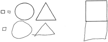
For each graphic reproduction, we recorded presence and type of CI, independently from accuracy (that was scored according to standardized procedures). CI was classified in two types (see Figure 1): adherent-CI (Conson et al., Reference Conson, Salzano, Manzo, Grossi and Trojano2009), when at least one element of the copy overlapped to at least one element of the model (e.g., a side of a square), and near-CI, when any point of the copy was within 10 mm from the lower side of the model (according to Ambron, McIntosh, Finotto, Clerici, Mariani, & Della Sala, Reference Ambron, McIntosh, Finotto, Clerici, Mariani and Della Sala2012). For the Rey Figure, the same method was adopted, but in this case near-CI was classified not only when the copy was produced within the model's page, but also when the figure was copied within 10 mm from the upper edge of the response sheet. Since presence of CI was assessed on 9 stimuli (the MMSE pentagons, the seven models of Spinnler and Tognoni's test, and the Rey complex figure), number of CI ranged 0–9 in each patient.
Examples of the two types of closing-in (CI) considered in the present study: on the left an example of Near-CI, in which the copy was within 10 mm from the lower side of the model; on the right, representative example of adherent-CI, in which the copy partly or totally overlapped the model (models are depicted in the upper row).

Statistical Analyses
Descriptive statistics
We computed number and type of CI in each patient. For descriptive purposes, we divided patients into two groups: AD patients without CI (no-CI group) and AD patients with CI (CI+ group), including in this last group all patients who presented occurrence of at least one type of CI (near or adherent) in at least one graphic task. We additionally divided CI+ patients into two groups (near-CI and adherent-CI), according to the specific type of CI shown in each graphic task (near-CI group included AD patients who showed only near type of CI, while adherent-CI group consisted of AD patients who presented adherent-CI, irrespectively).
Testing hypotheses
The compensation hypothesis would predict that CI is related to poor visuo-spatial skills, whereas according to the attraction hypothesis frontal/executive defects would be associated with CI. To investigate the contribution of visuo-spatial versus frontal/executive defects to CI, we developed composite indices as average Z-scores for relevant tests (high scores meaning better performance). In particular, we computed the following composite Z-scores: visuo-spatial score (including Corsi span forward and Clock drawing task); visuo-constructional score impairment (including the Rey Complex figure and Copying drawings tests); frontal/executive score (including FAB, FAS, Stroop color-word test – time, and Stroop color-word test – errors). Then, a series of hierarchical logistic regression models was run.
As a preliminary step, in the first model, we entered the presence or not of CI as the dependent measure (absence of CI coded as 0, and presence of CI coded as 1), and demographic data (age and education), MMSE, and CDR as independent variables to verify their possible role in predicting CI status.
Then, we run a second model in which the presence or not of CI was used as the dependent measure (coded as above), and the frontal/executive composite Z-score, the visuo-spatial composite Z-score, and the visuo-constructional composite Z-score were sequentially entered as independent variables.
In the third hierarchical logistic regression model, we explored if adherent-CI was qualitatively different from near-CI, or simply represented a more severe form of near-CI. In this case, we entered the type of CI (adherent or near) as the dependent measure (near-CI coded as 0, and adherent-CI coded as 1), whereas the independent variables were entered in the same order as above.
Last, to assess which frontal/executive test could be an independent predictor of adherent versus near type of CI, we run a logistic regression model entering in the same block the four frontal/executive measures as independent variables, with type of CI as the dependent variable (coded as above).
The McKelvey-Zavoina method was used to calculate the percentage of variance in the dependent measure accounted for by the model after adding each independent variable (DeMaris, Reference DeMaris2002). Bootstrap resampling method (1000×) was used to assess accuracy of predictors (Efron & Tibshirani, Reference Efron and Tibshirani1993). To avoid the risk of accepting Type 1 errors related to multiple comparisons, we corrected threshold for statistical significance using the False Discovery Rate (FDR) step-up procedure (Benjamini & Hochberg, Reference Benjamini and Hochberg1995).
All analyses were performed by SAS (version 9.1), complemented by an algorithm for computing McKelvey-Zavoina's R squared (courtesy of A. DeMaris).
Results
Descriptive Statistics
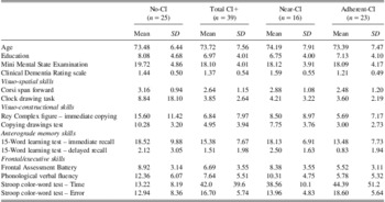
Twenty-five patients (39%) did not produce any CI (no-CI group in Table 1; age range, 58–86; education range, 2–17). CI was found in 179 out of a total of 576 drawings (31%). Thirty-nine AD patients (61%) showed at least one CI in at least one graphic task (CI+ group in Table 1; age range, 57–91; education range, 2–17); 10/39 (25.6%) patients showed CI in pentagon copying (MMSE), 38/39 (97.4%) in figure copying (Spinnler & Tognoni task), and 13/39 (33.3%) in copying Rey complex figure. Patients produced a mean number of 4 CI.
Mean and SD on demographic data, MMSE, CDR, and neuropsychological test scores in AD patients

Note: No-CI = AD patients without closing-in; CI+ = AD patients with CI; near-CI = AD patients with near type of closing-in; adherent-CI = AD patients with both near and adherent type of closing-in; CI = closing-in; AD = Alzheimer's disease; MMSE = Mini Mental State Examination; CDR = Clinical Dementia Rating scale.
With respect to the type of CI, near type only was found in 16/64 AD patients (25% of the total sample, near-CI in Table 1), whereas 3/64 (4.6%) produced the adherent type only, and 20/64 (31.2%) AD patients showed both near and adherent types of CI (we combined these two subgroups in the adherent-CI group in Table 1). Near-CI patients produced 50 errors out of 576 drawings across the three graphic tasks, whereas adherent-CI patients made 114 errors out of 576 drawings.
Means and SDs for demographic data, MMSE, CDR and neuropsychological test scores in each group are reported in Table 1.
Testing Hypotheses
The preliminary logistic regression model showed that demographic data, MMSE, and CDR were not significant predictors of presence or absence of CI (likelihood ratio = 2.86; p = .58; McKelvey-Zavoina's R2 = .058).
The second hierarchical model showed that the frontal/executive composite Z-score significant predicted the CI status (likelihood ratio = 20.72; p < .0001; McKelvey-Zavoina's R2 = .406); entering the visuo-spatial composite Z-score did not improve the model substantially (likelihood ratio = 21.09; p < .0001; McKelvey-Zavoina's R2 = .407), whereas a significant contribution was provided by the visuo-constructional composite Z-score (likelihood ratio = 27.07; p < .0001; McKelvey-Zavoina's R2 = .506). In the final model, only the frontal/executive composite Z-score (odds ratio = .181; 95% confidence interval = .043–.755; p = .019) and the visuo-constructional composite Z-score (odds ratio = .314; 95% confidence interval = .117–.840; p = .021) were significant independent predictors for CI. Bootstrapping produced substantially similar results (frontal/executive composite Z-score: p = .006; visuo-constructional composite Z-score: p = .021).
The third hierarchical logistic regression model showed that the frontal/executive composite Z-score was a significant predictor of adherent versus near type of CI (likelihood ratio = 14.31; p = .0002; McKelvey-Zavoina's R2 = .488); entering the visuo-spatial composite Z-score (likelihood ratio = 15.38; p = .0005; McKelvey-Zavoina's R2 = .514), and then the visuo-constructional composite Z-score (likelihood ratio = 16.44; p = .0002; McKelvey-Zavoina's R2 = .541) did not improve the model substantially. In the final model, only the frontal/executive composite Z-score was a significant independent predictor for type of CI (odds ratio = .020; 95% confidence interval = .001–.465; p = .015), a finding confirmed by the bootstrapping procedure (p = .009).
Last, we performed a logistic regression in which the four frontal executive measures were entered as independent variables in lieu of the frontal executive composite Z-score. Our goal was to determine which individual executive measure was most predictive of adherent versus near CI. As expected, the overall model was statistically significant (likelihood ratio = 19.72; p = .0006; McKelvey-Zavoina's R2 = .629). However, the only significant individual predictor was the number of errors on the Stroop color-word test (odds ratio = 11.05, 95% confidence interval = 1.916–64.001, p = .007); bootstrapping provided similar results (p = .002).
Probability levels of all the four significant predictors identified by regression models can be considered acceptable with respect to the FDR-determined threshold (p = .022)
Discussion
Patients affected by probable AD may show CI in copying drawings. In the present study, we observed a high prevalence of CI (61%), thus confirming that this clinical feature is common even in patients at a mild to moderate disease stage (Ambron et al., Reference Ambron, McIntosh, Allaria and Della Sala2009; Ober et al., Reference Ober, Jagust, Koss, Delis and Friedland1991; Rouleau et al., Reference Rouleau, Salmon and Butters1996).
Early studies considered CI to be related to constructional apraxia, suggesting that disorders of visuo-spatial cognition can account for CI (Critchley, Reference Critchley1953; Mayer Gross, Reference Mayer Gross1935). In our study, the logistic regression showed that the composite visuo-constructional index was significantly associated with presence of CI. This finding, however, does not allow concluding that a causal relationship exists between visuo-spatial disorders and CI. Actually, drawing and visuo-constructional tasks cannot be considered as a “pure” measure of visuo-spatial abilities, since they entail multiple cognitive abilities, including frontal/executive functions (Gasparini et al., Reference Gasparini, Masciarelli, Vanacore, Ottaviani, Salati, Talarico and Bruno2008; Solms, Turnbull, Kaplan-Solms, & Miller, Reference Solms, Turnbull, Kaplan-Solms and Miller1998; Trojano, Grossi, & Flash, Reference Trojano, Grossi and Flash2009); even copying is sensitive to defects in organization and planning abilities (Cosentino, Jefferson, Chute, Kaplan, & Libon, Reference Cosentino, Jefferson, Chute, Kaplan and Libon2004). Consistent with this general statement, some authors maintained that CI should be considered as a symptom distinct from constructional apraxia (Ambron, McIntosh et al., Reference Ambron, McIntosh, Finotto, Clerici, Mariani and Della Sala2012; Ambron et al., Reference Ambron, McIntosh, Allaria and Della Sala2009). In fact, Conson et al. (Reference Conson, Salzano, Manzo, Grossi and Trojano2009) reported a patient with CI occurring independently from severe visuo-spatial disturbances, in whom the tendency to be attracted toward the model, that is, CI, generated severe distortions in reproducing a graphic model. On the basis of these considerations, the association between CI and visuo-constructional impairments (that are not necessarily based on visuo-spatial defects only) cannot serve as a support for the compensation hypothesis.
These considerations are also reinforced by the finding that logistic regression did not show any association between poor scores on visuo-spatial tasks and CI. It is important to underline here that, to assess visuo-spatial abilities, we used two tasks that cannot be considered as “pure” measures, but these tasks have already been used in retrospective clinical studies on CI in AD patients (Ambron et al., Reference Ambron, McIntosh, Finotto, Clerici, Mariani and Della Sala2012; Serra et al., Reference Serra, Fadda, Perri, Caltagirone and Carlesimo2009), and have solid Italian normative data. The Corsi span forward is indeed considered as a test of visuo-spatial working memory, whereas the clock drawing task entails visuo-spatial, as well as semantic and planning abilities (Trojano & Conson, Reference Trojano and Conson2008). The two tests were not significantly associated with CI, thus suggesting that the defect of visuo-spatial abilities per se is not related to CI in our sample.
Recently, in their retrospective study on CI in AD, Serra et al. (Reference Serra, Fadda, Perri, Caltagirone and Carlesimo2009) found that CI patients achieved lower scores than patients without CI on two complex visuo-spatial tasks (Corsi span forward and Raven's Progressive Matrices), and on immediate reproduction of Rey complex figure, but not on one frontal/executive task (a modified card sorting test). Therefore, the findings of Serra et al. were consistent with the view according to which CI might result from an adaptive strategy to compensate spatial dysfunctions (Lee et al., Reference Lee, Chin, Kang, Kim, Park and Na2004). However, there are several differences between Serra et al.'s study and the present one: (i) Serra et al.'s patients had a more pervasive cognitive impairment (median MMSE score = 12.7; median CDR = 2) than the present sample (median MMSE score = 18; median CDR = 1); (ii) Serra et al.'s conclusions referred only to patients with adherent-CI; and (iii) in that retrospective study only a limited assessment of frontal functions was available, in which a floor effect was likely. Therefore, Serra et al.'s study is not directly comparable with the present one. It remains entirely possible that several mechanisms can contribute to the genesis of CI, and that not mutually exclusive defects may determine similar phenomenological manifestations in different stages of disease (see also Ambron et al., Reference Ambron, McIntosh, Allaria and Della Sala2009). Our results, indeed, are best consistent with the view that frontal/executive defects are related to presence of CI in mild to moderate AD patients, thus supporting the idea that a frontal/executive impairment might release an attraction of attention and action toward the visual model (Ambron et al., Reference Ambron, McIntosh, Allaria and Della Sala2009; Gainotti, Reference Gainotti1972; McIntosh et al., Reference McIntosh, Ambron and Della Sala2008), as recently shown in patients with mild cognitive impairment too (Ambron, McIntosh et al., Reference Ambron, McIntosh, Finotto, Clerici, Mariani and Della Sala2012).
Since we used several frontal/executive tasks, we could further refine the “attraction hypothesis” of CI by assessing if specific frontal/executive abilities are related to the different kinds of CI. The logistic regression models showed that adherent-CI was specifically related to more severe impairments of frontal/executive abilities with respect to near-CI, and that in particular the number of irrelevant responses in a test tapping susceptibility to cognitive interference (the Stroop color-word test) was the strongest significant independent predictor of adherent-CI. This pattern is compatible with the idea that AD patients tend to approach the model because of an impairment of their abilities to monitor and inhibit attraction toward salient visual stimuli. Likely, the specific impairment of cognitive control, that is, of the ability to inhibit irrelevant responses, might generate adherent-CI, that represents a strong violation of task instructions and leads patients to use parts of the model as a cue and produce their copies at least partly overlapping to the model.
The present findings provide a direct support to the “attraction” hypothesis and contribute to refine this interpretation of CI, but some limitations of the present study have to be taken into account. First, in the present study, we chosen to assessed visuo-spatial abilities by means of neuropsychological tests requiring a motor response; future studies might address the issue of visuo-spatial perception by means of tests more specifically devised to tap different aspects of visuo-spatial perception independently from motor output (e.g., judgment of line orientation, judgment of point position; Grossi et al., Reference Grossi, Fragassi, Chiacchio, Valoroso, Tuccillo, Perrotta and Trojano2002). Second, sample size was relatively small; it is well known that the lower the subjects-to-variables ratio the lower the generalizability of results to other populations. We tried to overcome this pitfall by bootstrapping methods, but further studies might consider to enroll larger patient samples to adopt more detailed neuropsychological test batteries.
In conclusion, the present prospective study contributes to the investigation of CI in AD patients, showing the prevalent involvement of frontal/executive impairments in its genesis. Moreover, we observed that specific types of CI are significantly associated with different patterns of impairments of frontal/executive functions, since adherent-CI was strongly associated with a defect of the ability to inhibit irrelevant responses. Taken together, the present findings would support the view according to which different manifestations of CI do not lay on simple continuum (Ambron et al., Reference Ambron, McIntosh, Allaria and Della Sala2009), but are related to distinctive cognitive impairments.
Acknowledgments
The authors have no conflict of interest to disclose. The study has not received any external financial support.