Background
Accessory mitral valve tissue is an infrequent congenital cardiac anomaly. The first reported case of this lesion and its first surgical repair were described in 1842 and 1963, respectively.Reference Yetkin, Cuglan, Turhan and Yalta1 Its prevalence is reported in 1 per 26,000 echocardiograms, and it is frequently associated with other congenital anomalies. Embryologically, accessory mitral valve tissue is due to an incomplete separation of the mitral valve from the endocardium cushions. Most of its echocardiographic, clinical, and anatomical descriptions are product of case reports among multiple centres especially in the United States, Canada, India, and China. Prifti et al. classified accessory mitral valve tissue according to its morphology in fixed and mobile.Reference Prifti, Bonacchi, Bartolozzi, Frati, Leacche and Vanini2 Yetkin et al. classified it as having attachments on the supra leaflets level (type I), attachments on the mitral leaflets (type II), and attachments below the mitral leaflets (type III).Reference Yetkin, Cuglan, Turhan and Yalta1 The researchers hereby present a case of an asymptomatic patient who was diagnosed with accessory mitral valve tissue.
Case presentation
A 10-month-old female patient with a medical history of myelomeningocele is referred to our centre due to a murmur detected at a control consult. At physical examination, we detected a III/VI, systolic murmur, in the aortic area radiating toward the suprasternal area, reduced S2 sound, normal pulses, and symmetric blood pressure. A chest radiography showed an abnormally large cardiac silhouette, predominantly due to large left cavities, lungs without infiltrates or pleural effusion. An electrocardiogram showed sinus rhythm, with a QRS axis + 30 degrees, and a prominent R and S in V1-V2, suggestive of dilation of the right ventricle (Supplementary Figure S1).
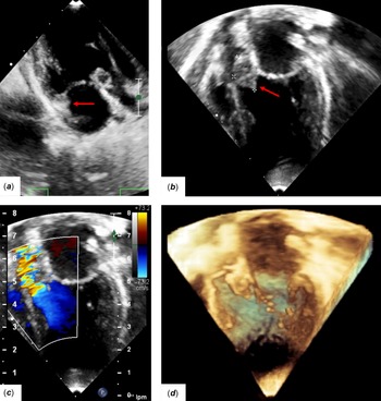
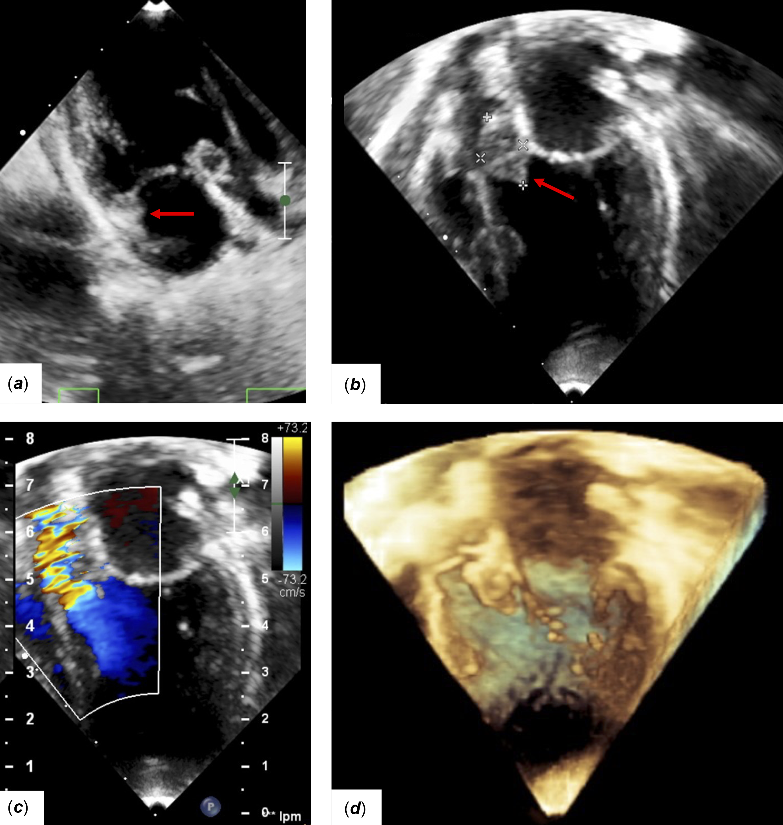
During admission, a transthoracic echocardiogram was performed. It reported a 5 mm patent foramen ovale, a 5 mm perimembranous ventricular septal defect with left-to-right shunt with a 32 mmHg gradient, dilation of left cavities and a 3 mm patent ductus arteriosus with a left-to-right shunt with an 80 mmHg gradient, biventricular function was preserved. A 16 × 9 mm cystic, spheric, mobile, pedunculated mass was observed (Fig. 1a, b). Color Doppler shows an obstruction of the left ventricular outflow, generating a 39 mmHg gradient (Fig. 1c). Three-dimensional reconstruction of the echocardiogram shows that the mass is attached to the subvalvular mitral apparatus and protrudes through the left ventricular outlet tract during systole (Fig. 1d).
Transthoracic echocardiogram (TTE) findings. (a) TTE images demonstrate a spheric pedunculated mass in the left ventricle, (b ) with cystic characteristics and a dimension of 16 x 9 mm. (c) Color doppler proves obstruction of the left outflow tract with a gradient of 39 mmHg. (d) Three-dimensional reconstruction of the echocardiogram illustrates that the mass is attached to the subvalvular mitral apparatus.
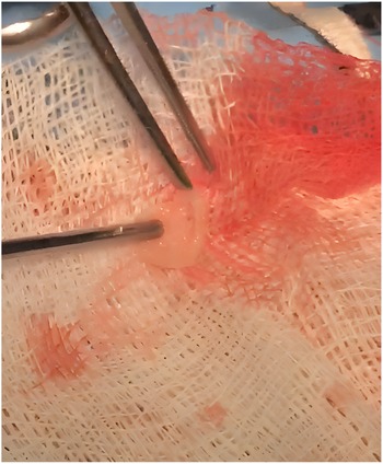
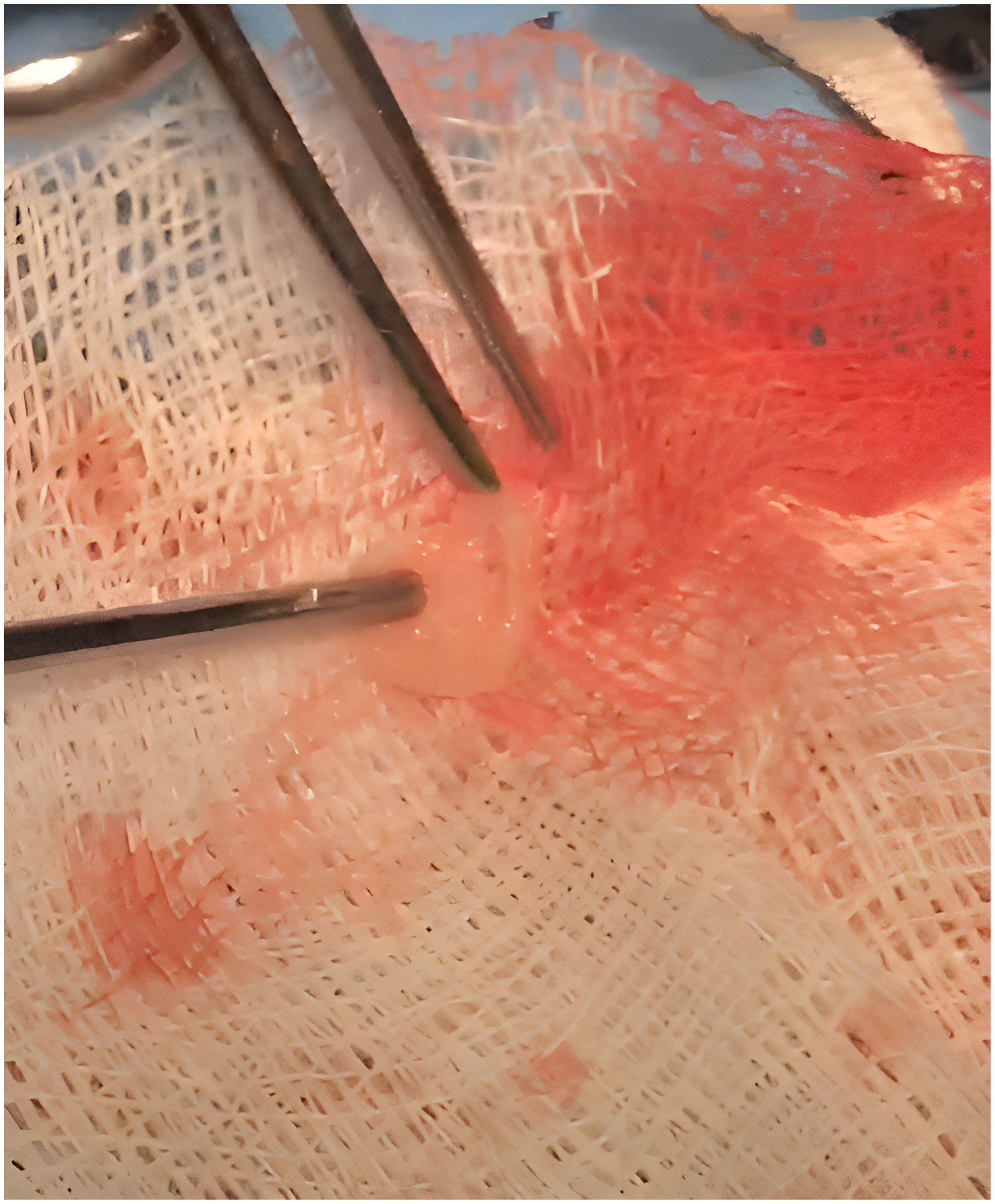
The surgery was programmed and performed with extracorporeal bypass, and the mass was removed and sent to pathology (Fig. 2). The patent foramen ovale, ventricular septal defects, and patent ductus arteriosus were closed with direct stitches to the atrial septum, a patch, and with a ligature, respectively. The surgeon describes the mass as with a membranous consistency. During the early immediate postoperative, the patient presents pleural effusion and an atrioventricular junctional tachyarrhythmia, both treated and resolved. A transthoracic echocardiogram was performed postoperatively demonstrating absence of the mass, and a left ventricular outlet tract without any significant gradient, a mild residual mitral regurgitation was found. An 11-month follow-up reported the patient asymptomatic. The histopathology study of the piece reports a fibrous tissue of mitral characteristics.
Surgical piece of the resected accessory mitral valve tissue.
Discussion
Accessory mitral valve tissue is a rare cause of obstruction of the left ventricular outflow tract, which usually presents as an asymptomatic heart murmur. It is described that patients present symptoms beyond 50 mmHg of gradient. The symptoms usually present after the first decade of life. The clinical manifestations have a wide spectrum, and the most common symptoms are syncope, shortness of breath, thoracic pain, and arrhythmias.Reference Panduranga, Eapen, Al-Maskari and Al-Farqani3 This pathology is commonly associated to other CHDs, usually of the left side of the heart, such as ventricular septal defects (19.21%), subaortic stenosis (9.6%), hypertrophy of the left ventricle (8.6%), transposition of the great arteries (7.7%), atrial septal defects (7.7%), aortic coarctation (4.8%), Ebstein’s anomaly (0.96%), and others.Reference Manganaro, Zito and Khandheria4
The key echocardiographic characteristics that suggest accessory mitral valve tissue are the protrusion and retraction of the mass through the aortic valve during systole and diastole, attachment to the mitral valve apparatus (subvalvular, anterior mitral valve, or a papillary muscle), and its appearance (cystic, pediculated, or valve-like form). There are other pathologies that resemble these characteristics. Three significant differential diagnoses are vegetations, intracardiac tumors, and thrombus. According to Prifti et al., our patient can be classified as Type IIA due to its mobile and pedunculated characteristics.Reference Prifti, Bonacchi, Bartolozzi, Frati, Leacche and Vanini2
Our patient was asymptomatic and eventually underwent surgery. The decision to resect the mass was taken since accessory mitral valve tissue is at potential risk of cardiac embolism, as well as the presence of associated CHD. Its mobile and membranous nature perpetuates adherence of platelets and fibrin making it an environment that favors the formation of thrombus. It has been described that this illness is at higher risk of sudden death. The indications for surgical resection of this disease are the gradient of obstruction, the presence of symptoms, associated HD, and the risk of thrombosis. Surgically, it is relevant to define adequately the relation between the anterior mitral valve and the accessory mitral valve tissue. The resection must be done cautiously to avoid lesions to the mitral valve.Reference Manganaro, Zito and Khandheria4 Postoperative mortality is reported in 9%, and the most common complications are residual stenosis (13%), reintervention (13%), mitral regurgitation (10%), and aortic regurgitation (10%).Reference Said5
Accessory mitral valve tissue should be included as a differential diagnosis when a mass is found in the left ventricular outflow tract. The description of its echocardiographic characteristics in the literature is fundamental for clinicians to suspect this pathology. This case also reviews its differential diagnoses and the need to contemplate surgical resection due to its association with other CHD, risk of sudden death, and thrombosis.
Supplementary material
The supplementary material for this article can be found at https://doi.org/10.1017/S1047951123003293.
Acknowledgements
None.
Author contribution
José M. Galindo-Hayashi: Conception and design, acquisition of data, analysis, and interpretation, revising the article.
Enrique G. Villarreal: Conception and design and revising the article.
Ely R Sanchez-Félix: Acquisition of echocardiographic images, drafting discussion, revising the article.
Financial support
This research received no specific grant from any funding agency, commercial, or not-for-profit sectors.
Competing interests
None.
Ethical standards
Since it is a descriptive study, with no intervention with humans or animals, it did not require ethics approval.