Introduction
Thrombotic and embolic complications are common after a Fontan palliation. The most common event is Fontan conduit/right atrial thrombus.Reference Egbe, Connolly and Niaz1 Thrombus in a rudimentary left ventricle is exceedingly rare and has portended poor neurological outcomes in previous reports.Reference Lee, Baek and Kim2,Reference Rosenthal, Friedman and Kleinman3 Here, we report an asymptomatic male teenager who was incidentally discovered to harbour an isolated, large, obliterative, and organised thrombus in situ within his rudimentary left ventricle cavity 15 years following his Fontan palliation. This complication developed despite primary prophylaxis with aspirin and in the absence of any additional identifiable pro-thrombotic milieu. To our knowledge, this is the first case of thrombus in rudimentary left ventricle without neurological sequelae.
Case report
A 17-year-old boy who had undergone a Fontan palliation presented for annual follow-up in our cardiology clinic. He was born at full term with no antenatal issues. He was diagnosed with hypoplastic left heart syndrome (aortic atresia with mitral stenosis) at 8 days of age when he presented in cardiogenic shock. He underwent staged palliation with an uneventful Norwood with Blalock–Taussig shunt, followed by hemi-Fontan and lateral tunnel fenestrated Fontan completion at ages of 2 weeks, 8 months, and 20 months, respectively. He did not have any surgery performed on the mitral valve during any of three stages of surgical palliation. His fenestration closed spontaneously at 5 years of age. His past history was pertinent for an episode of clinically symptomatic atrial flutter at 8 years of age requiring electric cardioversion followed by initiation of aspirin therapy. There was no recurrence of any tachyarrhythmia. His previous Holter monitors had yielded normal results without any silent atrial/ventricular arrhythmias. His exercise capacity, oxygen saturation, pulmonary function, chronotropic, and blood pressure responses were deemed normal based on a maximal cardiopulmonary treadmill exercise stress test performed 1 year previously at the age of 16 years. He remained compliant with aspirin therapy and was taking no other medications.
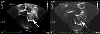
He was asymptomatic at presentation and denied any neurological complaints. The routine transthoracic echocardiogram yielded the unexpected finding of a large organised thrombus, completely filling the rudimentary left ventricle. The thrombus measured 3.5 × 3 cm in size (Fig 1a). Colour Doppler echocardiogram showed an evidence of mitral stenosis including moving mitral leaflets and trivial mitral valve regurgitation. It was difficult to assess mitral inflow with Doppler echocardiogram due to high left ventricular intracardiac pressures. The systemic right ventricular systolic function was subjectively normal. There was only mild tricuspid valve regurgitation. No other thrombus was identified in the intracardiac or Fontan pathway. The echocardiogram obtained 1 year ago did not reveal any thrombus and therefore this was new interval finding. The electrocardiogram demonstrated normal sinus rhythm without any changes compared to the previous year.
Left ventricular thrombus (arrow) in echocardiogram. (a) pre-treatment, (b) post-treatment.

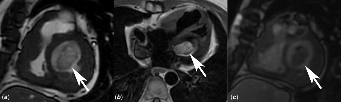
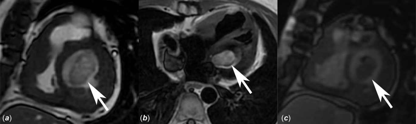
A cardiac MRI was subsequently obtained to assess the extent of left ventricle thrombus as well as to rule out any additional intracardiac thrombus. The sequence with steady-state free precession and T1-weighted double inversion recovery fast spin-echo showed filling defect in the rudimentary left ventricle compatible with thrombus, but no additional clots in the heart (Fig 2a and b). The Fontan pathway was wide open. The delayed gadolinium-enhanced inversion recovery imaging with 600 ms of inversion time nicely delineated a thrombus in the left ventricle, almost completely occupying the entire left ventricle (Fig 2c). Therapeutic oral anticoagulation with warfarin was promptly instituted at a dose of 5 mg/day. His International Normalised Ratio (INR) was maintained within the range of 2–3. Transthoracic echocardiogram demonstrated a significant decrease in the size (2.2 × 2.2 cm) of thrombus at a follow-up visit 3 months later (Fig 1b). At the time of follow-up, his INR was 2.1. His course was not complicated with any neurological sequelae at any point.
Left ventricular thrombus (arrow) in cardiac MRI. (a) Sagittal steady-state free precession imaging, (b) axial T1-weighted double inversion recovery fast spin-echo imaging, (c) delayed gadolinium-enhanced inversion recovery imaging with 600 ms of inversion time.
Discussion
The current literature suggests that 5–33% patients following a Fontan palliation may incur a thrombotic and embolic complication. The overwhelming majority of these are right atrial/Fontan conduit thrombus (38%), pulmonary embolus (32%), intracardiac thrombus (14%), and ischemic stroke (13%).Reference Egbe, Connolly and Niaz1 There are only four reported patients with left ventricle thrombus following a Fontan palliation.Reference Lee, Baek and Kim2–Reference Coon, Rychik and Novelle4 Of these, only Lee et al. have furnished a descriptive report of a male patient with double outlet right ventricle and recurrent thrombosis within the rudimentary left ventricle. This patient was diagnosed with cerebral infraction and presented 4 years (age of 7 years) pursuant to a lateral tunnel Fontan palliation with associated symptoms of syncope, motor weakness, and vomiting. The thrombus occurred despite continuous medication with warfarin. Subsequently, the patient required thrombectomy. The authors emphasised the clinical significance of thrombosis an intracardiac blind pouch, formed by the pulmonary stump or rudimentary ventricle, after a Fontan operation.Reference Lee, Baek and Kim2 Our patient may also require thrombectomy in the future in consideration of possible systemic embolisation from the thrombus in the rudimentary left ventricle. However, the plan is to continue warfarin therapy at present since there was an improvement in the size of thrombus over the 3 months of treatment. The other three reported patients are not well elaborated.Reference Rosenthal, Friedman and Kleinman3,Reference Coon, Rychik and Novelle4
In older patients following Fontan palliation, the images of echocardiogram are suboptimal due to multiple surgeries, scar formation, and change in the shape of chest. In addition, the slow passive flow within the Fontan conduit and pulmonary arteries makes visualisation of thrombus in these areas quite challenging. However, despite this low sensitivity (23%), the specificity of transthoracic echocardiogram remains high (96%).Reference Srichai, Junor and Rodriguez5 So given the low false positivity, a “possible” diagnosis when rendered even in an asymptomatic patient should mandate initiation of therapeutic anticoagulation to avoid serious complications. In such circumstances, it is always preferable to establish a confirmatory diagnosis utilising a cardiac MRI (sensitivity 88%; specificity 99%) rather than a transesophageal echocardiography (sensitivity only 40%).Reference Srichai, Junor and Rodriguez5
Cardiac MRI circumvents the issue of suboptimal images and can differentiate thrombus from tumours.Reference Slonimsky, Konen and Di Segni6 Although there was no suspicion of left ventricle tumour in our patient, the cardiac MRI more importantly excluded any additional intracardiac thrombus. The recommended imaging sequences pertinent for evaluation of intracardiac masses include post-contrast sequences such as steady-state free precession, T1, and delayed gadolinium enhancement. These methodologies are useful since thrombi are typically not enhanced after contrast administration.Reference Slonimsky, Konen and Di Segni6 A long inversion time (600 ms) for delayed gadolinium enhancement as employed in our case may increase in the sensitivity of this technique for thrombus detection.Reference Hoey, Mankad and Al-Chalabi7
In summary, this case highlights the perplexing clinical triad of (i) a late onset, but fortuitously identified, clinically silent thrombus, (ii) an unexpected location such as the rudimentary left ventricle, (iii) an unsuspecting patient with Fontan palliation devoid of the “usual” thrombogenic risk factors. Antithrombotic prophylaxis with aspirin failed in this patient, but warfarin therapy proved effective in reducing the size of thrombus. Cardiac MRI remains the gold standard for this diagnosis. However, the clinical caveat that a “positive” result noted on transthoracic echocardiogram merits prompt initiation of therapeutic anticoagulation should not be overlooked.
Financial support
This research received no specific grant from any funding agency, commercial or not-for-profit sectors.
Conflicts of interest
None.
Author contributions
D.T.: writing the manuscript; all authors: critically reviewing the manuscript.