INTRODUCTION
The species Echinococcus granulosus (Batsch, 1786) consists of a number of distinct entities, which have been distinguished on the basis of morphological, developmental, biochemical or epidemiological characteristics (Thompson and McManus, 2002). Today, these variants are referred to as strains, and they have been generally named after their typical intermediate hosts. Molecular studies have also revealed increasing evidence of considerable genetic variability in E. granulosus. To date, 10 genotypes (G1-10) have been identified, and the genotypic grouping follows closely the strain categorization (Bowles et al. 1992, 1994; Scott et al. 1997; Lavikainen et al. 2003).
Some of the E. granulosus strains differ biologically and genetically so clearly from each other, that a taxonomic revision of the genus Echinococcus has been proposed (Thompson et al. 1995; Thompson, 2002; Thompson and McManus, 2002; Jenkins et al. 2005). In that revision, only the common sheep strain (genotype G1) with closely related genotypes (G2-3) constitutes the species E. granulosus sensu stricto. Furthermore, the horse (G4) and cattle (G5) strains are evidently so distant, that they have been suggested to be distinct species. Based upon former morphological descriptions, the horse strain has been proposed to be named as E. equinus Williams et Sweatman, 1963, and the cattle strain as E. ortleppi López-Neyra et Soler Planas, 1943. The taxonomic status of the camel, pig and cervid strains has remained unclear, although they evidently belong to a single species (Xiao et al. 2005; Thompson et al. 2006).
In cervids, different variants of E. granulosus seem to exist. In 1963, 2 morphologically distinct subspecies were described in North America, namely E. granulosus canadensis in the reindeer-dog cycle and E. g. borealis in the moose-wolf/dog cycle (Sweatman and Williams, 1963). The former was suggested to be an introduced subspecies from Scandinavia, and the latter an indigenous North-American form. Later, 2 distinct genotypes were found. The cervid strain (G8) was characterized genetically by Bowles et al. (1994) from hydatid cysts of the North American moose (Alces alces). Recently, the E. granulosus strain in cervids in North-Eastern Finland was found to represent a distinct genotype, which was named as the genotype G10, the Fennoscandian cervid strain (Lavikainen et al. 2003).
The genetic variation of E. granulosus in cervids is still only partially known. According to a recent study, both G8 and G10 genotypes occur in North American cervids, and G10 appears to be the dominant one (Thompson et al. 2006). This finding questioned the existence of 2 geographically distinct subspecies or strains in cervids, and raised a need for further studies in Europe, where very few isolates have been genotyped. The original characterization of the G10 genotype was based on 5 Finnish isolates of cervid origin (Lavikainen et al. 2003). Recently, G10 was found from a wolf (Canis lupus) in Estonia (Moks et al. 2005). In Sweden, hydatid cysts have been found sporadically from cervids, but the genotype of Swedish E. granulosus has so far been unknown. The aim of the present study was to find out the degree of genetic variation of E. granulosus in cervids in Finland and Sweden, and to define the phylogenetic relationships of the Fennoscandian cervid strain with other strains of E. granulosus.
MATERIALS AND METHODS
Parasite material and DNA extraction
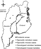
In the present study, 23 Finnish E. granulosus isolates (cysts) from reindeer (Rangifer tarandus) and 9 isolates from moose (designated also as ‘elk’ in Europe) were characterized. In addition, 1 Swedish reindeer E. granulosus isolate was genotyped. The Finnish reindeer isolates were collected in 2001–2003 from 22 individual animals (2 of the cysts were from a single animal), which came from 5 separate reindeer herding cooperatives. The Swedish E. granulosus isolate was from a reindeer, slaughtered in 1997 in Vilhelmina in the county of West Bothnia, over 450 km west of the westernmost cases of reindeer hydatidosis diagnosed in Finland. All the cysts of reindeer origin were found during routine meat inspection. Moose lungs were collected in collaboration with local Finnish hunters especially to search for echinococcal cysts. The isolates genotyped in this study were found from 9 moose shot in 2002–2004 in the regions of North Ostrobothnia, Kainuu and North Karelia. The geographical origins of the characterized cervid isolates are presented in Fig. 1.

Fig. 1. Cystic echinococcosis in reindeer (Rangifer tarandus) and moose (Alces alces) in Sweden and Finland in 1980–2004, and geographical distribution of the isolates, which were genetically characterized in the present study. In the endemic areas (shown in black) in Finland, altogether 85 reindeer cases and 9 moose cases were found during the 25-year period. The number in parenthesis indicates the number of genotyped reindeer isolates from 2 closely located reindeer herding cooperatives. One genotyped moose isolate is missing from the map due to insufficient data about the exact location. Data from Hirvelä-Koski et al. 2003; the database of the National Veterinary and Food Research Institute of Finland (EELA); the database of the National Veterinary Institute of Sweden (SVA); Dan Christensson, personal communication; Sauli Holmström, personal communication.
For phylogenetic analysis, mitochondrial genes were sequenced from certain G10 and G5 isolates of E. granulosus. The 4 DNA samples of the G10 genotype were from the same Finnish reindeer isolates that were used in the original genetic characterization of the Fennoscandian cervid strain (Lavikainen et al. 2003). DNA of the cattle strain (G5) was kindly provided by Dr Joke van der Giessen from the National Institute for Public Health and the Environment, Bilthoven, Netherlands.
For validation of new PCR primers (Table 1, see also section PCR and sequencing below), DNA from 3 E. granulosus sheep strain (G1) isolates from human patients were used. The patients were operated at the Helsinki University Central Hospital in 2002–2004. According to patient documents, the patients were classified as being infected in North Africa, Middle East and Eastern Europe.

The isolates were stored frozen at −20 °C. The Swedish isolate was fixed in 70% ethanol before it was sent to the University of Helsinki for analysis. DNA was extracted from 25–35 mg portions of protoscoleces or germinal layers from each isolate using the DNeasy™ Tissue Kit (Qiagen, Hilden, Germany).
PCR and sequencing
The strain determination of the Echinococcus isolates of cervid origin was based on sequencing of a region of the mitochondrial NADH dehydrogenase subunit 1 (ND1) gene. From the Swedish isolate, also complete ATPase subunit 6 (ATP6) and NADH dehydrogenase subunit 3 (ND3) genes, and partial cytochrome c oxidase subunit 1 (CO1) gene were sequenced to compare this geographically most distant isolate to the Finnish representatives of the genotype G10. For the phylogenetic analysis, ND1, ATP6, ND3 and CO1 were sequenced from representatives of the G10 and G5 genotypes.
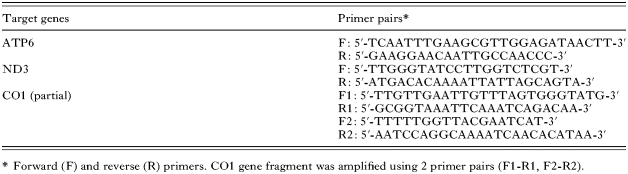
The ND1 gene fragment was amplified by PCR using previously published oligonucleotide primers (Bowles and McManus, 1993). To amplify the ATP6 and ND3 genes and a fragment of the CO1 gene, 4 new primer pairs (Table 1) were designed by selecting evolutionarily conserved regions of the published complete mitochondrial genomes of the sheep (G1) and horse (G4) strains of E. granulosus (Le et al. 2002) and E. multilocularis Leuckart, 1863 (Nakao et al. 2002), and published regions of mitochondrial genomes of the G6-8 genotypes of E. granulosus (McManus et al. 2002). DNA samples of the G1 isolates were used to verify that the correct areas could be amplified using the novel primers. Specificity of the primers was checked using reindeer and bovine DNA as negative controls.
PCR and sequencing were carried out as previously described (Lavikainen et al. 2003) with only minor modifications. Briefly, the PCR reaction mixture (100 μl) contained 2–5 μl of template DNA, 125 μM of each dNTP, 1 mM MgCl2, 10 pmol of each primer, 2 units of Taq DNA polymerase (Amersham Pharmacia Biotech, Piscataway, NJ, USA) and Taq PCR reaction buffer. The PCR conditions were as follows: an initial denaturing step at 94 °C for 10 min, followed by 35 cycles of 60 sec at 94 °C, 50 °C, and 72 °C, and a final extension step at 72 °C for 10 min. The samples were electrophoresed on a 1·5% (w/v) Tris-borate/EDTA agarose gel stained with ethidium bromide for DNA visualization. PCR products were purified by the QIAquick® PCR purification Kit (Qiagen), or excised from the agarose gels and purified by the QIAquick® Gel Extraction Kit. The sequencing was performed by using the ABI Prism® Dye Terminator Sequencing Kit (Applied Biosystems, Foster City, USA) and the ABI Prism® 3100 Genetic Analyzer. The sequencing primers were the same as used for the PCR.
Phylogenetic analysis
The Fennoscandian cervid (G10) and cattle strains (G5) of E. granulosus, and previously published sequences of 5 E. granulosus genotypes (G1, G4, G6, G7 and G8; Bowles and McManus, 1993; Le et al. 2002; McManus et al. 2002) and E. multilocularis (Nakao et al. 2002) were included into the phylogenetic analysis with Taenia crassiceps (Zeder, 1800) (Le et al. 2000) as an outgroup. Sequences of the mitochondrial genes ATP6, CO1, ND1 and ND3 were aligned using the ClustalW program (Thompson et al. 1994). The resulting alignments were refined manually and all ambiguous sites and gaps were removed before the alignments were concatenated.
All the phylogenetic analyses were performed using PAUP* 4.0b10 (Sinauer Associates Inc., Sunderland, MA, USA). For the analyses, an evolutionary model for the concatenated alignment was selected using program ModelTest v. 3.06 (Posanda and Crandall, 1998). Maximum parsimony (MP) and Maximum likelihood (ML) criterion tree searches were performed using the branch-and-bound algorithm. Relative support of the nodes was assessed using 100 (ML) or 1000 (MP) bootstrap replicates and the branch-and-bound algorithm. Phylogeny was also inferred with the neighbour-joining (NJ) method using 1000 bootstrap replicates.
The genetic divergence between strains and strain clusters of E. granulosus was compared to divergence between the 2 closely related species of Taenia, namely T. saginata Goeze, 1782, and T. asiatica Eom et Rim, 1993. For sequences of T. saginata and T. asiatica, see Gasser et al. (1999), Nakao et al. (2000), Yamasaki et al. (2004) and Jeon et al. (2005). The average sequence identity percentages of the aligned ND1, ATP6 and CO1 genes were used for comparison.
RESULTS
All the 33 cervid isolates from Finland and Sweden had ND1 (471 bp) sequences identical to the previously published G10 genotype (Lavikainen et al. 2003). In addition, the Swedish reindeer isolate and the original G10 reindeer isolates had identical ATP6 (513 bp), ND3 (348 bp) and CO1 (708 bp) sequences. The sequences have been submitted to the GenBank database with the Accession numbers DQ020475 (ATP6) and DQ021916 (ND3 and CO1). The ND1 sequence obtained for the Dutch cattle strain isolate in this study and that of the previously published G5 genotype (Bowles and McManus, 1993) differed in 2 positions (positions 126 and 420; see Bowles and McManus, 1993). The ND1, ATP6, ND3 and CO1 sequences of the cattle isolate have been submitted to the GenBank database with the Accession numbers DQ402037 (ND1), DQ318953 (ATP6) and DQ318954 (ND3 and CO1).
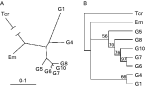
The concatenated sequence alignment comprised of 1548 unambiguously aligned positions, of which 592 were variable and 215 parsimony informative. The selected model for ML and NJ analyses was Tamura–Nei's model (Tamura and Nei, 1993) using invariable sites and gamma distribution with the following parameters: purine-purine change 11·7948, pyrimidine-pyrimidine change 15·9116, transversions 1·00, proportion of invariable sites 0·4272, and α-parameter of gamma distribution 1·9105. In ML, NJ and MP analyses, the cattle (G5), camel (G6), pig (G7), cervid (G8) and Fennoscandian cervid (G10) strains clustered similarly in a well-supported monophyletic group (ML tree presented in Fig. 2). This group became divided into 2 close evolutionary lineages, which were G5 and its sister group formed by G6-10. Concerning positions of the sheep (G1) and horse (G4) strains, and E. multilocularis, the phylogenetic resolution was less clear, and topologies of the ML, NJ and MP trees were slightly different.

Fig. 2. Maximum likelihood tree inferred from the alignment of concatenated sequences of ND1, ND3, CO1 and ATP6 genes of the selected Echinococcus granulosus genotypes (G1, G4, G5, G6, G7, G8, G10), E. multilocularis (Em) and Taenia crassiceps (Tcr), which was used as an outgroup. (A) Radial tree presentation of the phylogeny of E. granulosus genotypes, where the distance scale is proportional to 0·1 substitutions per site. (B) The phylogeny of E. granulosus genotypes shown as a cladogram. The tree was drawn using the 50% majority rule. The numbers at the nodes indicate percentage of bootstrap support for that node.
The average nucleotide sequence divergence of ND1, ATP6 and CO1 genes was 7% between the G6-10 cluster and G5, and 6% between T. saginata and T. asiatica. When the G6-10 cluster was compared to G1 and G4, the average differences were 14% and 13%, respectively. The G1 and G4 genotypes differed from each other by 12%.
DISCUSSION
Previous studies have shown that 2 different genotypes of E. granulosus (G8 and G10) occur in cervids (Bowles et al. 1994; Lavikainen et al. 2003), and they coexist in North America (Thompson et al. 2006). Phylogenetic analyses based on relatively short mitochondrial sequences have suggested close relationship of the cervid genotypes with the pig and camel strains.
In the present study, the existence of only 1 genotype (G10) of E. granulosus in Finnish cervids was demostrated. G10 is thus perhaps the sole genotype in the North-European wildlife. This hypothesis is supported by discovery of G10 from Sweden in this study and the previous finding of G10 from Estonia (Moks et al. 2005). On the other hand, genetic variation could be missing in our material just because the parasite is so uncommon in Finland, which is a result of rarity of definitive hosts. In Finland, E. granulosus occurs in the north-eastern and eastern parts of the country in the life-cycle between wolves and cervids (Hirvelä-Koski et al. 2003). According to limited evidence, the parasite has not spread to dogs (Maijala et al. 2002; Hirvelä-Koski et al. 2003; Lavikainen, 2005).
The present study also proved that G10 occurs in Sweden. The same genotype may have been the causative agent of former endemic human cases in Sweden. Human cystic hydatidosis was a significant public health problem among reindeer herding Sàmi population in Swedish Lapland until 1960's (Lavikainen, 2005). Later, the parasite was virtually eradicated from the reindeer-dog cycle, and endemic human cases have not been diagnosed any longer for several decades. The present-day life-cycle of E. granulosus in Sweden is unclear. Although the Swedish wolf population grew rapidly during the 1990's, it is still relatively small and has not spread to the same areas (Aronson and Sand, 2004), where the few infected cervids have been found. According to some reports, the fox (Vulpes vulpes) can be infected with Echinococcus of cervid origin (Söderhjelm, 1945; Sweatman and Williams, 1963; Kikot', 1980). Thus, foxes could possibly maintain the rather weak sylvatic life-cycle of E. granulosus in Sweden.
The current phylogenetic analysis of multiple locuses of mitochondrial DNA confirms previous findings (e.g. Thompson et al. 1995; Lavikainen et al. 2003; Xiao et al. 2005; Thompson et al. 2006) about the close affinity between the pig, camel and cervid strains. These strains cluster into a monophyletic group, and the cervid genotypes can not be separated out from this group. Furthermore, new molecular data of the cattle strain used in this study support the close relationship between the G6-10 cluster and the cattle strain. These sister groups differ clearly from the sheep and the horse strains. Echinococcus oligarthrus (Diesing, 1863), E. vogeli Rausch et Bernstein, 1972, and E. shiquicus Xiao et al. 2005, were not included in the present phylogenetic analysis due to insufficient molecular data. However, the lack of these taxa does not probably have any fundamental effects on the topology of the distal branches of the phylogenetic tree, concerning closely related E. granulosus strains.
The distinctiveness between the G6-10 cluster and the common sheep strain (‘E. granulosus s. s.’, see Introduction) is at least as obvious as it is between the sheep and the horse (‘E. equinus’) strains. Thus, the classification of this cluster as a distinct species is justified. A more arguable question is the relationship of the G6-10 group with its sister taxon, the cattle strain (‘E. ortleppi’). These closely related genotypes also share similar morphological characteristics (Thompson et al. 2006). In fact, these sisters with related taxonomical uncertainty resemble another pair of taeniid tapeworms: Taenia saginata and T. asiatica. Some authors have regarded T. asiatica only as a subspecies of T. saginata (e.g. Loos-Frank, 2000; Flisser et al. 2004), whereas others consider the species status of T. asiatica fully proven (Hoberg, 2006). Genetic divergence alone is insufficient to allow separation of sister taxa to distinct species, but also morphological comparison and evidence of reproductive and ecological isolation are required (Hoberg, 2006). The status and validity of T. asiatica and T. saginata as independent species are supported by numerous criteria including sympatry, and differences in morphology, epidemiology and life history (Hoberg, 2006). In the case of the G6-10 group and the cattle strain, further morphological studies, investigations of geographical distributions and occurrence in overlapping life-cycles, and detection of possible cross-breeding are required before the taxonomical revision can be made.
We are grateful to Joke van der Giessen for providing cattle strain DNA for phylogenetic analysis, and to Finnish hunters for collecting moose lungs for our Echinococcus research programme. This work was financed by Research Foundation of the University of Helsinki and the Academy of Finland.