INTRODUCTION
Traditionally, pain-alleviating therapies target neurons because they are the cells transmitting nociceptive signals. Glial cells in the peripheral and central (CNS) and peripheral nervous systems have long been recognized for their responses to injury, typified by the astrocytosis in the CNS and by Schwann cell activation in the periphery. It has also become clear, however, that glial cells also play a critical role in the genesis and persistence of pain (Watkins et al., Reference Watkins, Martin, Ulrich, Tracey and Maier1997; Sweitzer et al., Reference Sweitzer, Schubert and DeLeo2001; Raghavendra et al., Reference Raghavendra, Tanga and DeLeo2003).
There are far fewer satellite glial cells (SGCs) than astrocytes or Schwann cells, yet because of their unique location in sensory and autonomic ganglion, SGCs can strongly influence nociceptive sensation. Like other glial cells, the SGC responds to nerve injury by upregulating glial fibrillary acidic protein (GFAP) expression and undergoing division (Humbertson et al., Reference Humbertson, Zimmermann and Leedy1969; Elson et al., Reference Elson, Simmons and Speck2004b; Zhang et al., Reference Zhang, Mei, Zhang, Ma, White and Donnelly2009) in response to injury (Gunjigake et al., Reference Gunjigake, Goto, Nakao, Kobayashi and Yamaguchi2009). Evidence that SGCs proliferate comes from the observation of mitotic figures (Humbertson et al., Reference Humbertson, Zimmermann and Leedy1969) and BrdU positive cells surrounding ganglion neurons following the nerve damage (Cecchini et al., Reference Cecchini, Ferri, Ciaroni, Cuppini, Ambrogini and Papa1999; Elson et al., Reference Elson, Speck and Simmons2003, Reference Elson, Ribeiro, Perelson, Simmons and Speck2004a). One caveat to these studies is that specific markers were not used to unambiguously identify SGCs, rather they were recognized by morphological criteria alone. Given that invading immune cells that proliferate in the vicinity of sensory neurons are morphologically similar to SGCs (see below), this is an area requiring additional investigation. After nerve injury, resident macrophages and circulating white blood cells proliferate within the sensory ganglion (Hu and McLachlan, Reference Hu and McLachlan2003; Mika et al., Reference Mika, Rojewska, Makuch and Przewlocka2009; van Velzen et al., Reference van Velzen, Laman, Kleinjan, Poot, Osterhaus and Verjans2009), most likely in response to chemoattractants released by SCGs analogous to those released by Schwann cells (Campana, Reference Campana2007). The recent report that in the human trigeminal ganglion, cells surrounding the local neuron have many characteristics of macrophages and dendritic cells (van Velzen et al., Reference van Velzen, Laman, Kleinjan, Poot, Osterhaus and Verjans2009) makes it even more relevant to determine the nature of cells referred to in the above-mentioned published studies.
In addition to an increase in the number of SCGs following the nerve injury, there is also an increased coupling between SGCs, which is believed to result from an increase in the number of gap junctions (Huang and Hanani, Reference Huang and Hanani2005; Ledda et al., Reference Ledda, Blum, De Palo and Hanani2009; Zhang et al., Reference Zhang, Mei, Zhang, Ma, White and Donnelly2009). While it has been suggested that coupling between SGCs might play a role in the development or maintenance of neuropathic pain (Hanani et al., 2002; Cherkas et al., Reference Cherkas, Huang, Pannicke, Tal, Reichenbach and Hanani2004), it is unclear what the underlying mechanism is. One possibility is that the increase number of gap junctions allows recycling more glutamate (Ohara et al., Reference Ohara, Vit, Bhargava and Jasmin2008), which in turn acts to increase the pain behavior.
Glutamate is the principal neurotransmitter used by primary sensory neurons and is usually thought of in terms of release from central terminals in the spinal cord. There is circumstantial evidence that paracrine release of glutamate may occur within the ganglion given that glutamate receptors are expressed on the somata of primary sensory neurons (Carlton and Hargett, Reference Carlton and Hargett2007) and that SGCs contain all the protein necessary for the uptake and recycling of glutamate. Glial uptake of extracellular glutamate following neuronal release is the first step in glutamate recycling (i.e. the glutamate–glutamine cycle). The uptake is carried out by different transporters including the glutamate–aspartate transporter (GLAST) (Gadea and Lopez-Colome, Reference Gadea and Lopez-Colome2001). We previously showed that reducing GLAST expression in SGCs in the trigeminal ganglion of non-injured rats resulted in a decreased threshold to mechanical stimulation of the face (Jasmin et al., Reference Jasmin, Vit, Sundberg and Ohara2009). These data imply that there is a basal release of glutamate in the normal ganglion, and interrupting uptake by SGCs can alter the excitability of sensory neurons. One way to verify this hypothesis is to act on the glutamate–glutamine cycle downstream from the transporters. To this end, we chose to block the activity of the SGCs enzyme glutamine synthetase (GS). We also attempted reconstituting the GABA-glutamine cycle by making SGCs express the human transgene glutamine decarboxylase (GAD). Although there is no precedent of introducing the GAD gene in SGCs, primary sensory neurons have been transfected with GAD, resulting in analgesia caused by GABA being transported to the spinal cord (Liu et al., Reference Liu, Wolfe, Hao, Huang, Glorioso and Mata2004; Hao et al., Reference Hao, Mata, Wolfe, Huang, Glorioso and Fink2005; Lee et al., Reference Lee, Fink and Mata2006). In contrast, our approach involves GABA being released within the ganglion, around sensory neurons. As DRG neurons express GABA receptors, the hope was that inducing SGCs to release GABA would reduce nociceptive responses. At the same time it would confirm that SGCs can actively uptake and metabolize glutamate.
OBJECTIVE
Using the trigeminal system, we addressed the following questions regarding SGCs and neuropathic pain: (1) positively identify dividing cells surrounding neurons following nerve injury; (2) examine the consequences of decreasing coupling between SGCs after nerve injury; (3) examine the effect of blocking SGC glutamate metabolism on pain behavior; and (4) genetically modify SGCs to convert glutamate to GABA and examine the effects on nociceptive behavior.
METHODS
Animals and surgeries
ANIMALS
Adult male Sprague-Dawley weighing between 270 and 330 g were housed on a 12 h light–dark cycle and given food and water ad libitum. Procedures for the maintenance and use of the experimental animals conformed to the regulations of UCSF and Cedars-Sinai Medical Center Committees on Animal Research and were carried out in accordance with the guidelines of the NIH regulations on animal use and care (Publication 85-23, Revised 1996).
CHRONIC CONSTRICTION INJURY (CCI) OF THE INFRA-ORBITAL NERVE (ION)
The procedure for CCI of the ION is described in detail in a previous publication (Kernisant et al., Reference Kernisant, Gear, Jasmin, Vit and Ohara2008). Briefly, rats were anesthetized with mixture of ketamine (90 mg/kg; www.abbott.com) and xylazine (10 mg/kg; www.phoenixpharm.com). Once anesthetized, rats were placed in the stereotaxic head holder, and the skull and nasal bone were exposed through a 2-cm skin incision. The superficial muscle was detached from the rostral upper edge of the orbit. Gentle retraction of the orbital contents exposed the ION. Two 5-0 chromic gut ligatures (2 mm apart) were loosely tied around the exposed nerve. The incision was closed using 6-0 silk sutures or the CCI of the ION was followed by implantation of a guide cannula (see below).
CANNULA IMPLANT FOR INJECTION IN THE MAXILLARY DIVISION OF THE TRIGEMINAL GANGLION
The skull was exposed and a burr hole was drilled above the location of the maxillary division of the left trigeminal ganglion at 6.5 mm anterior to inter-aural zero and 2.3 mm lateral to the midline. A guide cannula pedestal (www.plastics1.com) was fixed to the skull over the burr hole using three stainless steel screws (www.aaronsmachinescrews.com) and dental acrylic cement. The guide cannula extended into the burr hole 1 mm below the pedestal but did not touch the surface of the cortex. At least 7 days were allowed for recovery from surgery before injection into the trigeminal ganglion.
INJECTION INTO THE TRIGEMINAL GANGLION
Each animal received a single injection of double-stranded RNA (dsRNA) in a lipid carrier (lipofectamine; Bhargava et al., Reference Bhargava, Dallman, Pearce and Choi2004). Prior to the injection, rats were lightly anesthetized with isoflurane. A 33-gauge beveled stainless steel cannula (www.plastics1.com) was inserted through the guide cannula (positioned over the maxillary division of the left trigeminal ganglion as described above) to 9.5 mm below the cortical surface. The injection cannula was connected to a 25-µl Hamilton syringe attached to a microinjection pump set to deliver 2 µl over a 1-min period.
Synthesis of dsRNA and preparation for injection
Total RNA was extracted from rat brain tissue. A reverse transcriptase reaction was set up using 1 mg of RNA. Complementary DNA (cDNA) of genes of interest produced by a 30-cycle PCR using gene-specific primers were cloned into pTOPO vector (Invitrogen). Rat β-globin sequences were used as nonspecific dsRNA control and were described previously (Bhargava et al., Reference Bhargava, Dallman, Pearce and Choi2004). Sense and antisense RNA were synthesized from cDNA inserts by using MegaScript RNA kit (Ambion) according to the manufacturer's specification.
Prior to the injection in the trigeminal ganglion, 15 µg of dsRNA were mixed with lipofectamine-2000 (www.invitrogen.com) in a final volume of 5 µl at room temperature. After a 30 min incubation, the red fluorescent marker DiI, DiIC18(3) or 1,1′-dioctadecyl-3,3,3′,3′-tetramethylindocarbocyanine perchlorate (www.probes.invitrogen.com) was added to the mixture at a final concentration of 10 mM.
BrdU injection
All animals received two intraperitoneal (i.p.) injections of BrdU (100 mg/kg each injection, 20 mg/ml in phosphate-buffered saline (PBS; pH 7.2) with 0.1% NaOH; Sigma), every 12 h for 2 days beginning at the time the CCI surgery was performed.
Behavioral testing
MECHANICAL (VON FREY HAIR) TESTING
Three von Frey hairs, 2, 10 and 50 g corresponding to log units 4.31, 5.07 and 5.88, respectively, were used in the study based on our own preliminary testing and the results of Ren and Dubner (Reference Ren and Dubner1999). During testing, mechanical stimulation was done with increasing intensities. Each filament was first applied on the side contralateral to the dsRNA injection or to the CCI of the ION. The stimulation consisted of five to six consecutive applications performed at 5-s intervals in slightly different area at the center and around the vibrissal pad, as well as in the perioral and perinasal territory. The scoring of the rat behavioral response was based on the method of Vos and colleagues (Vos et al., Reference Vos, Strassman and Maciewicz1994) as follows: 0 = no detection; 1 = detection and exploration of the von Frey hair; 2 = head withdrawal and/or single grabbing movement; 3 = attack and/or escape and/or multiple grabbing movements; and 4 = active asymmetrical grooming directed toward the stimulated facial area. For each hair, the highest score was recorded and the results are presented as the average of the highest score obtained from the three different hairs. The average value is presented because the analysis of scores from each hair separately gave the same results when compared between groups.
CONDITIONAL REWARD
Rats were tested for avoidance of an innocuous stimulus when attempting to obtain a reward of sugar-sweetened water (20% sucrose) delivered from a drinking tube. Rats that acclimated to sweetened water in their home cage quickly developed a preference for sweetened water and would drink in the test chamber without the need for water deprivation (Davis and Campbell, Reference Davis and Campbell1973). The testing apparatus (Med Associates) consisted of a chamber (20 cm × 20 cm × 20 cm) with a 5 cm × 5 cm × 3 cm alcove in one wall with a stainless steel drinking spout located at the back. A conductance-measuring device (Lickometer, Ugo Basile) attached to the drinking spout was used to count the number of licks when the rat drank from the spout. Brushes were positioned on the left side of the drinking alcove (ipsilateral to the injected trigeminal ganglion or to the CCI of the ION), so that the rats had to maintain contact with the brushes while drinking. The number and the position of the brushes were set up to stimulate the vibrissal pad as well as the perinasal and perioral area of the rat face. During testing, rats were placed in the chamber for a minimum of 5 min and returned to their homecage after 5 min or after the 10th attempt. The observer recorded the number of successful and unsuccessful licking attempts. A licking episode (successful attempt) was defined as an episode where the rat had a minimum of six licks before withdrawing its head from the drinking alcove. A licking episode could be one continuous series of licks or several periods of licking without removing the head from the drinking area. The number of licks was corrected for the weight of animals and the results of the licking behavior are expressed as number of licks per licking episode and per kg weight of rat.
TESTING SCHEDULE
For all experiments, a treatment-blinded observer conducted behavioral testing between 10:00 am and 4:00 pm. On each testing day, rats were brought into the behavior room at least 30 min prior to the test session in order to habituate them to the environment.
OROFACIAL FORMALIN TEST
One day prior to the formalin test, the rats were acclimated for 1 h to the testing chambers (44 × 24 × 24 cm). On the testing day, 50 µl of 2.5% formalin solution in saline was injected subcutaneously with a 30-gauge hypodermic needle into the left upper lip, lateral to the midline. The animal was then immediately placed into the testing chamber and continuously observed for 44 min, during which time the nociceptive behavior (i.e. face-rubs) was quantified. Data were collected using a computer program that records the pain behavior in successive 4-min bins. One hour after formalin injection the rats were euthanized and perfused for histological examination of the trigeminal ganglia.
Tissue processing
HISTOLOGY
Rats were deeply anesthetized with 100 mg/kg of pentobarbital (i.p.). For light microscopy, rats were perfused transcardially with 10% formalin. The left and right trigeminal ganglia were post-fixed in the same fixative for 30 min and then placed in 30% sucrose in PBS (pH 7.4) for 48 h. Left and right trigeminal ganglia from each animal were embedded together in Tissue Freezing Medium (www.trianglebiomedical.com) and cut longitudinally at 10 µm on a cryostat.
IMMUNOHISTOCHEMISTRY
Sections were blocked in 5% normal goat serum (NGS), 0.3% triton X-100 (www.sigmaaldrich.com) in PBS for 1 h and then incubated in the primary antiserum, BrdU (Sheep, 1:1000, US Biologicals), CD45 (Mouse, 1:750, BD Pharmingen,), Cx43 (Rb, 1:1000, Zymed and Rb, 1:2000, Sigma), ED1 (Mouse, 1:2000, Serotoec), ED2 (Mouse, 1:500, Serotec), glutamine synthase (Goat, 1:100, Santa Cruz), SK3 (Rabbit, 1:5000, Alamone) in 5% NGS and 0.3% Triton in PBS for 24 h. DAPI counterstaining was used on all sections to label all nuclei in order identify all cell whether they were immuno labeled or not.
For fluorescence analysis, a fluorescein (FITC)-tagged secondary antibody (www.vectorlabs.com), diluted 1:400 in 0.3% Triton/PBS was used for 1 h. Sections were then washed, mounted on slides and coverslipped with Vectashield (www.vectorlabs.com). For double immunolabeling, primary antibodies raised in different species were used and the primary antibodies and appropriate secondaries applied consecutively. To control for specificity of labeling, the primary antibody was absorbed with the peptide against which it was raised and we confirmed there was no tissue labeling following this procedure.
RESULTS
SGC division following nerve injury
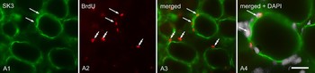
We used specific glial immunomarkers in conjunction with BrdU to positively identify trigeminal ganglion SGCs that have undergone division (Fig. 1) after trigeminal nerve injury. It should be noted, however, that not all dividing cells in the ganglia are SGCs (Fig. 1) although many of these dividing non-SGCs lie close to ganglion cells. Double labeling with markers of immune cells allowed us to determine they are not microglia and likely to be leukocytes (see below).
Cell division in SGCs following nerve injury. Four days following CCI of the ION and 2 days after BrdU injection, BrdU immuno-labeled nuclei (red, arrows) are present in SGCs identified by the SGC-specific marker SK3 (green; Vit et al., Reference Vit, Jasmin, Bhargava and Ohara2006). Other BrdU positive nuclei of an unidentified population of cells are also found in close proximity to SGCs. Scale bar, A1–A3 = 50 µm; A4 = 25 µm.

SGCs and immune response following nerve injury
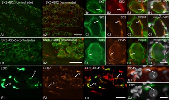
Neuropathic pain-like behavior was first induced by a CCI of the ION. Following nerve injury, we observed a marked increase in the number of ED1 (circulating macrophages), ED2 (resident macrophages) and CD45 (protein tyrosine phosphatase, receptor C, which is present in all circulating white blood cells) positive cells throughout the ganglion and frequently in close proximity to neurons. We were particularly interested in the identity of cells that were in close proximity to the neurons (Fig. 2), as previously reported (Hu and McLachlan, Reference Hu and McLachlan2002; Hu et al., Reference Hu, Bembrick, Keay and McLachlan2007). Some immune cells even become inserted between the SGCs and the neurons (Fig. 2B1–4). Similar to a previous report (Hu et al., Reference Hu, Bembrick, Keay and McLachlan2007) that described MHC-II immunopositive cell processes between SGCs and neurons 7 days after nerve injury, we saw ED2 immunopositive cell processes between SGCs and neurons as early as 4 days post-lesion. Similar to the recent report in humans (van Velzen et al., Reference van Velzen, Laman, Kleinjan, Poot, Osterhaus and Verjans2009), we found that some SGCs also expressed CD45 immunoreactivity (Fig. 2C,E). But unlike humans, we found CD45 positive cells were present in ganglia only on the side of the nerve injury, and not on the contralateral side. Interestingly, CD45 positive immune cells were not detectable in non-injured animals. Finally, some cells with the morphology and location typical of SGCs expressed both ED2 and CD45 immunoreactivity.
Immune response and SGCs following CCI of the ION. Following a CCI of the ION, there is an increase in number and staining intensity of both ED2- (red cells in A1–C4) and CD45- (red cells in D1–E4) immunopositive cells. It is common to see both ED2- and CD45 immunopositive cells lying closely apposed to SGCs. In some instances the immune cells appear to lie between the neuron cell body and the SGC (B3, arrow). In some cases, SK3 positive SGCs are also immunopositive for ED2 (C1–C4, arrow) or CD45 (E1–E4, arrow). In other cases, cells that appear to be SGCs based on morphological and location criteria (F1–G2), exhibit both ED2 and CD45 immunoreactivity. Letter a and b indicate the same element in F1–F3. N = nucleus. Scale bars, A1–A2 = 120 µm; B1–B4 = 10 µm; D1–D2 = 60 µm; E1–E4 = 30 µm; F1–F3 = 50 µm; G1–G2 = 20 µm.

Changes in gap junction coupling between SGCs can change nociception
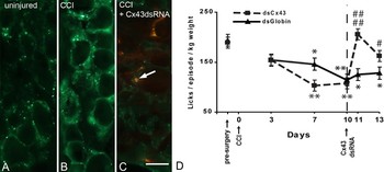
To investigate further what role gap junction might play in injury induced chronic pain, we used RNA interference (RNAi) to silence the expression of connexin (Cx43) in SGCs (Vit et al., Reference Vit, Jasmin, Bhargava and Ohara2006; Ohara et al., Reference Vit, Ohara, Bhargava, Kelley and Jasmin2008; Procacci et al., Reference Procacci, Magnaghi and Pannese2008). Following a CCI of the ION, the rats developed a long lasting decrease in sensory thresholds accompanied by an increase in Cx43 immunostaining in the trigeminal ganglion as determined by immunohistochemistry (Hanani et al., Reference Hanani, Huang, Cherkas, Ledda and Pannese2002; Pannese et al., Reference Pannese, Ledda, Cherkas, Huang and Hanani2003; Ohara et al., Reference Ohara, Vit, Bhargava and Jasmin2008). Cx43 dsRNA was then injected into the trigeminal ganglion 10 days after the placement of the ligature, and resulted in reduction of Cx43 expression as well as evoked pain behavior that lasted approximately 2 days (Fig. 3). In contrast, when Cx43 dsRNA was injected in rats without a CCI of the ION (Ohara et al., Reference Ohara, Vit, Bhargava and Jasmin2008) there was an increase in nociceptive responses that were equivalent to the values found following CCI of the ION (% change from baseline, licks/episode/kg body weight, CCI of ION, 11.73 ± 1.17; Cx43dsRNA, 10.66 ± 2.24; P > 0.05, n = 8 per group). Therefore the results of knocking down Cx43, and thereby reducing gap junction function, gave opposite results depending on whether or not nerve injury was present.
Cx43 dsRNA injection transiently reduces expression of Cx43 and increases nociceptive thresholds. (A) Cx43 immunostaining (green) is present throughout the trigeminal ganglia as a diffuse cytoplasmic staining together with many small brightly fluorescent puncta. (B) Following a CCI of the ION, there is a marked increase in both cytoplasmic staining and number of immunofluorescent puncta. The increase was confirmed by Western blot quantification (Cx43 expression density; contralateral side 7.3 ± 3.33; ipsilateral side 35.33 ± 2.66; P < 0.001. n = 8 per group). (C) Two days after injection of Cx43 dsRNA into the trigeminal ganglia on the side of the CCI, there is a marked reduction in Cx43 immunostaining to levels less than in uninjured animals. DiI (red) injected with the Cx43 dsRNA to confirm the injection site is indicated by the arrow. (D) When tested in the operant training apparatus there is a reduction in drinking following CCI of the ION (interrupted line). Between 1 and 3 days following Cx43 dsRNA injection the number of licks returns to baseline levels indicating analgesia on the operated side. * < 0.05, ** < 0.001 compared to baseline. # < 0.05, ## < 0.001 compared to Globin dsRNA control. n = 6. Scale bar, 30 µm.

Does intraganglionic glutamate play a role in neuronal excitability?
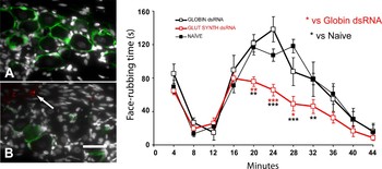
To investigate the role glutamate release might play in the trigeminal ganglion, we injected dsRNA against glutamine synthase (Fig. 4) into one trigeminal ganglion of uninjured rats to transiently silence its expression. Three days later, the rats injected with the glutamine synthase dsRNA showed significantly less nociceptive behavior to the injection of diluted formalin in the upper lip (orofacial formalin test), whereas rats injected with control dsRNA were not different from non-injected rats (Fig. 5).
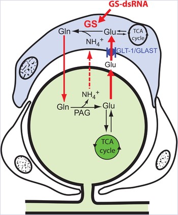
Diagram illustrating the role of SGCs in glutamate recycling. The glial glutamate transporter (GLAST) transports extracellular glutamate (Glu) into the SGC where glutamine synthetase (GS) converts the glutamate to glutamine (Gln) which is eventually recycled to the neuron for conversion into glutamate. Silencing GS using dsRNA-mediated RNAi will reduce the amount of glutamine available to neurons for conversion to glutamate.

Glutamine synthase (GS) dsRNA reduces glutamine synthase immunostaining in SGCs and results in analgesia in the second phase of the oro-facial formalin test. (A) GS immunoreactivity (green) is present in SGCs of the trigeminal ganglion. (B) Three days following injection of GS dsRNA into the trigeminal ganglion, GS immunoreactivity is greatly reduced. The arrow indicates DiI injected with the dsRNA. (C) Orofacial formalin tests were performed four days after the injection of GS dsRNA or control dsRNA. Compared to naive and control dsRNA rats, GS dsRNA resulted in significant decrease in pain behavior that occurred in the second phase of the formalin test. *P < 0.05, **P < 0.01, ***P < 0.001. n = 8. Scale bar, 30 µm.

Can SGCs be genetically modified to convert glutamate in GABA?
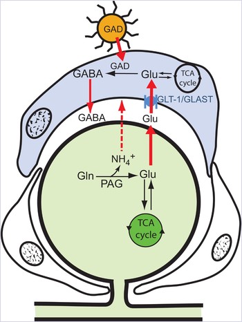
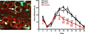
We injected an adenovector (Figs 6 and 7) harboring glutamic acid decarboxlylase (GAD65, one of the GAD isoforms), the enzyme responsible for synthesizing GABA (Vit et al., Reference Vit, Ohara, Sundberg, Rubi, Maechler and Liu2009) directly into on trigeminal ganglion using stereotaxic injection. We found that the adenovector preferentially targeted SGCs with only an occasional neuron showing expression of the GAD protein (Fig. 7). Six days after transduction of the viral vector, robust GAD65 immunostaining was present in the SGCs and coincided with a reduction in nociception as assessed by the orofacial formalin test (Fig. 7). Because a few neurons expressed GAD65, the antinociceptive effect could also be attributed to GABA being released by primary afferent terminals in the spinal cord. To determine that this analgesic effect occurred primarily at the ganglion level, and not at the spinal cord level, we injected the GABAA receptor antagonist, bicuculline directly into the trigeminal ganglion 6 days after viral vector delivery. Bicuculline abolished the antinociceptive effect of GAD65, strongly suggesting that the action of the GABA occurs at the ganglion level, rather than at the spinal level (Vit et al., Reference Vit, Ohara, Sundberg, Rubi, Maechler and Liu2009).
Diagram illustrating the synthesis of GABA from glutamate. Glutamate taken up by SGCs through the GLAST transporter is normally converted to glutamine via GS (see Fig. 4). Introducing the GAD65 transgene into the SGC via an adenovirus (AdGAD65) provides the pathway for the glutamate to be converted to GABA, which is then released by bulk transport to inhibit nearby primary sensory neurons by acting on GABAA receptors.

Injection of AdGAD65 into the trigeminal ganglion results in GAD65 expression in SGCs and produces analgesia in the second phase of the formalin test. (A) After AdGAD65 introduction into the trigeminal ganglion, GAD65 immunostaining (green) is found in SGCs (e.g. arrow) identified by GLAST immunostaining (red). (B) Six days after injection of AdGAD65, there is a significant reduction of the nociceptive response in second phase of the orofacial formalin compared to rats injected with a control virus (AdGFP) or naïve rats. AdGAD65 versus naïve: **P < 0.01, ***P < 0.001; AdGAD65 versus AdGFP: *P < 0.05, **P < 0.01, ***P < 0.001. n = 8. Bar = 30 µm.

DISCUSSION
A number of studies now show that CNS glia can profoundly affect the genesis and/or maintenance of pain (Adler et al., Reference Adler, Nico, VandeVord and Skoff2009; Carlton et al., Reference Carlton, Du, Tan, Nesic, Hargett and Bopp2009; McMahon and Malcangio, Reference McMahon and Malcangio2009). An outcome of this new understanding of glial cells function is the realization that glial cells may present targets for therapeutic intervention. Less attention has been given to the glial cells of the peripheral nervous system but as details of the biology of SGCs emerge, it would appear that these cells are involved in nociceptive processes and manipulated to provide analgesia in conditions of neuropathic pain.
SGCs do undergo phenotypic changes as evidenced by changes in GFAP expression and in cell coupling following nerve injury (Woodham et al., Reference Woodham, Anderson, Nadim and Turmaine1989; Gunjigake et al., Reference Gunjigake, Goto, Nakao, Kobayashi and Yamaguchi2009; Siemionow et al., Reference Siemionow, Klimczak, Brzezicki, Siemionow and McLain2009). The changes in SGC functional gap junctions (i.e. as evidenced by increased dye transfer between SGCs) are particularly well established and correlates with changes in nociceptive responses (Hanani et al., Reference Hanani, Huang, Cherkas, Ledda and Pannese2002; Huang et al., Reference Huang, Cherkas, Rosenthal and Hanani2005). We have shown that reducing the expression of gap junction protein using dsRNA changes nociceptive behavior. While the antinociceptive effect of reducing Cx43 expression was predictable, the same was not true when we injected Cx43 dsRNA into the trigeminal ganglion of non-injured rats. In this latter case, there was an appearance of nociceptive responses similar to those seen following the nerve injury. The most likely explanation is that gap junctions normally keep interactions between connected cells in a steady state. In the normal animal, gap junctions primarily allow the redistribution of potassium ions between adjacent SGCs, similar to what was described for CNS astrocytes (Wallraff et al., Reference Wallraff, Kohling, Heinemann, Theis, Willecke and Steinhauser2006). After neuronal injury, however, gap junctions increase in number in order to cope with the greater amounts of ATP and glutamate (Vidwans and Hewett, Reference Vidwans and Hewett2004; Takeuchi et al., Reference Takeuchi, Jin, Suzuki, Doi, Liang and Kawanokuchi2008). Silencing the expression of Cx43 is pronociceptive in the normal animal because it leads mainly to an increase in extracellular potassium. In the nerve-injured animal a different mechanism must be at work, antinociception from silencing Cx43 could result from a lack of glutamate because its recycling has been interrupted. Regardless of whether this is the explanation; the significance of our results is that alteration of a single protein in the SGCs can have significant behavioral consequences.
We have previously shown that reduction in a glutamate transporter (Jasmin et al., Reference Jasmin, Vit, Sundberg and Ohara2009), as well as a potassium channels Kir4.1 (Vit et al., Reference Ohara, Vit, Bhargava and Jasmin2008), and now a glutamate–glutamine cycle enzyme all change the pain behavior. For these latter molecules, we are not sure of their relative contribution to the altered nociceptive responses following nerve injury. There is accumulating evidence, however, that they each have a role: GLAST (Xin et al., Reference Xin, Weng and Dougherty2009); Kir4.1 (Zhang et al., Reference Zhang, Mei, Zhang, Ma, White and Donnelly2009); GS (Chen et al., Reference Chen, Muscoli, Doyle, Bryant, Cuzzocrea and Mollace2010; Hoffman and Miller, Reference Hoffman and Miller2010). We interpret these finding as showing that, SGCs scavenge extracellular glutamate, just as astrocytes do in the CNS (Zou et al., Reference Zou, Wang, Dou, Lu, Ma and Lu2010). Decreased expression of glutamate transporters results in an increase in extracellular glutamate (Han et al., Reference Han, Shioda, Moriguchi, Qin and Fukunaga2008), and consequently increased neuronal excitability. In contrast, knocking down GS results in less glutamine being released by SGCs and thus less available for uptake by neurons resulting in reduced conversion of glutamine to glutamate. While one could have predicted that the increased intra-SGC glutamate associated with the inhibition of GS was pronociceptive (by reverse transport through the glutamate transporters), we have no evidence for this. We were able to show that once in the SGCs glutamate can be converted to GABA, by transfecting SGCs with GAD65, just as it has been possible to transfect primary sensory neurons with GAD to release of GABA into the spinal cord (Liu et al., Reference Liu, Wolfe, Hao, Huang, Glorioso and Mata2004; Hao et al., Reference Hao, Mata, Wolfe, Huang, Glorioso and Fink2005; Lee et al., Reference Lee, Fink and Mata2006). SGCs are now believed to play a key role in the pathology of chronic pain and therefore are viewed as targets for therapy (Capuano et al., Reference Capuano, De Corato, Lisi, Tringali, Navarra and Dello Russo2009). The fact that the adenovirus serotype 5 preferentially infects SGCs means these vectors could be used to deliver other molecules to SGCs that would then produce factors that act on neurons. In some cases there may be advantages in infecting glial cells rather than neurons, as the normal cell biology and transmission properties of the neurons are left unchanged.
ACKNOWLEDGEMENTS
We acknowledge the expert assistance of Mr. Christopher Sundberg and Ms. Criselda Cua. Funded by NIH grants NS051336 and NS061241.
Statement of interest
None