Introduction
Endoscopic sinus surgery is a safe and reliable procedure for treating chronic rhinosinusitis resistant to medical treatment.Reference Jiang and Hsu1–Reference Watelet, Annicq, van Cauwenberge and Bachert3 Although the majority of maxillary sinus lesions can be removed through a widened natural ostium, some patients have extensive disease that is difficult to handle endoscopically, especially when located in the anterior and/or inferior regions of the antrum. In such cases, the canine fossa puncture is a useful alternative method for removing pathological mucosa or polyposis of the maxillary sinus which is difficult to access via a middle meatal antrostomy.Reference Sathananthar, Nagaonkar, Paleri, Le, Robinson and Wormald4, Reference Robinson, Baird, Le and Wormald5
Although several complications of the canine fossa puncture procedure have been reported, weakness of the buccal branch of the facial nerve has not been mentioned. To the best of our knowledge, the present case represents the first report of such a complication.
Case report
A 52-year-old woman presented with pain in the right cheek area which had begun a few months earlier. She also complained of purulent rhinorrhoea and nasal obstruction on the right side. Her symptoms did not improve despite several weeks of medication prescribed at local clinics. She had no past medical history or previous nasal surgery.
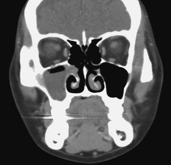
Endoscopic examination revealed polypoid changes around the right middle meatal area. Computed tomography of the paranasal sinus showed a soft tissue density within the right maxillary sinus, with obliteration of the natural ostium (Figure 1).
Pre-operative coronal computed tomography scan showing a soft tissue density within the right maxillary sinus, with obliteration of the natural ostium.

Endoscopic sinus surgery was performed under general anaesthesia. We could not completely remove the polyposis of the maxillary sinus via a middle meatal antrostomy, due to the anterior and inferolateral location of the lesion. We therefore decided to perform a canine fossa puncture. We did not use local infiltration around the puncture site before the procedure. As recommended by Sathananthar et al.,Reference Sathananthar, Nagaonkar, Paleri, Le, Robinson and Wormald4 the puncture entry point was the intersection of the midpupillary line and a horizontal line running along the lower border of the nasal alae. Under direct vision, and using a rigid, straight, 4-mm diameter, 70° endoscope (Karl Storz, Tuttlingen, Germany), the pathological mucosa of the maxillary sinus was removed with a curved, 4-mm microdebrider blade (Stryker Instruments, Kalamazoo, Michigan, USA) which was inserted through the puncture. During the procedure, traction of the upper lip was not necessary.
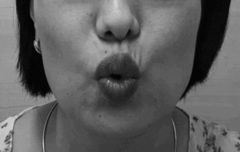
Immediately after the operation, the patient complained of motor weakness of the right upper lip and oral commissure in the area innervated by the buccal branch of the facial nerve. During phonation of the vowels [O] and [U], asymmetrical facial expression was observed owing to paralysis of the right upper lip (Figure 2).
Photograph of the patient taken immediately post-operatively, showing asymmetry of the superior lip and oral commissure during [O] phonation, due to paralysis of the right buccal branch of the facial nerve. Loss of wrinkles in the right upper lip area is also observed.

Electroneurography was performed five days post-operatively to estimate the degree of paralysis. The amplitude of response on the right side was 21 per cent of that on the left, normal side. Because electroneurography showed incomplete paralysis, we decided to prescribe short-term systemic corticosteroids and observe the patient.
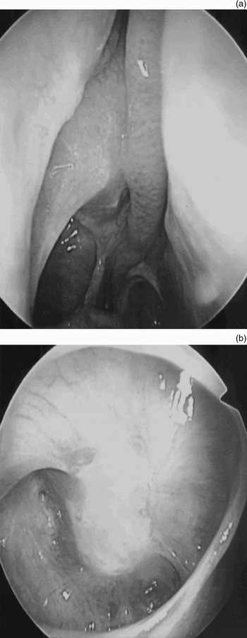
Fortunately, the patient's facial weakness had recovered spontaneously by three months post-operatively (Figure 3); furthermore, she demonstrated no evidence of recurrent sinusal pathology (Figure 4).
Photograph of the patient three months post-operatively, showing symmetry of the upper lip and oral commissure.

Post-operative nasal endoscopy showing (a) a patent maxillary ostium and (b) sinus mucosa free from pathology.

Discussion
The common complications associated with the canine fossa puncture procedure are cheek swelling, facial pain, facial numbness, dental numbness and facial tingling.Reference Robinson, Baird, Le and Wormald5, Reference Ferekidis, Tzounakos, Kandiloros, Kaberos and Adamopoulos6 Most of these problems resolve spontaneously within one to three months. However, persistent or permanent complications may occur, such as facial numbness and tingling. Injury to the branches of the infraorbital nerve is thought to be the main cause of these side effects.Reference Robinson and Wormald7
By contrast, injury of the buccal branch of the facial nerve following canine fossa puncture has not previously been reported.
Anatomically, the buccal branch of the facial nerve separates from the pes anserius and courses superiorly and anteromedially, passing 1 cm inferior to the inferior border of the zygomatic arch.Reference Gosain8 It innervates the orbicularis oris and levator labii superioris alaeque nasi muscles. Its injury limits upward movement of the superior lip and oral commissure. Such weakness may increase with laughing.
• Canine fossa puncture is a useful alternative method for enabling removal of maxillary sinus pathological mucosa or polyposis which is difficult to access via a middle meatal antrostomy
• Although several complications of canine fossa puncture have been reported, weakness of the buccal branch of the facial nerve is not among them
• In the presented patient, the terminal branch of the buccal nerve may have been injured because the puncture site was more lateral than intended; alternatively, the patient may have had anatomical variation of the facial nerve, with a longer course of the buccal branch to the upper corner of the lip, placing the nerve very close to the puncture site
The cause of injury in our patient was not clear. Local infiltration of adrenaline and lidocaine, or excessive traction of the upper lip, may cause transient paralysis of the surrounding sensory and motor nerves. However, our patient did not receive local infiltration around the puncture site, and traction of the upper lip was not necessary. Moreover, paralysis caused by local infiltration or traction is generally transient and would not be expected to last for a few months. Therefore, we postulate that the terminal branch of our patient's buccal nerve was injured, because the puncture site was more lateral than intended. Alternatively, the patient may have had an anatomical variation of the facial nerve, with a longer course of the buccal branch to the upper corner of the lip, placing the nerve very close to the puncture site.
It has been reported that 70 to 90 per cent of facial nerve buccal branches communicate with adjacent branches of the facial nerve, especially the zygomatic branch. Therefore, injury to the buccal branch has been reported to recover faster than injury to other branches of the facial nerve, because this branch generally receives cross-innervation from adjacent branches of the facial nerve.Reference Gosain8 As we expected, our patient recovered completely within three months of her initial procedure.
Conclusion
To the best of our knowledge, our patient represents the first reported case of an injury of the buccal branch of the facial nerve sustained during canine fossa puncture. Although the incidence of this complication is very low, surgeons should inform their patients of the possibility of this rare complication, and should take care to locate the canine fossa puncture site not too far lateral to the recommended puncture point.