Introduction
Facial nerve paralysis is a common problem with significant morbidity related to functional and aesthetic issues. Traumatic facial paralysis is the most common cause of peripheral facial palsy in children and is the second most common cause in adults after Bell's palsy.Reference Fisch 1 Traumatic facial nerve paralysis can occur after blunt and penetrating head or neck trauma, or surgical trauma such as tympanoplasty, tympanomastoidectomy, acoustic tumour surgery or parotid surgery.
Many surgical and medical treatment approaches have been described for the treatment of traumatic facial nerve paralysis and rehabilitation of facial function.Reference Yian, Paniello and Spector 2 – Reference Nachemson, Lundborg, Myrhage and Rank 5 Despite these treatments, poor facial nerve function and aesthetic problems may result. Incomplete recovery of the facial nerve fibres and longer regenerative course are the most important causes of incomplete facial nerve function.Reference Fu and Gordon 6
Facial nerve injuries may cause a series of biochemical and molecular cascades. Axonal damage occurs in a time-dependent manner, and involves increased polyamine metabolism and increased structural proteins.Reference Gilad and Gilad 7 – Reference Tetzlaff, Graeber and Kreutzberg 9 Some studies have suggested that treatment with polyamines and proteins increases nerve regeneration.Reference Dornay, Gilad, Shiler and Gilad 10 – Reference Oble, Burton, Maxwell, Hassard and Nathaniel 12
Agmatine is an aminoguanidine molecule, which is formed by decarboxylation of L-arginine by arginine decarboxylase.Reference Regunathan and Reis 13 Agmatine production is very low in the nervous system, but is present at very high levels after facial nerve injury. This finding indicates the potential effects of agmatine for repair and regeneration of the nerve.Reference Gilad, Gilad and Rabey 14 Agmatine therapy was found to be effective in the treatment of nerve injuries in some reports.Reference Gilad, Salame, Rabey and Gilad 15 – Reference Gilad and Gilad 17 The effect of agmatine on the facial nerve is still unclear, but it may accelerate facial nerve regeneration.
This study aimed to evaluate the effect of agmatine sulphate on facial nerve regeneration after facial nerve injury using electron and light microscopy.
Materials and methods
This study was performed at Cukurova University Experimental Research and Application Center of Medical Sciences, Adana, Turkey, and was approved by the Committee of Cukurova University on Animal Research.
Thirty male Wistar albino rats, with a mean weight of 250 g (range, 230–275 g), were used for the study. The rats were kept in separate cages at a constant temperature (20 ± 2 °C) and humidity (55.5 per cent), in 12-hour light/dark cycles, and were fed with regular commercial rat food and tap water from drinking bottles. This study was performed according to the Guide for the Care and Use of Laboratory Animals. 18
This study was planned as a pilot study. The animals were divided into five groups, with six rats in each group. Group 1 was the normal (intact, untreated) group – no surgical or medical therapies were given. Group 2 was the sham-treated group – the nerve blood supply was interrupted (only a skin incision was made). Group 3 was the study control group – the nerve was sectioned and closed end to end, and no therapy (such as anastomosis and agmatine) was given; the rats were sacrificed at the end of the second month. Group 4 was subjected to anastomosis – the nerve ends were sutured, with size 9-0 suture primarily, and no medical treatment was given; the rats were sacrificed at the end of the second month. Group 5 was subjected to anastomosis with suturing, and treated with agmatine sulphate (Sigma-Aldrich®); the rats were sacrificed at the end of the second month.
Agmatine sulphate was injected intraperitoneally (100 mg/kg/day) and administered at the 15th minute post-operatively, for 5 successive days, in group 5. Groups 3 and 4 received saline intraperitoneally at the 15th minute post-operatively, for 5 successive days. This protocol was designed according to Berenholz et al.Reference Berenholz, Segal, Gilad, Klein, Yehezkeli and Eviatar 19 The animals were anaesthetised with 5 mg/kg xylazine (Rompun; Bayer, Leverkusen, Germany) and 80 mg/kg ketamine (Ketalar; Eczacibasi, Istanbul, Turkey), administered via intraperitoneal injection before the surgical procedure.
Under an operating microscope, the right mandibular branch of the facial nerve was carefully dissected and cut with micro scissors. A piece of the mandibular branch of the facial nerve measuring approximately 1 mm was excised. The proximal and distal stumps were closed end to end. The proximal and distal stumps of the marginal branch of the facial nerve were produced by suturing to the epineurium of the nerve using size 10-0 nylon thread. The anastomosis area was marked with silk suture. All incisions were then closed.
The animals were sacrificed on the 60th day after the initial procedure. The mandibular branch of the facial nerve was dissected, and the reconstructed nerve segment was removed for histological and electron microscopic examination.
For histomorphometric evaluation, 1 µm semi-thin sections were taken from the facial nerve blocks. The semi-thin blocks taken formed the six facial nerve blocks prepared for electron microscopic evaluation. Six fields out of 10 sections were counted from each group (400× objective magnification). The sections were stained with toluidine blue and evaluated under a light microscope (BX53; Olympus, Tokyo, Japan). The average number and diameter of axons were measured with the CellSens™ Standard software analysis program (Olympus).
For electron microscopic evaluation, the tissue specimens were fixed in 2.5 per cent glutaraldehyde with phosphate buffered saline (pH 7.2). Following a 24-hour fixation procedure, the tissues were rinsed with phosphate buffered saline. They were fixed for a second time with 1 per cent osmium tetroxide and then rinsed again with phosphate buffered saline. Dehydration was performed with a graded alcohol series. The tissues were embedded in an Araldite® CY212 (epoxy resin) plus dodecenyl succinic anhydride (DDSA, hardener) plus benzyl dimethylamine (BDMA, accelerator) plus dibutyl phthalate mixture. Blocks were prepared, and thin sections were examined using a JEOL™ JEM 1400 transmission electron microscope and digital photographs were taken. Samples were evaluated by the same experienced author (LS).
Statistical analysis was performed using SPSS® version 20.0 statistical software package. Normality was checked for each variable. Continuous variables were compared using the Kruskal–Wallis test for non-parametric data, with post-hoc analysis performed using a Mann–Whitney U test. Bonferroni's correction was applied (p < 0.05/n; where n = number of comparisons) when multiple comparisons were made. Results are presented as means ± standard deviations and medians (ranges).
Results
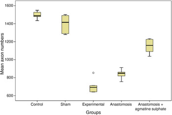
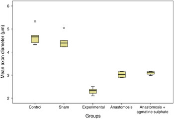
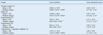
Electron microscopic evaluations of the facial nerve in group 1 (normal control) and group 2 (sham-treated) showed that the nerve fascicles consisted of myelinated and unmyelinated nerve fibres. These nerve fibres were covered with Schwann cells and, in turn, the Schwann cells were covered by a basal lamina. Mitochondria, Golgi apparatus and lysosomes in the cytoplasm of the Schwann cells were seen to have a normal appearance. In group 1, the median axon diameter was 4.7 µm (range, 4.3–5.3 µm) and the median axon number was 1488.2 (range, 1435–1549) (Table I; Figures 1 and 2). In group 2, the median axon diameter was 4.4 µm (range, 4.2–5.1 µm) and the median axon number was 1415.4 (range, 1279–1502).
Comparison of axon numbers between groups.

Comparison of axon diameter between groups.

Number and diameter of axons in each group

*p < 0.004 between anastomosis plus agmatine sulphate (group 5) and the other groups. † p < 0.004 between normal control (group 1) and the other groups. SD = standard deviation
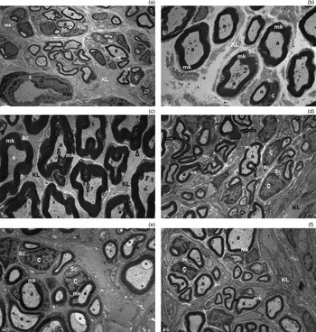
There were important structural changes in the myelin sheaths of the myelin nerve fibres in group 3 (study control group). The concentric lamellar structure of the myelin sheaths was damaged. Various sizes of vacuoles were present between axons and myelin sheaths in most areas. Different sizes of oil droplets were observed in fibroblast cytoplasm in areas where degeneration was obvious. The median axon diameter in group 3 was 2.3 µm (range, 2.1–2.5 µm) and the median axon number was 692.4 (range, 641–854).
Myelin sheath defects and axonal degenerative changes were seen in some areas examined in group 4 (anastomosis group). Increased heterochromatin in the nucleus of the Schwann cells and vacuolisation were observed. In some areas, degenerative changes were seen on the lamellar structure of the nerve fibre myelin sheaths. The median axon diameter in group 4 was 3 µm (range, 2.9–3.2 µm) and the median axon number was 849 (range, 756–912).
In electron microscopic evaluation, the best regeneration occurred in group 5 (anastomosis plus agmatine sulphate group), and regeneration was better compared with group 3 (study control group). Slight changes were observed in the axon myelin sheaths, but, in many areas, these were seen to have an almost normal appearance. Mitochondria, endoplasmic reticulum cisternae, and lysosome and ribosome structures, located in the Schwann cells, were generally considered to have a normal appearance. There were also fibroblasts, which had normal features, and collagen fibres. The median axon diameter in group 5 was 3.1 µm (range, 3.0–3.2 µm) and the median axon number was 1159.1 (range, 1038–1234).
Regeneration was better in group 4 (anastomosis group) than in group 3 (study control group), but not as good as in group 5 (anastomosis plus agmatine group), the latter of which had the best regeneration findings (Figure 3). There was a statistically significant difference in the number of axons between the agmatine group and all other groups (p < 0.004). The median diameter of the axons was found to be statistically significantly different in the agmatine group (group 5) compared with the other groups (Table I).
Electron microscopic images: (a) normal (scale, 2 µm); (b) sham (scale, 2 µm); (c) study control (scale, 1 µm); (d) anastomosis (scale, 1 µm); (e) anastomosis plus agmatine sulphate with treatment (scale, 0.5 µm); and (f) only anastomosis plus agmatine sulphate (scale, 1 µm). Degenerative changes are shown with arrows. mk = myelin shield; a = axon; ma = unmyelinated nerve; Sc = Schwann cell; Ç = nucleus; E = endothelium cell; KL = collagen; Kap = capillary; m = macrophage; * = gaps between myelin sheats

Discussion
Peripheral motor nerve fibres have a greater regenerative capability than central nerves. Schwann cells play a very important role in the repair of peripheral nerve injuries. Local injuries may activate Schwann cell proliferation, and promote axon sprouting and elongation.Reference Siemionow and Sari 20 Axon recovery is related to local oxidative factors, molecular transportation, calcium and sodium inflow, free radicals, phospholipase activity, and genetic factors.Reference de Faria, Testa, Borin and Toledo 21
Oedema, oxidative stress, ischaemia and inflammatory reactions can occur as a result of trauma. These changes lead to severe damage to cell and cytoplasm structure.Reference Stoll and Muller 22 – Reference Hirata and Kawabuchi 24 Many therapeutic approaches have been described to enhance regeneration of the facial nerve after injury. Facial nerve anastomosis may be performed or cable grafts (with nervus suralis or auricularis magnus) may be used for the repair of facial nerve defects. Direct anastomosis with suturing is a well-known and effective method for the repair of small defects if there is no tension between the sides of the facial nerve. After trauma to or oncological surgery performed on the facial nerve region, direct suturing of the facial nerve may not be possible. In these cases, cable grafts may offer an effective method for facial nerve repair.Reference Spector, Lee, Peterein and Roufa 25
The current study has shown that anastomosis can accelerate and increase regeneration. Several pharmacological agents have been used to achieve functional recovery of the facial nerve after injuries. Despite promising findings with various agents, no pharmacological agent (with the exception of corticosteroids) has been deemed safe or sufficiently efficacious to be used in clinical practice. This study demonstrated that agmatine sulphate is an effective agent for facial nerve injuries. Regenerative findings were better for the facial nerve treated with agmatine sulphate, and axon numbers and diameters were statistically significantly different in the agmatine group. These findings indicate the positive effect of agmatine on facial nerve injuries.
-
• Many surgical and medical approaches have been described for traumatic facial nerve paralysis treatment and rehabilitation
-
• However, poor function and aesthetic problems may result
-
• Incomplete facial nerve fibre recovery and longer regenerative course are the most important causes of incomplete facial nerve function
-
• Agmatine therapy can be effective in nerve injury treatment
-
• Although its effects on the facial nerve are unclear, agmatine may accelerate facial nerve regeneration
This is the second study in the English-language literature to evaluate the effects of agmatine sulphate on facial nerve injuries. Facial nerve regeneration and the positive effects of agmatine sulphate have been shown with electron microscopic evaluation. Berenholtz et al. reported that reconstruction of a facial nerve gap may be successful with an autogenous jugular vein graft, and that agmatine sulphate treatment together with the vein graft may be an advantageous alternative to other treatments for facial nerve injuries.Reference Berenholz, Segal, Gilad, Klein, Yehezkeli and Eviatar 19 They found a statistically significant difference in the number of axons between nerve transection with saline and an unsutured vein graft with saline and agmatine treatment. Agmatine may have a positive effect on peripheral targets, but this has not been explored extensively. Some studies have suggested that agmatine sulphate treatment can raise insulin levels and reduce catecholamine discharge.Reference Pfeiffer, Sarrazin and Weitzel 26 – Reference Raasch, Schäfer, Chun and Dominiak 28
Conclusion
This experimental study showed that agmatine sulphate treatment with anastomosis may increase nerve regeneration in traumatic facial paralysis. These effects were demonstrated using electron microscopy and histomorphometric evaluation. However, further studies are required to establish their definitive actions on healing.