Introduction
Bilateral vestibular schwannomas (also known as acoustic neuromas) are the hallmark of neurofibromatosis type two. Although benign, these tumours cause significant morbidity due to their location, with progressive hearing loss, tinnitus and pressure effects.Reference Lanser, Sussman and Frazer1 Vestibular schwannomas usually grow slowly, with a mean growth rate of 1.9 mm per year reported in two meta-analyses.Reference Yamakami, Uchino, Kobayashi and Yamaura2, Reference Smouha, Yoo, Mohr and Davis3 Gadolinium-enhanced magnetic resonance imaging (MRI) is the investigation of choice.Reference Plotkin, Stemmer-Rachamimov, Barker, Halpin, Padera and Tyrrell4
Treatment of vestibular schwannomas aims to preserve function and quality of life. Standard treatment options include active surveillance, surgical resection and radiation therapy, including gamma knife radiosurgery. In one meta-analysis, 43 per cent of patients had reported tumour growth, 20 per cent required treatment, and 51 per cent experienced hearing loss, over a mean follow-up period of 3.2 years.Reference Smouha, Yoo, Mohr and Davis3
Microsurgery offers excellent tumour control, with only 1.8 per cent recurrence and low morbidity and mortality.Reference Yamakami, Uchino, Kobayashi and Yamaura2 However, hearing outcomes are poor, with only 44 per cent preservation of serviceable hearing (i.e. Gardner–Robertson class one or two), and side effects include cerebrospinal fluid leakage (3–15 per cent), facial nerve injury (2.5–7 per cent) and meningitis (1–3 per cent).Reference Yamakami, Uchino, Kobayashi and Yamaura2 Contemporary studies have shown that gamma knife radiosurgery results are comparable to microsurgery results for small and medium-sized vestibular schwannomas. However, the efficacy of radiosurgery and microsurgery in neurofibromatosis type two patients is inferior in comparison to sporadic patients, with 50 per cent eight-year control rates for gamma knife radiosurgery.Reference Rowe, Radatz, Walton, Soanes, Rodgers and Kemeny5 Furthermore, morbidity has been noted to be higher in neurofibromatosis type two patients, for both gamma knife radiosurgery and microsurgery, which is of concern given these patients’ bilateral disease.
Ongoing follow up with MRI has become the accepted standard of care for monitoring treatment response in vestibular schwannoma patients. Although some tumours shrink with time following gamma knife radiosurgery, a significant proportion remain static or even expand transiently before shrinking.Reference Nakamura, Jokura, Takahashi, Boku, Akabane and Yoshimoto6 In most patients, MRI scans show residual, enhancing tumour which persists indefinitely.
Biological pathways regulating cell-cycle progression, apoptosis, and intercellular and intracellular interactions are now understood to be involved in vestibular schwannoma growth and treatment response (or resistance) to radiosurgery.Reference Yeung, Sughrue, Kane, Tihan, Cheung and Parsa7 Advances in our understanding of vestibular schwannoma molecular biology have expanded the therapeutic role of targeted growth inhibition. Vascular endothelial growth factor pathways are now implicated in the growth of vestibular schwannomas.Reference Plotkin, Stemmer-Rachamimov, Barker, Halpin, Padera and Tyrrell4, Reference Yeung, Sughrue, Kane, Tihan, Cheung and Parsa7 A recent study of bevacizumab, an anti-angiogenesis monoclonal antibody, achieved tumour stabilisation and hearing recovery in a subset of neurofibromatosis type two patients with progressive disease.Reference Plotkin, Stemmer-Rachamimov, Barker, Halpin, Padera and Tyrrell4 This further confirms the important role of vascular endothelial growth factor in tumour growth, and its clinical potential.
In the case of vestibular schwannomas which progress despite gamma knife radiosurgery or microsurgery, treatment decisions are often very complex. Systemic treatment may be the most appropriate option, especially for bilateral vestibular schwannomas. The two cases reported in this paper strongly support an earlier study's conclusion that bevacizumab treatment may be useful, not only in previously untreated patients but also in those relapsing after prior non-surgical therapy.
Case reports
Patient one
A 37-year-old man with neurofibromatosis type two and bilateral vestibular schwannomas noticed deterioration of his hearing on the right side, and was referred for gamma knife radiosurgery.
The right-sided tumour received a marginal dose of 14 Gray to the 45 per cent isodose, using standard gamma knife technology.
Following this radiosurgery, further significant deterioration in right-sided hearing was documented.
Thirteen months later, MRI demonstrated a stable appearance of the right vestibular schwannoma but an increase in the size of the left schwannoma. At this point, it was paramount to avoid compromising the patient's hearing any further.
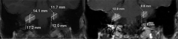
Therefore, the patient was commenced on fortnightly intravenous bevacizumab (450 mg; 5 mg/kg), which achieved a good clinical response. After six injections (over 12 weeks), another MRI scan confirmed an unequivocal decrease in all dimensions of both tumours; before bevacizumab treatment, the left tumour had measured 12.9 × 11.7 × 12 mm (a volume of 0.9 cm3) and the right tumour 14.9 × 14.1 × 17.2 mm (1.9 cm3), while after treatment the left tumour measured 12.2 × 9.8 × 10.9 mm (0.7 cm3) and the right tumour 11 × 10.9 × 14.2 mm (0.9 cm3) (Figure 1). The enhancement characteristics of the schwannomas also changed following treatment, with decreased central enhancement particularly on the left side (which had not received gamma knife radiosurgery).
Axial magnetic resonance imaging scans of patient one, taken before (left half of figure) and after (right half of figure) bevacizumab treatment.

The patient went on to receive a total of nine bevacizumab injections. At the time of writing, he had been clinically stable for eight months without further treatment.
Patient two
This woman was diagnosed with bilateral vestibular schwannomas, and subsequently neurofibromatosis type two, at 31 years of age, after suffering hearing loss in the right ear.
She underwent trans-cranial resection of the right vestibular schwannoma, whilst the left was observed. This rendered her deaf in the right ear. At this time, her hearing on the left was normal.
However, five years later the patient's hearing on the left had gradually diminished. She was entered into an oral vascular endothelial growth factor inhibitor trial at Boston Massachusetts General Hospital; this used PTC299, which targets the post-transcriptional processes regulating vascular endothelial growth factor.
However, MRI scanning showed an increase in tumour volume over the following four months.
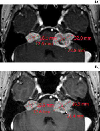
Six months after entering the PTC299 trial, this patient was started on bevacizumab. She responded clinically to this treatment. After eight fortnightly injections of intravenous bevacizumab (310 mg; 5 mg/kg), MRI scanning confirmed an unequivocal reduction in the size of the left vestibular schwannoma and a modest reduction in the size of the right schwannoma; before bevacizumab treatment, the right tumour had measured 17.7 × 12.6 × 18.1 mm (volume 2.2 cm3) and the left tumour 21.2 × 23.8 × 32.0 mm (8.4 cm3), while after treatment the right tumour measured 17.0 × 12.6 × 16.9 mm (2.0 cm3) and the left tumour 16.4 × 21.4 × 28.5 mm (5.2 cm3) (Figure 2).
Axial magnetic resonance imaging scans of patient two, taken (a) before and (b) after bevacizumab treatment.

At the time of writing, this patient was continuing to receive fortnightly bevacizumab injections.
Discussion
Both of these cases support the role of vascular endothelial growth factor in vestibular schwannoma growth in patients with neurofibromatosis type two, and the efficacy of bevacizumab therapy. Previous reports have demonstrated the therapeutic efficacy of bevacizumab in neurofibromatosis type two patients. These results are supported by our own findings. Furthermore, we also report a reduction in tumour size following bevacizumab treatment in patients who had previously been treated variously with gamma knife radiosurgery and another vascular endothelial growth factor targeted therapy.
In our first patient, the option of bevacizumab treatment was considered when there were signs of radiological and clinical progression of the contralateral tumour. The alternative to bevacizumab would have been microsurgery or gamma knife radiosurgery, with the associated risk of hearing loss in the dependent ear, which had relatively normal function. In this case, it is fascinating to note the rapid shrinkage of both vestibular schwannomas, especially the right tumour, which had remained stable in volume for 13 months following earlier radiosurgical treatment.
• Bilateral vestibular schwannomas in neurofibromatosis type two (NF2) are difficult to manage
• Current options are conservative treatment, surgery and gamma knife radiosurgery
• Morbidity includes hearing loss and facial nerve damage; the risk is significant with surgery and radiotherapy
• Vascular endothelial growth factor plays an important role in NF2-associated vestibular schwannoma growth
• Bevacizumab effectively treats selected NF2 patients
Our second case involved cross-over treatment. Notably, this case demonstrates that not all inhibitors of the vascular endothelial growth factor receptor pathway are equivalent in terms of therapeutic potency.
Centres using bevacizumab for vestibular schwannoma treatment use the same dosage schedule, 5 mg/kg fortnightly, and treat patients either continuously or for 12 months, unless unacceptable toxicity or tumour growth occurs.Reference Plotkin, Stemmer-Rachamimov, Barker, Halpin, Padera and Tyrrell4, Reference Mautner, Nguyen, Kutta, Fuensterer, Bokemeyer and Hagel8 Our first case completed nine injections, and at the time of writing was being monitored with six-monthly MRI scans; it was planned that any regrowth would be treated with bevacizumab. Our second case was receiving ongoing fortnightly treatment at the time of writing, and this was planned to continue unless unacceptable toxicity or progression occurred (as detected via MRI undertaken every three to four months).
Both our patients’ contrast-enhanced MRI scans revealed an unequivocal response to treatment, with a significant decrease in maximum tumour dimension on both sides, comparing pre- and post-bevacizumab scans. In the study by Plotkin et al. a reduction in tumour volume was noted in nine patients; six of which achieved a radiological response, defined as a decrease in volume of greater than or equal to 20 per cent.Reference Plotkin, Stemmer-Rachamimov, Barker, Halpin, Padera and Tyrrell4 In their case report of two patients, Mautner et al. described tumour volume reduction in both patients; in one patient, the cystic part of the tumour regressed more than the solid part.Reference Mautner, Nguyen, Kutta, Fuensterer, Bokemeyer and Hagel8 Given the extremely slow growth of these tumours over many years, the observations of rapid volume reduction after bevacizumab therapy are remarkable, and contrast with the effect of gamma knife radiosurgery.
An interesting new observation in our first patient is the central signal change seen on MRI. This is well known in the subacute period following gamma knife radiosurgery, and is thought to be due to central tumour damage or necrosis. Interestingly, this phenomenon was more marked in the tumour which had not received gamma knife radiosurgery. We suggest that this change is due to rapid inhibition of internal tumour vasculature development and/or maintenance. This change occurred more quickly than that reported following gamma knife radiosurgery – an added point of interest.
In our two cases, we did not note any change in meningiomata. This is of interest, as vascular endothelial growth factor mediated inhibition of the blood supply of neurofibromatosis type two associated meningiomata may differ from the effect of vascular endothelial growth factor on the blood supply of vestibular schwannomas.
We are still in the early phase of experience with bevacizumab, and several questions are currently unanswered, including the expected duration of treatment response and the role of other treatment modalities such as gamma knife radiosurgery or microsurgery. Plotkin et al. have reported a sustained response to bevacizumab (11–14 months) in a small patient cohort.Reference Plotkin, Stemmer-Rachamimov, Barker, Halpin, Padera and Tyrrell4 In our two cases, more time is needed to evaluate treatment response. Following bevacizumab treatment, gamma knife radiosurgery of residual disease is possible, but it may be less effective due to diminished tumour oxygenation. Surgery performed immediately after bevacizumab treatment carries the risk of haemorrhage and poor wound healing, but this risk can be minimised by allowing a four-week ‘wash-out’ period.Reference Plotkin, Stemmer-Rachamimov, Barker, Halpin, Padera and Tyrrell4, Reference Mautner, Nguyen, Kutta, Fuensterer, Bokemeyer and Hagel8 However, a significant risk of hearing loss would still remain. More experience is needed to help determine which patients will benefit most from such combined treatment.
Our experience, together with previously published literature, supports the use of bevacizumab in previously treated, complex neurofibromatosis type two patients with vestibular schwannomas. Bevacizumab should be considered as a viable treatment option in patients with bilateral vestibular schwannomas or progressive, post-treatment vestibular schwannomas.