Case report
A 44-year-old man was referred for a T-wave abnormality on an electrocardiogram. As he had no symptoms, he underwent treadmill echocardiography, which identified hypokinesia of the apical anterior segment of the left ventricle. To rule out coronary artery disease, a 256 multi-detector CT coronary angiography was performed.
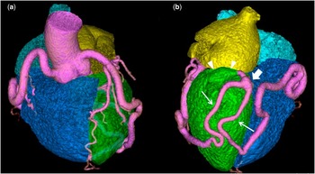
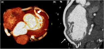
His three-dimensional volume-rendering images showed a diffusely tortuous right coronary artery and ramus intermedius with a 7–8-mm diameter (Fig 1a). In addition, two aberrant communicating arteries, respectively, originated from the posterior descending artery and the ramus intermedius, creating a dilated common channel, which drained into the left ventricular cavity (Fig 1b). A thick-slab image demonstrated the common channel draining to the left ventricle at the base of the heart, posterior to the postero-lateral mitral valve annulus (Fig 2a). In addition, unusual myocardial bridging of the distal ramus intermedius was shown on a curved multiplanar re-formatted image (Fig 2b). The patient had no symptoms, and he was reluctant to proceed with further investigation or intervention; therefore, we elected for conservative management with regular follow up.
( a ) A three-dimensional volume-rendering image shows diffuse fistulous dilatation of the right coronary artery and ramus intermedius. ( b ) A three-dimensional volume-rendering image shows two anomalous communicating coronary arteries (arrows) form a common channel with aneurysmal dilatation (thick arrow) draining into the left ventricular cavity at the base of the heart (arrow heads).

( a ) Thick-slab image demonstrates a coronary to left ventricular fistulous tract (thick arrow), which drains into the left ventricular base posterior wall, at the level of mitral valve leaflets (arrows). ( b ) A curved multiplanar re-formatted image shows deep myocardial bridging of the distal ramus intermedius (arrows). Dilatation of the left ventricle was not seen in this case.

Discussion
Coronary artery fistula is a rare anomaly, affecting 0.002% of the general population.Reference Podolsky, Ledley and Goldstein 1 Left ventricular drainage is the most infrequent form, and multiple origins of coronary arterial fistulae are even more rare.Reference Podolsky, Ledley and Goldstein 1 We found only one case described in the literature in which bilateral coronary artery to left ventricular fistula via a common channel was depicted by cardiac angiography.Reference Podolsky, Ledley and Goldstein 1
In our case, the coronary CT proved to be a useful and less-invasive diagnostic option compared with coronary angiography, allowing clear visualisation of the origin and drainage site of the coronary artery fistula as well as concurrent anomalous communicating coronary arteries.
Acknowledgement
None.
Financial Support
This research received no specific grant from any funding agency or commercial or non-for-profit sectors.
Conflicts of Interest
None.