




No CrossRef data available.
Article contents
Myotonic dystrophy presenting as atrial flutter in childhood
Published online by Cambridge University Press: 20 January 2005
Abstract
A 12-year-old boy presented with tachycardia and cardiomegaly. An electrocardiogram showed atrial flutter with a ventricular rate of 170 beats/min. The echocardiogram showed left ventricular dilation, with decreased contractility. Electromyography confirmed the diagnosis of myotonic dystrophy. The atrial flutter was converted to sinus rhythm by electroversion, leading to restoration of normal cardiac function. Our case illustrates that atrial flutter can be an early sign of the cardiac complications of myotonic dystrophy in childhood.
Information
- Type
- Brief Report
- Information
- Copyright
- © 2004 Cambridge University Press
Myotonic dystrophy is a genetic muscular disease that rarely presents in childhood outside the neonatal period.1 Cardiac involvement includes cardiomyopathy and arrhythmia,2 the latter usually represented by atrioventricular block or ventricular premature contractions. On rare occasions, nonetheless, patients with myotonic dystrophy may present with tachyarrhythmias.3–5 We describe a schoolboy who presented with atrial flutter in association with cardiomegaly, and was diagnosed as having myotonic dystrophy.
Case report
A 12-year-old boy complained of a frequent cough 3 days prior to admission, and was treated by a local physician with the tentative diagnosis of common cold. The day before referral to us, he presented with tachycardia, the rate being 160 beats/min. Cardiomegaly and hypotension were noted, so he was transferred to a regional public hospital. There, he was diagnosed with atrial flutter and dilated cardiomyopathy, and treated with an intravenous infusion of digoxin, which decreased the heart rate to 120 beats/min. For further investigation and treatment, he was transferred to our hospital.
When he presented to us, his body weight was 50 kg, and his height was 160.5 cm. The blood pressure was 100/80 mmHg. The liver was felt 1 cm below the right costal margin, and he showed a mild precordial bulge. No significant enlargement of any cervical lymph nodes or goiter was appreciated. Auscultation revealed a gallop rhythm, but without any cardiac murmur. The deep tendon reflexes were mildly diminished, but no abnormal tendon reflex was evoked. His grasping power was 12 kg in both hands. Percussion myotonia of the hand was easily induced at a later examination.
An electrocardiogram showed atrial flutter, with an atrial cycle length of 190 ms, and with either 2:1 or 3:1 atrioventricular conduction, giving a ventricular rate varying between 150 and 170 beats/min. (Fig. 1A). A chest X-ray film showed mild cardiomegaly, with a cardiothoracic ratio of 55%, and mild pulmonary congestion.

Figure 1. Panel A, electrocardiogram showed atrial flutter with 2:1 or 3:1 atrioventricular conduction giving a rate of 150 to 170 beats/min. Panel B, The 12-lead electrocardiogram after direct-current electrical conversion showed evidence of mild left atrial and ventricular hypertrophy.
Blood chemistry showed mildly increased plasma lactic and pyruvic acids. Creatinine kinase and thyroid hormones were within the normal range. Types A and B natriuretic peptides, however, were remarkably increased, at 380 and 620 pg/ml, respectively.
A careful interrogation revealed that he had been suffering from muscular weakness since the age of 10 years, when he joined a baseball team. At that time, he could not throw a ball straight to friends. After he entered junior high school, he started playing table tennis. After the regular training session, he got exhausted and required a long rest. In the second semester, he was frequently absent from the school because of easy fatigue. In addition, his mother showed easy fatigue and required long rests for these several years.
An electromyogram showed the “diving bomber” phenomenon, confirming that he had been suffering from myotonic dystrophy. Later, DNA analysis confirmed ×367 of CGT repetition.
His atrial flutter was converted to sinus rhythm by direct-current electrical conversion. At this point, a 12-lead electrocardiogram showed left atrial and ventricular hypertrophy, and inverted T waves in leads V4 and V5 (Fig. 1B). The echocardiogram now showed left ventricular dilation, with a left ventricular end-diastolic dimension of 67 mm, and decreased contractility, the left ventricular shortening fraction being no more than 7% (Fig. 2A). Since then, he has been treated with digoxin, lasix, and enarapril.

Figure 2. Composite panels of M-mode echocardiogram of the left ventricle. Soon after direct-current electrical conversion (A), the left ventricle was severely dilated and poorly contractile. After a period of 18 months subsequent to the electrical conversion (B), the left ventricle had decreased in size, with restoration of its function.
At the most recent follow-up, 18 months after the initial presentation, his general condition has improved gradually, though he suffers daytime sleepiness. A chest X-ray film revealed a normal cardiac silhouette, with a cardio-thoracic ratio of 41%. The echocardiogram showed a remarkable improvement in cardiac function, with left ventricular end-diastolic dimension now of 48 mm, and a left ventricular shortening fraction of 35% (Fig. 2B).
Discussion
Our patient illustrates that atrial flutter can be an early sign of the cardiac complications of myotonic dystrophy in childhood, and that conversion to sinus rhythm can normalize cardiac function. As a cardiac complication of myotonic dystrophy, however, atrial flutter is not frequent, with a reported frequency ranging from 0 to 16%.2 As evidenced by our search of the literature, however, no child has previously been reported with atrial flutter as such a complication. Indeed, Morgenlander et al.6 found no patient with atrial flutter among their 17 children with myotonic dystrophy over a period of follow-up ranging from 1 to 9 years.
We presume that the atrial flutter in our patient was the consequence of pathological change in the atrial muscular wall establishing the substrate for reentry. Pickering et al.5 reported focal right dysplasia and atrial flutter in a 22-year-old patient with myotonic dystrophy. This patient presented with a single syncopal episode associated with atrial flutter and exercise-induced 1:1 atrioventricular conduction, sinus nodal dysfunction, and mild prolapse of the mitral valve. In this patient, only right atrial dysplasia was found as a pathological finding, with no ventricular myocardial abnormality. Although we did not have any pathological examination, our patient might have the same focal myocardial disease localized to the atriums. The fact that we could improve his cardiac function remarkably by converting atrial flutter to normal sinus rhythm supports the possibility that he was suffering from arrhythmia-induced cardiomyopathy, his ventricular myocardium being spared from any pathological change.
It is also possible, nonetheless, that our patient has atrioventricular block or ventricular premature contractions, the commoner cardiac complications of myotonic dystrophy, and may suffer real cardiomyopathy later in his life with the progression of the disease. In addition, it must be kept in mind that digoxin may contribute to the progression of atrioventricular block. We are well aware, therefore, that he needs careful follow-up with echocardiography and electrocardiography.
In contrast to infants, atrial flutter and fibrillation as seen in school children occur predominantly in association with underlying congenital cardiac diseases, most often following operative treatment.7 It is rare, therefore, to see atrial flutter in school children with a structurally normal heart. In future, we need to keep myotonic dystrophy in mind as one of the causes of atrial flutter in school children.
Acknowledgement
We thank Dr Julien I. E. Hoffman, Professor of Pediatrics, University of California, San Francisco, for his kind assistance with the manuscript.
References
Hageman AT, Gabreels FJ, Liem KD, Renkawek K, Boon JM. Congenital myotonic dystrophy: a report on thirteen cases and a review of the literature. J Neurol Sci 1993; 115: 95–101.Google Scholar
Phillips MF, Harper PS. Cardiac disease in myotonic dystrophy. Cardiovasc Res 1997; 33: 13–22.Google Scholar
McLay JS, Norris A, Kerr F. Could it be myotonic dystrophy? Myotonic dystrophy presenting with atrial flutter. Scott Med J 1992; 37: 149–150.Google Scholar
Rechavia E, Strasberg B, Agmon J. Unusual cardiac manifestations in a patient with myotonic muscular dystrophy. Int J Cardiol 1986; 11: 349–352.Google Scholar
Pickering JG, Guiraudon C, Klein GJ. Focal right atrial dysplasia and atrial flutter in a patient with myotonic dystrophy. Pacing Clin Electrophysiol 1989; 12: 1317–1323.Google Scholar
Morgenlander JC, Nohria V, Saba Z. EKG abnormalities in pediatric patients with myotonic dystrophy Pediatr Neurol 1993; 9: 124–126.Google Scholar
Fish FA, Benson DW Jr. Disorders of cardiac rhythm and conduction. In: Allen HD, Gutgesell HP, Clark EB, Driscoll DJ (eds). Moss and Adams' Heart disease in infants, children, and adolescents. 6th edn. Lippincott Williams & Wilkins, Philadelphia, PA, 2001: pp 482–533.
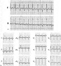
Panel A, electrocardiogram showed atrial flutter with 2:1 or 3:1 atrioventricular conduction giving a rate of 150 to 170 beats/min. Panel B, The 12-lead electrocardiogram after direct-current electrical conversion showed evidence of mild left atrial and ventricular hypertrophy.
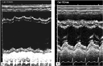
Composite panels of M-mode echocardiogram of the left ventricle. Soon after direct-current electrical conversion (A), the left ventricle was severely dilated and poorly contractile. After a period of 18 months subsequent to the electrical conversion (B), the left ventricle had decreased in size, with restoration of its function.
 You have
Access
- 6
- Cited by