




No CrossRef data available.
Article contents
Antioxidant enzymes in intramolluscan Schistosoma mansoni and ROS-induced changes in expression
Published online by Cambridge University Press: 06 May 2004
Abstract
Killing of intramolluscan schistosomes by host haemocytes is mediated by reactive oxygen metabolites. Hence, defence against oxidative damage is essential for the parasite to survive. In this study, expression of three key antioxidant enzymes, superoxide dismutase (EC 1.15.1.1), glutathione peroxidase (EC 1.11.1.9) and glutathione-S-transferase (EC 2.5.1.18) was determined in Schistosoma mansoni miracidia, sporocysts and cercariae. Stage-dependent expression of these enzymes was shown to be regulated at the transcriptional level. Second, the influence on enzyme expression of reactive oxygen species (ROS) and of haemocytes from schistosome-resistant and -susceptible host snails was determined. Generation of ROS by xanthine/xanthine oxidase resulted in increased transcript levels for all three enzymes. Addition of hydrogen peroxide induced a significantly increased expression of GPx and SOD but not GST. Snail haemocytes induced an up-regulation of SOD and GPx at 12 and 18 h post-exposure, respectively. Susceptible haemocytes elicited a stronger induction of transcript expression than resistant haemocytes. After 36–48 h, SOD remained up-regulated in sporocysts encapsulated by haemocytes from susceptible hosts, whereas a down-regulation of SOD and GPx occurred in schistosomes encapsulated by haemocytes from resistant snails. These observations indicate that schistosomes express elevated levels of antioxidant enzymes in interaction with haemocytes from susceptible snail hosts in which they survive. On the other hand, haemocytes of resistant snails may interfere with reactive oxygen detoxification via down-regulation of schistosome antioxidant enzymes, thus shifting the balance towards parasite killing.
Keywords
Information
- Type
- Research Article
- Information
- Copyright
- 2004 Cambridge University Press
INTRODUCTION
Freshwater snails are essential intermediate hosts in the schistosome life-cycle. Miracidia penetrate an appropriate snail species and asexually generate two generations of sporocysts before developing into cercariae, the infective larvae for the final, vertebrate host. The parasite develops in susceptible snail hosts without being eliminated by the immune system but the defence cells, the haemocytes, of resistant snails, rapidly kill it. Snail haemocytes usually kill invading pathogens and parasites by phagocytosis or encapsulation (Bayne, Hahn & Bender, 2001). During these processes oxygen is consumed by the haemocytes followed by an oxidative burst and the production of a variety of reactive oxygen species (ROS). Thus, schistosomes are exposed to ROS not only through their own respiratory processes but also as a result of the host immune responses, and must possess adequate mechanisms of ROS detoxification. Several enzymes form a crucial function of antioxidant defence (Cervi, Rossi & Masih, 1999; Henkle-Dührsen & Kampkötter, 2001). Superoxide dismutase (SOD, EC 1.15.1.1) catalyses the dismutation reaction of the toxic superoxide radical to molecular oxygen and hydrogen peroxide through the alternate reduction and oxidation of the active-site metal ions. On the other hand, SOD generates another toxic oxygen species, hydrogen peroxide, which is uncharged and diffuses freely across cell membranes. Hydrogen peroxide was shown to be highly toxic for helminths such as Onchocerca volvulus (Callahan, Crouch & James, 1988), Trichobilharzia ocellata (Adema, van der Knaap & Sminia, 1991) and Schistosoma mansoni (Hahn, Bender & Bayne, 2001).
The selenoprotein glutathione peroxidase (GPx, EC 1.11.1.9) detoxifies hydrogen peroxide via the oxidation of glutathione (Arthur, 2000). Glutathione-S-transferases (GST, EC 2.5.1.18) effectively neutralize cytotoxic by-products of lipid peroxidation arising from reactive oxygen species acting on cell membranes. This ability provides evidence that GSTs have the potential to protect the parasite against the host immune response (Campbell et al. 2001). Schistosome GST has been extensively investigated with respect to its biochemistry and independently has been identified as a potential vaccine candidate (Mitchell, 1989; Boulanger et al. 1999; Lebens et al. 2003).
The aim of our work is to understand the mechanisms used by schistosomes to survive in immunologically hostile body compartments of the snail host. We report herein the developmental expression of the antioxidant enzymes SOD, GPx and GST in non-mammalian stages of Schistosoma mansoni. Changes in transcript abundance of these enzymes induced by reactive oxygen species (ROS) and the effects of snail immune cells derived from schistosome-resistant or -susceptible host strains were also analysed.
MATERIALS AND METHODS
Parasites
Schistosoma mansoni (PR1 strain) miracidia were collected from eggs derived from livers of infected Syrian hamsters. Miracidia were collected under sterile conditions, washed and transferred to Medium F containing 2 μl/ml gentamycin for overnight transformation into mother sporocysts (Stibbs et al. 1979). Sporocysts were cultured for 14 days in unconditioned sporocyst medium SM under 8% O2 according to Bixler et al. (2001).
Cercariae were collected from infected snails after 1 h exposure to light.
Chemical stimulation
Stimulation of miracidia and cercariae was performed in sterile water (ASW, Ulmer, 1970) and sporocysts were stimulated in serum-free Medium F. A total of 5000 individuals were used for each treatment (4·5 mM xanthine, 4·5 mM xanthine/10 mU xanthine oxidase or 100 μM hydrogen peroxide at 26 °C for 1 or 3 h) or control. Controls were incubated in water or medium under identical conditions.
Stimulation by host haemocytes
Biomphalaria glabrata haemolymph of resistant (13-16-R1) and susceptible (M-line) strains was isolated by heart puncture as previously described (Zelck, Becker & Bayne, 1995) and transferred to individual wells of a poly-L-lysine (100 μg/ml) coated 48-well microtitre plate (800 μl of M-line haemolymph and 500 μl of 13-16-R1 haemolymph, since this strain has higher haemocyte numbers). Cells were allowed to settle for 15 min, plasma was carefully exchanged with iso-osmotic buffer according to the method described by Hahn et al. (2001) and schistosome sporocysts were added to the cells in a ratio of approximately 1[ratio ]150 (i.e. 900–1100 sporocysts, calculated based on cell counts in a Neubauer chamber). Control sporocysts were incubated in buffer without haemocytes, control haemocytes were incubated in buffer without schistosomes. RNA was extracted from unstimulated and encapsulated sporocysts as well as from haemocytes after 6, 12, 18–24, 36 and 48 h.
RNA preparation and OneStep-RT-PCR analysis
Total cellular RNA was isolated from non-stimulated or stimulated schistosomes as well as from haemocytes using the RNeasy Kit (Qiagen, Hilden) according to the manufacturer's instructions. Prior to the RT-PCR experiments, a DNA digestion was performed using 1U DNase I (Gibco BLR) for up to 2 μg RNA according to the manufacturer's instructions.
Gene-specific primers were designed based on Schistosoma sequences published in the database. The expected size of the amplified fragment is given in parenthesis. CT-SOD sense: ACT GTT TCA CAG GGA GGA TAG and CT-SOD antisense: ACG AGG ATG AAA GCT GTT TG (500 bp), GPx sense: GTT CAT AAG TTC CAT TGA TCA C and GPx antisense: ATG TCT TCA TCT CAC AAG TC (519 bp), GST sense: TCA GGA TAC TTG CCA GTT AG and GST antisense: ATG GCT GGC GAG CAT ATC (551 bp), tubulin sense: GAA GTG GAT ACG AGG ATA AGG TAC CAG and tubulin antisense: TGG AAC TTA TCG TCA ACT TTT CCA TCC (561 bp), Tubulin IN sense: GGC GGT GGT ACT GGT TCT GGG TTC (207 bp), GAPDH sense: AAT GCC TCC TGC ACC ACC and GAPDH antisense: ATG CCA GTG AGC TTC (316 bp). PCR mixtures contained 70 ng schistosome RNA, 2 μl enzyme mix, 2 μM each primer, 400 μM dNTPs and 1·25 mM MgCl2 (OneStep-RT-PCR Kit, Qiagen, Hilden). The PCR consisted of an initial round at 50 °C for 30 min and 95 °C for 15 min followed by 35 cycles of 94 °C for 1 min, 55 °C for 1 min and 72 °C for 2 min. The final extension time at 72 °C was lengthened to 10 min. All PCR reactions were carried out in an OmnE Thermocycler (Thermo Life Sciences, Egelsbach). The resulting PCR products were cloned into the pCR4 vector (TOPO-TA cloning Kit, Invitrogen) and sequenced using the sequence-specific primers listed above with the BigDye Terminator Kit (Applied Biosystems) on an Applied Biosystems 373 DNA sequencer.
Semi-quantitative duplex RT-PCR
The relative levels of transcript expression throughout schistosome development, after stimulation by ROS and in the encapsulation with haemocytes from different snail strains were compared in semi-quantitative duplex-PCR reactions utilizing normalization to β-tubulin. A series of preliminary experiments was performed to establish and optimize the semi-quantitative PCR conditions by determining the exponential phase for PCR amplification. Using different amounts of RNA as a template, PCR amplification curves were generated. When 40–50 ng of total RNA from schistosomes or encapsulations was used in RT-PCR amplification system, cDNA amplification of all target genes remained in the exponential phase until 32 cycles of amplification. This cycle number resulted in a linear relationship between the amount of cDNA input and PCR product, thus a saturation plateau in detecting amplification product could be avoided. Using β-tubulin as an internal standard, these studies determined that the amplification cycle number was optimal at 29 cycles (data not shown).
PCR mixtures contained 50 ng (SOD) or 40 ng (GST and GPx) RNA from schistosomes or encapsulations, 2 μl enzyme mix, 2 μM each enzyme primer, 1 μM tubulin primers, 400 μM dNTPs and 1·25 mM MgCl2 (OneStep-RT-PCR Kit, Qiagen, Hilden). The initial and final elongation steps of the PCR were carried out as for OneStep-PCR, and 29 cycles of 94 °C for 1 min, 53 °C for 1 min and 72 °C for 2 min were completed. All PCR reactions were carried out in an OmnE Thermocycler (Thermo Life Sciences, Egelsbach). The resulting PCR products were visualized on a 1·8% agarose gel. Gel images were digitally captured and fragment intensity was determined using the Easy Win 32 software (Herolab, Wiesloch) or in a 2100 Bioanalyzer (Agilent Technologies, Waldbronn/Palo Alto). Values are presented as a ratio of the specific gene's signal in the selected linear amplification cycle divided by the β-tubulin signal. Fragment identity was confirmed after cloning into the pCR4 vector (TOPO-TA cloning Kit, Invitrogen) followed by sequencing using the respective gene specific primers with the BigDye Terminator Kit (Applied Biosystems) on an Applied Biosystems 373 DNA sequencer.
Sequence analysis
General homology searches were performed with the Blast software on the NCBI home page (BLASTn, http://www.ncbi.nlm.nih.gov./BLAST/). Deduced amino acid sequences were aligned by Clustal W (http://www.ebi.ac.uk/clustalw/), searches for homologies were accomplished by Fasta3 (http://www.ebi.ac.uk/fasta33/) and PropSearch (http://www.embl-heidelberg.de/prs.html).
RESULTS
Expression of antioxidant enzymes in unstressed Schistosoma mansoni
Superoxide dismutase (SOD), glutathione-peroxidase (GPx) and glutathione-S-transferase (GST) are expressed in unstimulated miracidia, mother and 14-day sporocysts and cercariae (Fig. 1). All RT-PCR products yielded a single band of the expected size on agarose gels (500 bp for SOD, 519 bp for GPx and 551 bp for GST), and sequencing analysis showed the primers were amplifying the expected products. Sequences were 99–100% identical to the respective stretches in those reported for adult schistosomes in the database with GenBank Accession nos M97298, M86510 and M98271. Enzyme expression is developmentally regulated: miracidia, mother sporocysts and cercariae are most susceptible to immune killing and exhibit higher transcript levels compared to daughter sporocysts which develop within the lobules of the digestive gland.

Fig. 1. Representative gel image of relative RT-PCR for superoxide dismutase (SOD), glutathione peroxidase (GPx), glutathione-S-transferase (GST) and β-tubulin (TUB) expression in unstressed Schistosoma mansoni miracidia, mother sporocysts, 14-day sporocysts and cercariae.
Effect of ROS on enzyme expression
Xanthine/xanthine oxidase stimulation (Fig. 2) resulted in increased transcription expression for SOD (5·2-fold), GPx (3·2-fold) and GST (1·8-fold). Results are the mean of 10 independent experiments. Incubation with xanthine alone decreased the expression of GPx and GST.

Fig. 2. Effect of ROS on enzyme expression. (A) SOD, GPx and GST expression in Schistosoma mansoni miracidia, mother sporocysts, 14-day sporocysts and cercariae stimulated with xanthine/xanthine oxidase (X/XO) for 3 h. The values on the ordinate represent the mean (±S.E.) amount of expression relative to non-treated controls from 10 independent experiments, respectively. Gene transcript quantity was measured by densitometry using the internal standard β-tubulin signal as the denominator as described in the Materials and Methods section. (B) Representative gel image of relative RT-PCR for SOD, GPx, GST and β-tubulin (TUB) expression in S. mansoni cercariae either un-stimulated (control) or stimulated with xanthine (X) or xanthine/xanthine oxidase (X/XO) for 3 h. Target genes were cycled within the pre-determined linear amplification range.
Stimulation with hydrogen peroxide induced an increased transcript abundance of SOD and GPx in all larval schistosomes, whereas the intensity of the GST amplification product did not change significantly. Figure 3 shows the H2O2-induced 2·2- and 4·8-fold excess for SOD and GPx, respectively (mean from 10 experiments).

Fig. 3. Effect of H2O2 on enzyme expression. (A) SOD, GPx and GST expression in Schistosoma mansoni miracidia, mother sporocysts, 14-day sporocysts and cercariae stimulated with 100 μM H2O2 for 1 h. The values on the ordinate represent the mean (±S.E.) amount of expression relative to non-treated controls from 10 independent experiments, respectively. Gene transcript quantity was measured by densitometry using the internal standard β-tubulin signal as the denominator as described in the Materials and Methods section. (B) Representative gel image of relative RT-PCR for SOD, GPx, GST and β-tubulin (TUB) expression in S. mansoni cercariae either un-stimulated (control) or stimulated with 100 μM H2O2 for 1 h. Target genes were cycled within the pre-determined linear amplification range.
Exposure to H2O2 resulted in stronger GPx expression responses compared to exposure to ROS generated by the xanthine/xanthine oxidase system, whereas SOD expression was higher after stimulation by X/XO.
Effect of host haemocytes on enzyme expression
Schistosoma mansoni mother sporocysts were surrounded in vitro by haemocytes of susceptible and resistant B. glabrata snails within 60 min (Fig. 4). Haemocytes from resistant snail strains efficiently kill sporocysts of the PR1 strain in vitro in the absence of plasma molecules within 48–72 h, whereas haemocytes from susceptible strains leave the parasite unharmed (Bayne, Buckley & DeWan, 1980; Hahn et al. 2001). Figure 5 shows the relative intensities of enzyme expression levels in schistosome sporocysts during encapsulations in the time-course experiment. SOD and GPx are up-regulated during 12–24 h upon stimulation by either host haemocytes, with the maximal induction of SOD and GPx expression at 24 h. In S-encapsulations at 48 h, expression of SOD remained up-regulated and GPx expression dropped to the levels in unstimulated sporocysts. In R-encapsulations, no SOD and only minimal GPx expression was detectable at 48 h whereas amplification of housekeeping genes β-tubulin and GAPDH (not shown) was not impaired. GST expression remained unchanged during the first 24 h, but decreased at 48 h in R-encapsulations. Amplification was schistosome-specific since no products were amplified from host haemocyte RNA.

Fig. 4. Encapsulation of Schistosoma mansoni mother sporocysts (Sp) by haemocytes (H) of the snail host Biomphalaria glabrata. Initially, single haemocytes adhere to the sporocyst surface (A). After 60 min, a cellular aggregate has formed around the sporocyst (B). (Magnification 20×.)

Fig. 5. Effect of host haemocytes on enzyme expression. Relative intensities of SOD, GPx and GST expression during in vitro encapsulation of Schistosoma mansoni mother sporocysts by haemocytes from susceptible (SHc) or resistant (RHc) intermediate hosts. Results are the mean from 12 independent experiments. The values on the ordinate represent the mean (±S.E.) amount of expression relative to non-encapsulated controls (N.S.; set as 1·0). Gene transcript quantity was measured by densitometry using the internal standard β-tubulin signal as the denominator as described in the Materials and Methods section. Gel images show the amplification of the respective enzyme and tubulin in R-haemocyte-encapsulated schistosomes at 36 h and 48 h.
DISCUSSION
The classic example of metabolic production of reactive oxygen species (ROS) is the production of O2− and H2O2 by activated phagocytes when they contact foreign particles, pathogens or immune complexes. The abundant sources leading to free radical generation have required all aerobic organisms to develop intra- and extracellular mechanisms of detoxification. The first line of cellular antioxidant defence is the superoxide dismutase (SOD). SODs catalyse the reaction 2O2−+2H+→H2O2+O2. Hydrogen peroxide is then inactivated by glutathione peroxidase which catalyses the reduction of peroxides into alcohols using the reducing potential of glutathione. Phase II detoxification enzymes include the glutathione S-transferases that neutralize cytotoxic by-products of lipid peroxidation arising from reactive oxygen species acting on cell membranes. In order to identify these antioxidant enzymes and analyse their gene expression, different stages of S. mansoni were subjected to exogenous oxidative stress. SOD, GPx and GST were expressed in unstressed miracidia, sporocysts and cercariae. Sequences were identical to those reported in the database for adult schistosomes (GenBank Accession nos. M97298, M86510 and M98271) indicating an expression throughout schistosome development. However, the expression is developmentally regulated. High transcript expression correlates with higher exposure of the parasite to oxygen and potentially toxic oxygen metabolites. Previous studies have shown that antioxidant enzymes were more highly expressed in adult schistosomes in line with their higher resistance to the host immune defence compared with larvae (Nare, Smith & Prichard, 1990; Mei & LoVerde, 1997). In the present study, the highest enzyme expression was shown in free-swimming miracidia and cercariae with an aerobic metabolism (von Kruger et al. 1978; Tielens et al. 1991) and, after penetration, with direct exposure to the immediate immune response of their respective hosts. Transcript expression was lower in daughter sporocysts, which live under less aerobic conditions within the digestive gland lobules of the snail intermediate host. This supports the earlier observation that axenic culture of sporocysts requires low oxygen conditions (Bixler et al. 2001). Sporocysts developing in the gland are supposedly better protected from host immune responses. Haemocytes are rarely seen between the gland lobules and immune reactions against daughter sporocysts in the snail host were never observed (de Souza, Cunha Rde & Andrade, 1995; Zelck, unpublished observations). Thus, lower enzyme expression in these sporocysts might be due to lower exposure to oxygen and/or to potentially toxic oxygen metabolites produced by the host.
Our results show that schistosomes can up-regulate the respective antioxidant enzyme(s) depending on the ROS to be detoxified. Stimulation with the primarily superoxide generating xanthine/xanthine oxidase system resulted in elevated expression of SOD, GPx and GST in miracidia, sporocysts and cercariae. Stimulation with hydrogen peroxide resulted in significantly increased transcript expression for GPx and SOD. Longer exposure to H2O2 had an inhibitory effect on the SOD expression (results not shown). Hydrogen peroxide is known to be highly toxic for schistosomes and other helminths (Callahan et al. 1990; Hahn et al. 2001). Consequently, exposure to this agent induced higher GPx expression levels in schistosomes compared with the X/XO system.
Stimulation of snail host immune cells by schistosomes leads to a massive local production of ROS during the encapsulation process (Adema et al. 1994; Hahn et al. 2001). While attachment of haemocytes to the sporocyst surface and the formation of multiple cell layers occur after 60 min, ROS production and killing by haemocytes start after 10 h and continue to increase for 48 h. We show here that haemocyte stimulation caused an ROS scavenging response in the sporocysts, i.e. a boost in antioxidant enzyme expression. The time-course experiments showed that induction of SOD transcript levels occurred 12 h after treatment, peaking at 24 h. Up-regulation of GPx was first observed at 18 h, and maximum expression occurred at 24–36 h. This response correlates with the increasing release of superoxide and hydrogen peroxide by haemocytes over a period of 2–48 h, also peaking at 24 h (Zelck, unpublished observations). However, the parasite's response to encapsulating haemocytes varied depending upon the strain of host snail from which they were derived. Haemocytes from susceptible snails induced an up-regulation that lasted for 36–48 h, whereas haemocytes from resistant snails induced an up-regulation, which was lower compared to S-encapsulations and lasted only for 24–36 h. Thereafter SOD and GPx were down-regulated. This correlates with increased sporocyst mortality in these encapsulations (Hahn et al. 2001). Quantification of individual ROS production by haemocytes of susceptible and resistant hosts is needed to correlate the differentially induced expression of schistosome antioxidant enzymes and other potential target genes. Published studies on ROS production by haemocytes from different snail strains show differing results (Connors & Yoshino, 1990; Hahn et al. 2001) and need verification. We propose that susceptible haemocytes produce moderate amounts of ROS, which are not lethal because ROS can be detoxified within a few hours and is evident in the strong induction of antioxidant enzymes in the schistosomes. If ROS production is high as suggested for resistant host haemocytes, it not only causes irreversible degradation of cellular macromolecules, but can also repress gene expression. Haemocytes from resistant hosts induced an initial moderate increase in antioxidant enzyme expression followed by a down-regulation. ROS were previously shown to induce or repress the expression of a variety of genes (Morel & Barouki, 1999; Finkel & Holbrook, 2000) such as transcription factors and molecules involved in signal transduction and intra- or intercellular signalling mechanisms (Hancock, Desikan & Neill, 2001; Reth, 2002). Possible mechanisms of gene repression are DNA methylation and histone deacetylation (El-Osta & Wolffe, 2000; Curradi et al. 2002) but need to be determined in this system.
In conclusion, S. mansoni was able to up-regulate antioxidant enzymes under exogenous oxidative stress and when encapsulated by host haemocytes. Haemocytes from susceptible hosts induced higher levels of enzyme expression in schistosome sporocysts compared to haemocytes from resistant hosts. Our results indicate that haemocytes from resistant host snails are able to directly or indirectly down-regulate antioxidant enzyme expression. Suboptimal expression, especially of GPx, in the parasite results in impaired H2O2 detoxification and enhanced cytotoxic effects caused by haemocytes from resistant snails.
We are grateful to Ulrike Müller for excellent technical assistance, to Lia Bixler for cultivation of the schistosome 14-day sporocysts and to Kathy Astrahantseff for critical reading of the manuscript.
References
REFERENCES
ADEMA, C. M., VAN DER KNAAP, W. P. W. & SMINIA, T. (1991). Molluscan haemocyte-mediated cytotoxicity: the role of reactive oxygen intermediates. Reviews in Aquatic Sciences 4, 201–233.Google Scholar
ADEMA, C. M., VAN DEUTEKOM-MULDER, E. C., VAN DER KNAAP, W. P. W. & SMINIA, T. (1994). Schistosomicidal activities of Lymnaea stagnalis haemocytes: the role of oxygen radicals. Parasitology 109, 479–485.CrossRefGoogle Scholar
ARTHUR, J. R. (2000). The glutathione peroxidases. Cellular and Molecular Life Science 57, 1825–1835.Google Scholar
BAYNE, C. J., BUCKLEY, P. M. & DEWAN, P. C. (1980). Schistosoma mansoni: cytotoxicity of hemocytes from susceptible snail hosts for sporocysts in plasma from resistant Biomphalaria glabrata. Experimental Parasitology 50, 409–416.CrossRefGoogle Scholar
BAYNE, C. J., HAHN, U. K. & BENDER, R. C. (2001). Mechanisms of molluscan host resistance and of parasite strategies for survival. Parasitology 123 (Suppl.), S159–S167.CrossRefGoogle Scholar
BIXLER, L. M., LERNER, J. P., IVANCHENKO, M., McCORMICK, R. S., BARNES, D. W. & BAYNE, C. J. (2001). Axenic culture of Schistosoma mansoni sporocysts in low O2 environments. Journal of Parasitology 87, 1167–1168.Google Scholar
BOULANGER, D., SCHNEIDER, D., CHIPPAUX, J. P., SELLIN, B. & CAPRON, A. (1999). Schistosoma bovis: vaccine effects of a recombinant homologous glutathione S-transferase in sheep. International Journal for Parasitology 29, 415–418.CrossRefGoogle Scholar
CALLAHAN, H. L., CROUCH, R. K. & JAMES, E. R. (1988). Helminth anti-oxidant enzymes: a protective mechanism against host oxidants? Parasitology Today 4, 218–225.Google Scholar
CALLAHAN, H. L., CROUCH, R. K. & JAMES, E. R. (1990). Hydrogen peroxide is the most toxic oxygen species for Onchocerca cervicalis microfilariae. Parasitology 100, 407–415.CrossRefGoogle Scholar
CAMPBELL, A. M., TEESDALE-SPITTLE, P. H., BARRETT, J., JEFFERIES, J. R. & BROPHY, P. M. (2001). A glutathione S-transferase class common to Caenorhabditis elegans and parasitic nematodes. Comparative Biochemistry and Physiology 128B, 701–708.CrossRefGoogle Scholar
CERVI, L., ROSSI, G. & MASIH, D. T. (1999). Potential role for excretory-secretory forms of glutathione-S-transferase (GST) in Fasciola hepatica. Parasitology 119, 627–633.CrossRefGoogle Scholar
CONNORS, V. A. & YOSHINO, T. P. (1990). In vitro effect of larval Schistosoma mansoni excretory-secretory products on phagocytosis-stimulated superoxide production in haemocytes from Biomphalaria glabrata. Journal of Parasitology 76, 895–902.CrossRefGoogle Scholar
CURRADI, M., IZZO, A., BADARACCO, G. & LANDSBERGER, N. (2002). Molecular mechanisms of gene silencing mediated by DNA methylation. Molecular Cell Biology 22, 3157–3173.CrossRefGoogle Scholar
DE SOUZA, C. P., CUNHA RDE, C. & ANDRADE, Z. A. (1995). Development of Schistosoma mansoni in Biomphalaria tenagophila, Biomphalaria straminea and Biomphalaria glabrata. The Revistado Instituto de Medicinia Tropical de Sao Paulo 37, 201–206.CrossRefGoogle Scholar
EL-OSTA, A. & WOLFFE, A. P. (2000). DNA methylation and histone deacetylation in the control of gene expression: basic biochemistry to human development and disease. Gene Expression 9, 63–75.Google Scholar
FINKEL, T. & HOLBROOK, N. J. (2000). Oxidants, oxidative stress and the biology of ageing. Nature, London 408, 239–247.CrossRefGoogle Scholar
HAHN, U. K., BENDER, R. C. & BAYNE, C. J. (2001). Killing of Schistosoma mansoni sporocysts by haemocytes from resistant Biomphalaria glabrata: role of oxygen species. Journal of Parasitology 87, 292–299.CrossRefGoogle Scholar
HANCOCK, J. T., DESIKAN, R. & NEILL, S. J. (2001). Role of reactive oxygen species in cell signalling pathways. Biochemical Society Transactions 29, 345–350.CrossRefGoogle Scholar
HENKLE-DÜHRSEN, K. & KAMPKÖTTER, A. (2001). Antioxidant enzyme families in parasitic nematodes. Molecular and Biochemical Parasitology 114, 129–142.CrossRefGoogle Scholar
LEBENS, M., SUN, J. B., SADEGHI, H., BÄCKSTROM, M., OLSSON, I., MIELCAREK, N., LI, B. L., CAPRON, A., CZERKINSKY, C. & HOLMGREN, J. (2003). A mucosally administered recombinant fusion protein vaccine against schistosomiasis protecting against immunopathology and infection. Vaccine 2003 21, 514–520.CrossRefGoogle Scholar
MEI, H. & LOVERDE, P. T. (1997). Schistosoma mansoni: the developmental regulation and immunolocalisation of antioxidant enzymes. Experimental Parasitology 86, 69–78.CrossRefGoogle Scholar
MEI, H., THAKUR, A., SCHWARTZ, J. & LOVERDE P. T. (1996). Expression and characterisation of glutathione peroxidase activity in the human blood fluke Schistosoma mansoni. Infection and Immunity 64, 4299–4306.Google Scholar
MITCHELL, G. F. (1989). Gluthatione-S-transferases – potential components of anti-schistosoma vaccines? Parasitology Today 5, 34–37.Google Scholar
MOREL, Y. & BAROUKI, R. (1999). Repression of gene expression by oxidative stress. The Biochemical Journal 342, 481–496.CrossRefGoogle Scholar
NARE, B.,
SMITH, J. M. &
PRICHARD, R. K.
(1990).
Schistosoma mansoni: levels of antioxidants and resistance to oxidants increase during development.
Experimental Parasitology 70, 389–397.CrossRefGoogle Scholar
RETH, M.
(2002).
Hydrogen peroxide as second messenger in lymphocyte activation.
Nature Immunology 3, 1129–1134.CrossRefGoogle Scholar
STIBBS, H. H.,
OWCZARZAK, A.,
BAYNE, C. J. &
DEWAN, P.
(1979).
Schistosome sporocyst-killing amoebae isolated from Biomphalaria glabrata.
Journal of Invertebrate Pathology 33, 159–170.CrossRefGoogle Scholar
TIELENS, A. G.,
VAN DE PAS, F. A.,
VAN DEN HEUVEL, J. M. &
VAN DEN BERGH, S. G.
(1991).
The aerobic energy metabolism of Schistosoma mansoni miracidia.
Molecular and Biochemical Parasitology 46, 181–184.CrossRefGoogle Scholar
ULMER, M. J. (1970). Notes on rearing snails in the laboratory. In Experiments and Techniques in Parasitology (ed. MacInnis, A. J. & Voge, M.), pp. 143–144. W. H. Freeman and Company, San Francisco.
VON KRUGER, W. M.,
GAZZINELLI, G.,
FIGUEIREDO, E. A. & PELLEGRINO, J.
(1978).
Oxygen uptake and lactate production by Schistosoma mansoni cercariae, cercarial body and tail, and schistosomule.
Comparative Biochemistry and Physiology, B 60, 41–46.Google Scholar
ZELCK, U. E., BECKER, W. & BAYNE, C. J. (1995). The plasma proteins of Biomphalaria glabrata in the presence and absence of Schistosoma mansoni. Developmental and Comparative Immunology 19, 181–194.CrossRefGoogle Scholar

Fig. 1. Representative gel image of relative RT-PCR for superoxide dismutase (SOD), glutathione peroxidase (GPx), glutathione-S-transferase (GST) and β-tubulin (TUB) expression in unstressed Schistosoma mansoni miracidia, mother sporocysts, 14-day sporocysts and cercariae.

Fig. 2. Effect of ROS on enzyme expression. (A) SOD, GPx and GST expression in Schistosoma mansoni miracidia, mother sporocysts, 14-day sporocysts and cercariae stimulated with xanthine/xanthine oxidase (X/XO) for 3 h. The values on the ordinate represent the mean (±S.E.) amount of expression relative to non-treated controls from 10 independent experiments, respectively. Gene transcript quantity was measured by densitometry using the internal standard β-tubulin signal as the denominator as described in the Materials and Methods section. (B) Representative gel image of relative RT-PCR for SOD, GPx, GST and β-tubulin (TUB) expression in S. mansoni cercariae either un-stimulated (control) or stimulated with xanthine (X) or xanthine/xanthine oxidase (X/XO) for 3 h. Target genes were cycled within the pre-determined linear amplification range.
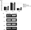
Fig. 3. Effect of H2O2 on enzyme expression. (A) SOD, GPx and GST expression in Schistosoma mansoni miracidia, mother sporocysts, 14-day sporocysts and cercariae stimulated with 100 μM H2O2 for 1 h. The values on the ordinate represent the mean (±S.E.) amount of expression relative to non-treated controls from 10 independent experiments, respectively. Gene transcript quantity was measured by densitometry using the internal standard β-tubulin signal as the denominator as described in the Materials and Methods section. (B) Representative gel image of relative RT-PCR for SOD, GPx, GST and β-tubulin (TUB) expression in S. mansoni cercariae either un-stimulated (control) or stimulated with 100 μM H2O2 for 1 h. Target genes were cycled within the pre-determined linear amplification range.

Fig. 4. Encapsulation of Schistosoma mansoni mother sporocysts (Sp) by haemocytes (H) of the snail host Biomphalaria glabrata. Initially, single haemocytes adhere to the sporocyst surface (A). After 60 min, a cellular aggregate has formed around the sporocyst (B). (Magnification 20×.)
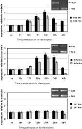
Fig. 5. Effect of host haemocytes on enzyme expression. Relative intensities of SOD, GPx and GST expression during in vitro encapsulation of Schistosoma mansoni mother sporocysts by haemocytes from susceptible (SHc) or resistant (RHc) intermediate hosts. Results are the mean from 12 independent experiments. The values on the ordinate represent the mean (±S.E.) amount of expression relative to non-encapsulated controls (N.S.; set as 1·0). Gene transcript quantity was measured by densitometry using the internal standard β-tubulin signal as the denominator as described in the Materials and Methods section. Gel images show the amplification of the respective enzyme and tubulin in R-haemocyte-encapsulated schistosomes at 36 h and 48 h.
 You have
Access
- 56
- Cited by