INTRODUCTION
Pelagic copepods of the genus Calanus are among the most dominant members in marine zooplankton both in abundance and biomass, and play important roles in the food web and matter cycling in the sea (e.g. Marshall & Orr, Reference Marshall and Orr1955; Conover, Reference Conover1988; Mauchline, Reference Mauchline1998). While a large body of knowledge has accumulated on the biology and ecology of Calanus species, relatively little is known of the structure and function of the organs that they possess (e.g. Lowe, Reference Lowe1935; Nishida, Reference Nishida1989; Boxshall, Reference Boxshall and Harrison1992). Among these organs are the large glands in their caudal rami, hereafter referred to as the ‘caudal glands’. During the course of our study on the life history of the Calanus species in Sagami Bay, central Japan, we noted the presence of these glands in C. sinicus Brodsky and C. jashnovi Hulsemann but have been unable to find any mention of these glands in the literature. Since the glands are massive and suspected to be common in the genus, we investigated their structure, spatiotemporal variability of secretions and taxon specificity, and discuss their possible function.
MATERIALS AND METHODS
Stratified zooplankton samples were collected in the central part of Sagami Bay (35°00′N 139°20′E) on 7–9 May, 2001, both day and night from 4 discrete layers at 250-m intervals in the upper 1000 m by using a Vertically-towed Multiple Plankton Sampler (VMPS, mesh size = 100 µm; Terazaki & Tomatsu, Reference Terazaki and Tomatsu1997). The original samples were split into 1/2 aliquots with a box-type splitter (Motoda, Reference Motoda1959b). One aliquot was fixed and preserved in 2% formaldehyde for observation of general morphology by light- (LM) and scanning electron microscopy (SEM), while the other was fixed in 2% glutaraldehyde + 2.5% paraformaldehyde for fine-structural observation in a transmission electron microscope (TEM). Calanus copepodids were sorted from the formaldehyde-preserved samples, their species and stages identified and enumerated, and the gross morphology of the caudal glands examined. Some of the sorted specimens were post-fixed in 1% OsO4, dehydrated through ethanol series to 100% ethanol, freeze-dried in t-butyl alcohol, then examined in a Hitachi-4500 SEM for the external morphology of the caudal rami. The formaldehyde-fixed samples were also examined for day–night, and depth occurrences of granules in the caudal glands. The TEM samples were post-fixed with OsO4, embedded in epoxy resin, thin-sectioned, stained with uranyl acetate and lead citrate, and examined in a Hitachi-7100 TEM for the internal fine structure of the glands and granules.
Another series of monthly samples was collected from May 2002 to January 2004 at the same location as above in the daytime in the upper 200 m by vertical tows either of a VMPS or a Norpac net (Motoda Reference Motoda1959a; mesh size = 100 µm). The samples were immediately fixed in 2% formaldehyde and examined for presence/absence of granules in the caudal glands.
The ability of the caudal glands of Calanus sinicus to produce luminescence was examined during the May 2001 cruise. Copepods were collected by a vertical tow of the Norpac net from 50 m to the surface. Ten adult females were immediately sorted from the original sample into 500 ml of GF/F filtered seawater in a glass vial, and kept in darkness at 15°C for 2 hours. The vial was then introduced to a dark room and hand-shaken to mechanically disturb the copepods, and incidence of luminescence was examined by eye. Another 5 adult females, collected as above, were each put on a glass slide alive in a drop of filtered seawater and examined under an epi-fluorescence microscope with UV excitation for autofluorescence from the secreted granules following Bannister & Herring (Reference Bannister and Herring1989).
The zooplankton samples archived in the Ocean Research Institute were also examined for the presence of the caudal glands in other Calanus species and other calanid genera.
RESULTS
Macrostructure and external morphology
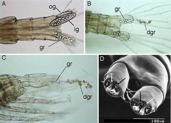
The specimens of Calanus from Sagami Bay included stages IV–VI copepodids (CIV–CVIs) of Calanus sinicus, CIV–CVs of C. jashnovi, and CI–CIIIs of unidentified Calanus. All these specimens had a gland-like structure occupying nearly the whole space of the caudal ramus and extending to the anterior part of the anal somite, regardless of the species and stage (Figure 1A–C).
Abdominal somites and caudal rami of Calanus showing inner gland (ig), outer gland (og), granules in inner gland (gr) and discharged granules (dgr). (A) Calanus sinicus, adult male, dorsal view; (B) C. sinicus, adult female, dorsal view; (C) unidentified copepodid stage-II, lateral view; (D) C. sinicus, scanning electron microscopy photograph of adult female, ventral view, showing pores of inner gland (black arrows) and outer gland (white arrows).

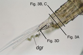
According to the LM- and SEM observations, there are two types of glands in each of the paired caudal rami, and each gland opens to the environment via a pore located on the ventral side of the base of the two medial-terminal caudal setae (Figures 1D & 2). These glands and pores are hereafter referred to as ‘inner/outer gland’ and ‘inner/outer pore’, respectively. Of these, the inner gland had numerous transparent granules in their cavities in most of the specimens both day and night, while the outer gland had no such granules (Figure 1A, B). In some specimens, the granules were apparently discharged from the gland through the pore into the environment (Figures 1B, C & 2).
Abdominal somites and caudal rami of adult female Calanus sinicus; lateral view, showing granules in inner gland and discharged granules (dgr). Lines indicate locations and directions of transmission electron microscopy sections as shown in Figure 3.

Fine structure
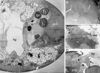
The cross-sections of the anal somite (Figures 2, & 3A, B) show different types of granules between the inner- and outer gland cells. The granules in the inner gland (Figure 3A) are much denser than those of the outer gland and fairly homogenous devoid of any visible structure (note that the striated appearance in the granules labelled ‘gri’ is an artefact from thin-sectioning), while the granules in the outer gland (Figure 3B) are sparser and have a fine texture. Other regions of the cells contain smaller vesicles that appear to be precursors of the granules (Figure 3A). The section containing the anterior region of the inner gland cell (Figures 2 & 3C) shows presence of a nucleus, well-developed rough endoplasmic reticulum, mitochondria, Golgi-bodies and vesicles, indicating that the cell comprises an exocrine gland. The section crossing the pore of the inner gland (Figure 3D) shows pre-discharge and discharged granules, a collar of the pore, and a valve-like structure that appears to regulate the discharge.
Transmission electron microscopy sections from adult female Calanus sinicus. (A) Cross-section of anal somite, showing granules in inner cavity (gri), those in outer cavity (gro), vesicles (ves), and hind-gut lumen (hg); ventral side to the top; (B) enlargement of granules in outer cavity; (C) anterior region of inner-gland cell, containing nucleus (n), rough endoplasmic reticulum (rer), mitochondrion (arrow), and vesicle (ves); (D) longitudinal section of pore of inner gland, showing pre-discharge (pdgr) and discharged granules (dgr), collar (arrows), and valve-like structure (stars).

Vertical, diel and seasonal occurrence of granules
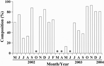
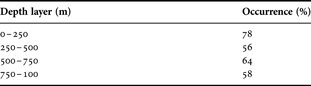
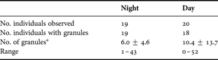
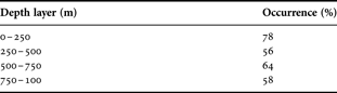
To get insights into the function of the glands and secretions, we investigated the occurrence of the granules in the populations of Calanus in Sagami Bay. While the granules might be discharged by the disturbances of sampling and other artefacts associated with sample processing, the presence of granules would also be an indicator of the activity of the glands. Table 1 shows the percentage of the CVs that had granules in the caudal glands to the total CVs in the 1000-m water column. It is evident that more than 50% of C. sinicus CVs had granules throughout the water column. It should be noted that the CVs in the mesopelagic zone (>250 m), which are probably in diapause as suggested by Nonomura et al. (Reference Nonomura, Machida and Nishida2008), had granules as well as those in the epipelagic population. The day–night comparison in the upper 200 m (Table 2) shows that equally large proportions (>90%) of C. sinicus had granules both day and night, with the number of granules per individual ranging from 0 to 52. The seasonal variation of the percentage of adult females having granules to all adult females in the upper 200 m in the daytime (Figure 4) shows presence of the granules throughout the year with no apparent seasonality but with considerable temporal fluctuation (13–95%). All these observations show that the granules are produced actively by C. sinicus copepodids in Sagami Bay, regardless of the stages, depth, season, or diel period.
Seasonal variation of percentage of adult female Calanus sinicus having granules to all adult females in the upper 200 m of central Sagami Bay. All samples were collected in the daytime. Asterisks denote no data due to small sample sizes (<10 individuals) or specimen damage.

Occurrence of granules (% of 50 copepods) in caudal glands of stage-V Calanus sinicus from different depth layers in central Sagami Bay, May 2001.
Day/night occurrence of granules (number per copepod) in the inner caudal gland in adult female Calanus sinicus from the upper 200 m of central Sagami Bay in May 2001.
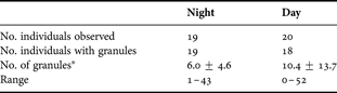
*, sum of granule from both sides (number/copepod), mean ± SD.
Luminescence ability
The mechanical disturbance of the live Calanus sinicus females kept in filtered seawater failed to excite luminescence from the copepods. No autofluorescence was observed from the caudal glands of live C. sinicus females under UV excitation.
Occurrence in other regions and species
An examination of Calanus specimens that are archived in the Ocean Research Institute indicated the presence of similar types of caudal glands in C. sinicus from the Inland Sea of Japan and the East China Sea, C. pacificus Brodsky from the Oyashio Region off north-eastern Japan, C. jashnovi from the Kuroshio Extension area and C. helgolandicus (Claus) from the North Sea.
Among the other genera of Calanidae, the glands were also present in Cosmocalanus darwinii (Lubbock), Mesocalanus tenuicornis (Dana), and Nannocalanus minor (Claus), but were absent in Neocalanus robustior (Giesbrecht), N. plumchrus (Marukawa), N. flemingeri Miller, N. cristatus (Kröyer), Canthocalanus pauper (Giesbrecht) and Undinula vulgaris (Dana).
DISCUSSION
The present observations demonstrated that the copepodids of Calanus species possess caudal exocrine glands that were hitherto unknown. In a large proportion of the copepod population the glands secrete numerous granules regardless of season, depth, sex, stage, or diel period. We consider that this excludes a possible function in pheromone secretion and suggesting their fundamental role in the copepods' life. In addition, there are two types of glands (inner and outer) suggesting that different substances must be mixed after discharge to be functional.
We have been unable to find reports of similar caudal glands in Calanidae in the literature, except the paper by Chiba (Reference Chiba1953) who described numerous granules in the caudal rami of the adult female Calanus darwinii (=Cosmocalanus darwinii). He interpreted them as eggs in a chamber that may have functioned as a brood pouch. We re-examined the species and confirmed that the structure is indeed an exocrine gland.
The gross- and fine structure of the present caudal glands is reminiscent of some luminous glands in copepods. Well-known examples are the luminous glands in the Metridiniidae and Augaptillidae (e.g. Herring, Reference Herring1988; Bannister & Herring, Reference Bannister and Herring1989). Some of the luminous glands in the genera Pleuromamma and Metridia are paired and present in the caudal rami, as well as in the prosome and swimming appendages. The luminous glands in many species are fluorescent, being excited with UV radiation (Herring, Reference Herring1988), although some luminous glands such as those in Megacalanus and Heterorhabdus are non-fluorescent (Herring, Reference Herring1988; Bannister & Herring, Reference Bannister and Herring1989). The present glands did not fluoresce under UV excitation, and the mechanical disturbance of the Calanus in bottles of seawater excited no luminescence. In addition, there is no previous report of confirmed luminescence in Calanidae, as far as we are aware, except for the unconfirmed reports on Nannocalanus minor, Neocalanus gracilis (Dana) and Undinula vulgaris (Herring, Reference Herring1988). These observations suggest that the caudal glands in Calanus species are non-luminous, although a possibility that special, but still-unknown, conditions may be required to stimulate luminescence cannot totally be ruled out. An alternative function might be secretion of defensive substances against predators, but this appears to contradict with the fact that Calanus species are equipped with myelinated axons (Lenz et al., Reference Lenz, Hower and Hartline2004) and are fast swimmers (Davis et al., Reference Davis, Weatherby, Hartline and Lenz1999), both of which facilitate their quick escape from predators, and might discount the adaptive significance of the presumed secretion of defensive substances. Another possibility may be secretion of chemicals that facilitate formation of swarms by conspecific copepods, which have often been observed in the populations of C. sinicus and C. jashnovi in Sagami Bay and the area of Kuroshio Extension (Nonomura, unpublished observation).
Interestingly, the distribution of the caudal glands in the calanid genera is consistent with the phylogenetic relationships among them proposed by Bucklin et al. (Reference Bucklin, Frost, Bradford-Grieve, Allen and Copley2003), in which the genera with the caudal glands, i.e. Calanus, Mesocalanus, Nannocalanus and Cosmocalanus, comprise a monophyletic group. This suggests that the caudal glands first appeared in the ancestor of this clade. The suggested role of the caudal glands in predator avoidance may partly explain the proliferation of these copepods over a wide range from coastal to oceanic waters (e.g. Conover, Reference Conover1988; Bucklin & Wiebe, Reference Bucklin and Wiebe1998). However, the definitive function of the caudal glands is still an open question, which would invite further studies on their chemical ecology, involving behavioural, histochemical and biochemical approaches.
ACKNOWLEDGEMENTS
We thank the captain and crew members of the RV ‘Tansei Maru’ and TV ‘Seiyo Maru’ for their help in sampling at sea. We also thank Takashi Ishimaru, Jun Nishikawa, and Keisuke Yoshida for their support in monthly sample collections in Sagami Bay. Special thanks are due to Hiroshi Itoh for information and discussion on the morphology and distribution of the Calanus species. This study has been supported by the Ministry of Education, Science, Arts and Sports of Japan (S.N., Grant-in-Aid for Creative Basic Research, No. 12NP0201); the Japan Society for the Promotion of Science (S.N., Multilateral Cooperative Research Program, Coastal Oceanography); and Alfred P. Sloan Foundation (S.N., Census of Marine Zooplankton).