Sequestration is classified as intralobar or extralobar on the basis of whether or not there is separation from normal lung tissue by its own visceral pleura. There is only one previously reported case of a patient with combined intralobar and extralobar sequestration, treated with surgical resection. We present a case of multivessel, combined intralobar and extralobar pulmonary sequestration that was treated with transcatheter embolisation using Type 2 and Type 4 Amplatzer Vascular Plugs.
Case report
A 5-year-old Chinese female presented at 19 months of age with mild mitral valve insufficiency. Her echocardiogram showed no anatomical aetiology for the mitral regurgitation. The left atrium and left ventricle were dilated with an end-diastolic dimension Z score of +2.8.
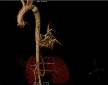
The patient’s left heart dilation was deemed out of proportion to her mitral regurgitation; therefore, chest CT was performed to evaluate for other aetiologies. CT angiography showed extralobar left lower lobe pulmonary sequestration with feeding arteries from the abdominal aorta to the sequestration. There was systemic venous drainage via a left paralumbar venous structure that ultimately drained into the inferior caval vein (Fig 1).
Reconstructed image of CT angiogram showing feeding vessels to the sequestered lung segment off of the thoracic aorta.

Cardiac catheterisation was performed with plans for device occlusion of the feeding vessels. Haemodynamic evaluation showed left ventricle end-diastolic pressure and pulmonary artery wedge pressure of 13 mmHg. Angiography of the aorta showed three feeding vessels arising from the descending aorta at the level of the diaphragm. Selective angiography performed in the left lower pulmonary artery showed paucity of blood supply to the posteromedial bronchopulmonary segment but otherwise normal distal arborisation.
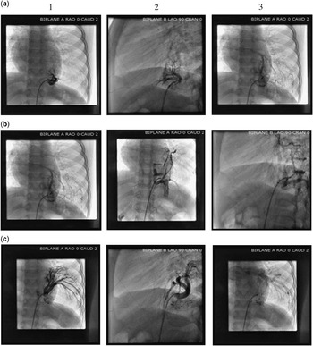
Selective angiograms were performed in the caudal feeding artery (Fig 2a). A 7-mm Amplatzer Vascular Plug 4 (St. Jude Medical, St. Paul, Minnesota, United States of America) was deployed into the superior branch using a 4-Fr angled Glidecath (Terumo Medical Corporation, Somerset, New Jersey, United States of America ), and an 8-mm Amplatzer Vascular Plug 4 was used to occlude the inferior posterolateral branch. The proximal portion of the inferior posterolateral branch was occluded with an 8-mm Amplatzer Vascular Plug 2 (St. Jude Medical). Selective angiograms performed in the middle feeding artery showed venous drainage to both the left atrium via the left lower pulmonary vein and to the inferior caval vein via the paravertebral venous system, consistent with intralobar and extralobar sequestration (Fig 2b). The vessel was completely occluded with an 8-mm Amplatzer Vascular Plug 4. The cranial feeding artery had two prominent branches (Fig 2c). The superoposterior branch was occluded with a 6-mm Amplatzer Vascular Plug 2, and the anterolateral branch was occluded with a 10-mm Amplatzer Vascular Plug 2. Post-occlusion angiograms in the descending aorta showed no significant aortopulmonary flow to the sequestered lung segment. The total procedure time was 3 hours and 28 minutes, and fluoroscopy time was 46.3 minutes with a radiation dose of 171.87 mGy (total dose area product of 1137 mGy cm2).
( a1 ) Anteroposterior and lateral ( a2 ) projection of selective angiogram in the caudal feeding vessel. ( a3 ) Levophase showing pulmonary venous return from the feeding vessel. ( b1 ) Anteroposterior projection of selective angiogram in the middle feeding vessel. ( b2 ) and ( b3 ) Levophase angiogram showing venous drainage to the pulmonary veins as well as to the paravertebral venous plexus that drains to the inferior caval vein. ( c1 ) Anteroposterior and ( c2 ) lateral projection of selective angiogram in the cranial feeding vessel. ( c3 ) Levophase showing pulmonary venous return from the feeding vessel.

At the 2-month follow-up, the patient’s energy level was significantly improved. Repeat echocardiogram showed mild left atrial dilation, mild mitral regurgitation, and normalisation of her left ventricular end-diastolic Z score to +0.77.
Discussion
Alternative sources of pulmonary blood supply should be considered in a patient with a dilated left heart without an alternative aetiology.Reference Hubail, Eapen and Lemler 1
Pulmonary sequestration is a rare congenital lung malformation, with an estimated incidence of 0.15–1.8% and a male to female predominance of 1.58:1.Reference Wei and Li 2 In intralobar sequestration, the arterial supply is derived from the thoracic aorta and venous drainage occurs through the pulmonary veins with resultant left-to-left shunt. Symptomatic patients often present with chronic cough, fever, haemoptysis, chest pain, or shortness of breath. Rarely, they present with evidence of congestive heart failure secondary to excessive blood flow through the sequestered lobe.Reference Levine, Nudel and Gootman 3
Extralobar sequestrations are generally asymptomatic and are often incidentally diagnosed. The venous drainage is usually via the inferior caval vein, portal venous system, or azygous vein, resulting in a left-to-right shunt. Extralobar sequestrations have been associated with CHD including truncus arteriosus, total anomalous pulmonary venous return, pulmonary atresia with ventricular septal defect, and dextrocardia.Reference Wei and Li 2 , Reference Nakata, Ikeda and Doi 4
Sequestration most commonly occurs in the left lower lobe. The majority of sequestered lung segments are supplied by a single artery with greater than two feeding arteries seen <5% of the time.Reference Wei and Li 2 , Reference Nakata, Ikeda and Doi 4 To our knowledge, there is only one other case report of pulmonary sequestration with triple arterial supply and venous drainage via the pulmonary veins and the inferior caval vein.Reference Theodoropoulos and Schwartz 5 Classically, sequestered lung segments have been resected via thoracotomy or with thorascopic surgery. There are multiple reports of use of transcatheter embolisation of systemic arterial supply of sequestered lung segments using embolisation coils and devices, microcoils, particles, absolute alcohol, or histoacryl.Reference Castaldi, Santoro and Gaio 6 – Reference Curros, Chigot and Emond 10 Early complications in a small number of patients from the procedure have included transient ischaemic limb changes, sepsis, and renal abscess in one patient from thrombus.Reference Leoncini, Rossi and Ferro 8 , Reference Curros, Chigot and Emond 10
Most devices used for successful embolisation of pulmonary sequestrations require a long sheath or guide catheter for delivery.Reference Castaldi, Santoro and Gaio 6 – Reference Curros, Chigot and Emond 10 The feasibility of utilising the delivery long sheaths or guide catheters can be limited in tortuous vessels. The technical difficulties can lead to prolonged procedure times, increased radiation dose, incomplete occlusion, or limit the patient population for which interventional approach is suitable. A recent addition to our device armamentarium is the St. Jude Medical Amplatzer Vascular Plug 4 that can be delivered via any 0.038″ guide wire-compatible catheter. This is a major advantage over other devices, as Amplatzer Vascular Plug 4 devices can be delivered through the same flexible catheters used to achieve access into the feeding vessels and perform selective angiography, obviating the need for exchanging to a long sheath or guide catheter. There is only one other case report of utilising an Amplatzer Vascular Plug 4 for embolisation for lung sequestration.Reference Castaldi, Santoro and Gaio 6 We used six devices for closure of multiple feeding branches to achieve complete occlusion of the sequestered lung segments. The Amplatzer Vascular Plug 4 proved useful as this device was easily advanced to distal tortuous sites not amenable to long sheath placement. In addition, this device offered the advantage of a controlled release, unlike standard embolisation coils that go through the same catheters, making embolisation of the device less likely.
Conclusion
Our case demonstrates the importance of evaluating for alternate sources of shunting when the degree of left-sided dilation is out of proportion to what would be expected. This case is unusual in that the sequestered segment has combined intralobar and extralobar components with both pulmonary venous and systemic venous drainage. Pulmonary sequestration, particularly with multiple feeding vessels, is a rare congenital lung anomaly that has been predominantly ameliorated by surgical resection; however, we have demonstrated that Amplatzer Vascular Plugs can be used safely to occlude multiple feeding vessels with systemic and pulmonary venous drainage of a sequestered lung segment, resulting in significant clinical improvement of the patient.
Acknowledgement
None.
Financial Support
This research received no specific grant from any funding agency, commercial, or not-for-profit sectors.
Conflicts of Interest
None.