Introduction
Neuroendocrine head and neck carcinomas are rare tumours occurring predominantly in the larynx and paranasal sinuses. Ultrastructural detection of dense core granules is a hallmark of these lesions. Neuroendocrine carcinomas frequently express chromogranin A, synaptophysin, somatostatin and somatostatin receptors.
Chromogranin A is a glycoprotein consisting of 439 amino acids, which is stored in the secretory granules of neuroendocrine cells. It is the precursor of several functional peptides, including vasostatin. Chromogranin A has been widely used as a general neuroendocrine cell marker in histopathological diagnosis.Reference Modlin, Gustafsson, Moss, Pavel, Tsolakis and Kidd1
Synaptophysin is a 38-kDa, transmembrane glycoprotein and was the first protein to be described in presynaptic vesicles. It has important functions in synaptic vesicle exocytosis. Synaptophysin serves as a molecular marker in the diagnosis of neuroendocrine and neuroectodermal tumours.Reference Erickson and Lloyd2, Reference Valtorta, Pennuto, Bonanomi and Benfenati3
Somatostatin (also known as somatotropin release inhibiting factor) is produced by neuronal, neuroendocrine, inflammatory, immune and tumour cells. It was discovered in 1973 as an inhibitory hypothalamic neuropeptide.Reference Brazeau, Vale, Burgus, Ling, Butcher and Rivier4 Somatotropin release inhibiting factor regulates various physiological functions via specific cell surface receptors, of which five have been cloned (and named somatostatin receptors 1 to 5). These receptors have high affinity for the naturally occurring peptides somatotropin release inhibiting factor 14 and somatotropin release inhibiting factor 28, and the related cortistatin peptides, which are specifically produced in the brain. All five somatostatin receptors deliver an antiproliferative signal, either by inhibition of mitogenesis or by stimulation of apoptosis.Reference Grozinsky-Glasberg, Shimon, Korbonits and Grossman5 Somatostatin receptors have been detected in small cell lung cancer (SCLC), which is considered a neuroendocrine tumour.Reference Rekhtman6 Higher levels of expression of somatostatin receptor 1 messenger RNA (mRNA) have been found in both SCLC and squamous cell carcinoma (SCC), compared with adenocarcinoma cell lines.Reference Fujita, Yamaji, Sato, Murao and Takahara7
Interestingly, neuroendocrine differentiation has also been found in some tumours not considered to be of neuroendocrine origin, including SCC of the lung and of the oesophagus.Reference Nisman, Heching, Biran, Barak and Peretz8, Reference Yuan, Liu, Liu and Cui9 Somatostatin receptors have been detected in laryngeal SCC and epithelial precursor lesions.Reference Stafford, Condon, Rogers, MacDonald and Atkin10, Reference Condon, Stafford, Bedford, MacDonald and Atkin11 However, the biological significance of tumour cells with neuroendocrine differentiation in SCC remains elusive. It has been hypothesised that tumour cells with neuroendocrine characterisation may produce peptides to stimulate tumour growth via autocrine or paracrine mechanisms.Reference Yuan, Liu, Liu and Cui9 In one study, neuroendocrine differentiation in SCC was associated with a poor prognosis.Reference Nisman, Heching, Biran, Barak and Peretz8
The occurrence and possible role of neuroendocrine differentiation in head and neck SCC has not yet been systematically analysed. The immunohistochemical detection of neuroendocrine markers in head and neck SCC tumour cells, and the electron microscope identification of dense core granules, would indicate the possibility of neuroendocrine differentiation within head and neck SCC.
In this study, we analysed the immunohistochemical expression pattern of chromogranin A, synaptophysin and somatostatin receptors in head and neck SCC. Moreover, we questioned whether: (1) chromogranin A, synaptophysin and somatostatin receptor expression differs in head and neck SCC and normal pharyngeal mucosa; (2) chromogranin A, synaptophysin and somatostatin receptor expression levels correlate with each other in head and neck SCC; (3) chromogranin A, synaptophysin or somatostatin receptor expression correlates with tumour stage; (4) chromogranin A, synaptophysin and somatostatin receptor expression is detectable at the RNA level; (5) chromogranin A expression is associated with the ultrastructural detection of secretory granules; and (6) somatostatin is expressed in head and neck SCC.
Materials and methods
Patients
We evaluated tumour specimens from 78 patients with head and neck SCC treated at the departments of otorhinolaryngology and maxillofacial surgery of Medical University Innsbruck.
Of these specimens, 23 were conventionally embedded in paraffin and 55 were prepared as tissue microarrays.Reference Laimer, Blassnig, Spizzo, Kloss, Rasse and Obrist12
One normal human pancreas and two SCLC specimens were used as positive controls, while five normal oropharyngeal mucosa specimens (obtained during uvulopalatopharyngoplasty) were used as negative controls.Reference Fujita, Yamaji, Sato, Murao and Takahara7, Reference Sagman, Mullen, Kovacs, Kerbel, Ginsberg and Reubi13
Clinical data were obtained from the clinical tumour registry.
Permission for the study was obtained from the local ethics committee (permission grant number UN3328-268/4.623).
Immunohistochemical analysis and in situ hybridisation
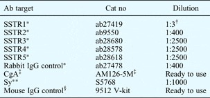
Two-micrometre thick paraffin sections were used for immunohistochemical analysis. Sections were dewaxed and antigen was retrieved using a Discovery automated staining system (Ventana, Tucson, Arizona, USA). Rabbit polyclonal primary antibodies (Abcam, Cambridge, UK) or mouse monoclonal antibodies (Biogenex, San Ramon, California, USA and Sigma, Vienna, Austria) were added to the sections by manual titration (antibody dilutions are given in Appendix 1), and staining was completed by the Discovery automated staining system using universal secondary antibody solution, haematoxylin counterstaining and the diamino-benzamidine Map Kit (all Ventana products). The immunohistochemical staining reaction was confirmed in the pancreas sections and SCLC samples, which served as positive controls.Reference Fujita, Yamaji, Sato, Murao and Takahara7, Reference Sagman, Mullen, Kovacs, Kerbel, Ginsberg and Reubi13 Normal oropharyngeal mucosa and normal tissue from within the same tissue section were used as negative controls. In addition, all sections were stained with control mouse and rabbit immunoglobulins, using the same highest concentration as for the primary antibodies. Only those immunohistochemical reactions which were clearly different from the non-specific reactions were analysed in the study.
In addition, 2-μm thick paraffin sections from five head and neck SCC samples and two pancreas samples were used for in situ hybridisation with somatostatin sense (AGCAGGATGAAATGAGGCTTGAGCTGCAGA, where A = adenine, G = guanine, C = cytosine and T = thymine) and antisense (TCTGCAGCTCAAGCC TCATTTCATCCTGCT) oligonucleotides. One hundred picomoles of oligonucleotide was Digoxigenin-labelled in 20 µl reaction volume using the Dig Oligonucleotide Tailing Kit (Roche, Mannheim, Germany), and 50 ng (1.2 µl) oligonucleotide was used per slide in 100 µl Ribohyb (Ventana). The sections were dewaxed and prepared using a Ventana in situ hybridisation protocol. Oligonucleotide was added to the sections by manual titration; all the rest of the hybridisation procedure was automatic. The hybridisation and wash temperatures were both 58°C. The Dig signal was developed with an alkaline phosphatase labelled anti-Dig FAB-antibody (Roche; 1:2000 dilution) using nitro-blue tetrazolium and bromo-chloro-indoxylphosphate substrates. Pancreas sections were used as positive controls.
Gene expression analysis
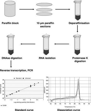
Gene expression of chromogranin A and somatostatin receptors was analysed at mRNA level by reverse transcription and quantitative real-time polymerase chain reaction. For this purpose, 10-µm paraffin sections were collected (5 to 8 pieces depending on the surface area of the sections) in 1.5 ml reaction tubes from 11 head and neck SCC samples, 2 SCLC samples and 1 normal mucosa sample. The PureLink Ffpe RNA Isolation Kit (Invitrogen, Darmstadt, Germany) was used for deparaffinisation, lysis and RNA isolation, following the provider's instructions. Reverse transcription of total RNA was performed using the iScript cDNA Synthesis Kit (Bio-Rad, Munich, Germany), and quantitative real-time polymerase chain reaction was performed using the iQ Sybr Green Supermix system (Bio-Rad) in a MyiQ cycler (Bio-Rad; see Appendix 2). Alternatively, non-real-time polymerase chain reaction was performed under the same conditions, using the Go-Taq Master Mix system (Promega, Madison, Wisconsin, USA). The primers used had all been described, tested and published, except for the self-designed chromogranin A primers (forward, CGAGCTGAAAGGTCGGTCGGA; reverse, TCCTCAGCCCCAGGCTTCCC), which produced a polymerase chain reaction product of 63 base pairs.Reference Li, Li, Kim, Yao, Chen and Fisher14
Semiquantitative evaluation
The immunohistochemical reactions in tumour cell nests were evaluated independently by 2 blinded observers, who collected 10 areas from each specimen. These areas were analysed using an Olympus BX50 light microscope (Tokyo, Japan). Whole numbers of total cells and positive cells were counted in 10 high-power fields. The mean ratio between positive cells and total cells was calculated to give a positive cell percentage, termed the staining index. Staining index values were then converted to labelling scores as follows: 0 = no staining; 1 = 0.1–29 per cent positive cells; 2 = 30–59.9 per cent; and 3 = 60–100 per cent. The score results of the two observers were significantly correlated (Cohen's weighted kappa = 0.83; p < 0.001).
Electron microscopy
Ultrathin sections were prepared for transmission electron microscopy, using a Reichert Microtome (Vienna, Austria). Chromogranin A immunostaining was performed using the same primary antibody (see Appendix 1) together with a biotin-labelled secondary anti-mouse antibody (Vectastain Kit; Vector, Burlingame, California, USA), utilising the substrate diamino-benzamidine (Polysciences, Eppelheim, Germany) and contrasting with cobalt chloride (Sigma, Vienna, Austria). Contrasted sections were visualised using a Zeiss Libra FTM transmission electron microscope (Zeiss, Jena, Germany). Detailed preparation protocols have been described previously.Reference Glueckert, Bitsche, Miller, Zhu, Prieskorn and Altschuler15, Reference Schmutzhard, Glueckert, Bitsche, Abraham, Falkeis and Schwentner16
We used PC-12 rat pheochromocytoma cells as a positive control.
Statistical analysis
Frequency tables of staining scores (0 to 3) were used for analysis of immunohistochemical data. For data analysis, staining indices were generally recorded as either 0 or 1, defined respectively as no reaction products detectable or at least some reaction products detectable. However, the staining index of somatostatin receptor 5 was recorded as either 0 or 1, indicating an original immunohistochemical staining index of either less than 60 per cent, or 60 per cent or more, respectively. Results for normal mucosa and for head and neck SCC were compared using Fisher's exact test. Kendall's tau was used to assess the correlation between protein expression and tumour stage. Survival was analysed using Kaplan–Meier plots and compared using the log-rank test. All calculations were performed using PASW Statistics 18 software (Chicago, Illinois, USA).
Results
Patient, clinical and pathological data
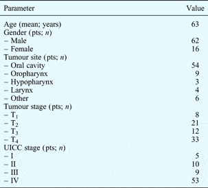
The 78 included patients comprised 62 males and 16 females, with a mean age of 63 years. The oral cavity was the most common tumour site in this patient group (Table I). Local disease was observed in 15 patients and advanced head and neck SCC in 63, but none had distant metastasis.
Patient characteristics

Pts = patients; T = tumour stage; UICC = Union for International Cancer Control
Protein reaction and expression
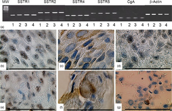
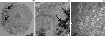
Chromogranin A reaction products were found in 41 per cent of head and neck SCC specimens. In most specimens, chromogranin A reaction products were sparse (Table II). Scattered chromogranin A reaction products were seen in epithelial cells with polymorphic nuclei. Tumour cell nodules were often recognised using this marker. Chromogranin A was typically cytoplasmic, although intracellular granules were also seen (Figure 1f and Figure 2b). Chromogranin A was positive in one of four normal pharyngeal mucosa specimens and in both of the two positive controls. Chromogranin A positivity was not influenced by gender (p = 0.6), tumour site (p = 0.2) or Union for International Cancer Control stage (p = 0.17). Chromogranin A mRNA expression was exemplarily evaluated, and was detectable in five chromogranin A positive head and neck SCC specimens investigated. Moreover, using the chromogranin A peroxidase reaction and transmission electron microscopy, dense core granules were detected in chromogranin A positive head and neck SCC cells (Figure 3b).
(a) Reverse transcription polymerase chain reaction analysis of messenger RNA for somatostatin receptors (SSTRs) 1, 2, 4 and 5 and chromogranin A (CgA) (RNA isolated from paraffin sections of head and neck squamous cell carcinoma (SCC)), showing amplified polymerase chain reaction products for SSTR1 at 184 base pairs, for SSTR2 at 274 base pairs, for SSTR4 at 143 base pairs, and for SSTR5 at 233 base pairs (CgA reaction products were expected at 63 base pairs and β-actin reaction products at 139 base pairs). 1 = oropharynx SSC, 2 = oral cavity SSC, 3 = hypopharyngeal SSC, 4 = small lung cell cancer (a positive control). Figure also shows photomicrographs following immunohistochemical staining for (b) SSTR1, (c) SSTR2, (d) SSTR4, (e) SSTR5 and (f) CgA in oral cavity SCC (all specimens from the same case), and for (g) CgA in small cell lung cancer. (Original magnification ×1000; bar = 100 µm)

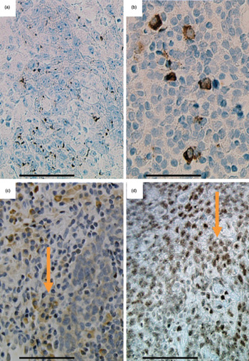
Photomicrographs showing immunohistochemical staining for (a) synaptophysin, (b) chromogranin A, (c) somatostatin receptor 2 and (d) somatostatin receptor 5 in oropharyngeal squamous cell carcinoma. Arrows indicate co-localisation. (Original magnification ×400; bar = 100 µm)

Transmission electron micrographs of (a) PC-12 cells, and of (b) chromogranin A positive and (c) chromogranin A negative head and neck squamous cell carcinoma cells. Chromogranin A was detected using immuno-electron microscopy: arrows indicate secretory granules.

Immunohistochemical staining

Data represent percentage of specimens. Specimen numbers: somatostatin receptor 1 (SSTR1) = 64; SSTR2 = 74; SSTR3 = 64; SSTR4 = 65; SSTR5 = 69; chromogranin A (CgA) = 71; synaptophysin (Sy) = 72.
Synaptophysin was found in 18 per cent of head and neck SCC specimens. Noteworthy levels of synaptophysin expression (i.e. >30 per cent positive cells) were observed in 7 per cent of head and neck SCC specimens (Table II). As with chromogranin A, scattered synaptophysin reactions were seen in epithelial cells with polymorphic nuclei. Synaptophysin was seen both within the cytoplasm and in intracellular granules (Figure 2a). Synaptophysin was negative in five of five normal pharyngeal mucosa specimens and in one of two positive controls. Synaptophysin positivity was not influenced by gender (p = 0.1), tumour site (p = 0.9) or Union for International Cancer Control stage (p = 0.1).
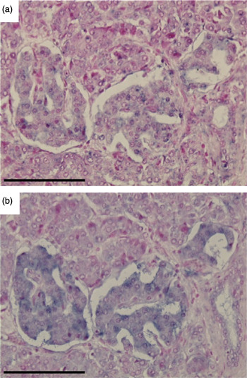
In situ hybridisation revealed no somatostatin expression in five head and neck SCC specimens, but positive expression in pancreatic island cells (Figure 4). In contrast, somatostatin receptors and somatostatin receptor mRNA were frequently observed in head and neck SCC specimens (Table II). Somatostatin receptor 1 was detectable in 69 per cent of the head and neck SCC specimens, somatostatin receptor 2 in 54 per cent, somatostatin receptor 3 in 42 per cent, somatostatin receptor 4 in 53 per cent and somatostatin receptor 5 in 82 per cent. Somatostatin receptor 1 showed diffuse reaction in tumour cells (Figure 1b). Somatostatin receptor 2 showed membranous, cytoplasmic or perinuclear reaction in tumour cells, while in basal and suprabasal cells it showed light reactivity, and in tumour nodules it showed a heterogeneous reaction pattern (Figure 1c, Figure 2c). Somatostatin receptor 3 was recognised only in a few specimens, and its intracellular distribution was similar to that of somatostatin receptor 2. The reaction pattern of somatostatin receptor 4 within tumour cells was scattered and heterogeneous, showing mainly membranous, submembranous and perinuclear configurations (Figure 1d). Somatostatin receptor 5 was mainly recognised in the nuclei of tumour cells, often accompanied by diffuse cellular staining (Figures 1e and 2d).
Photomicrographs showing in situ hybridisation of human somatostatin (a) sense and (b) antisense oligonucleotide probes hybridising to messenger RNA (only the antisense probe) at nucleotide positions 355–384 in the human pancreas. The hybridisation reaction shows as purple. Note that only the antisense probe revealed a selective positive reaction in the islet cells. (Original magnification ×400; bar = 100 µm)

Somatostatin receptors 1 to 5 showed no reaction in any of the negative control specimens. In head and neck SCC, there was significantly different expression of somatostatin receptor 1 (p = 0.05), somatostatin receptor 2 (p = 0.021), somatostatin receptor 4 (p = 0.027) and somatostatin receptor 5 (p < 0.01), compared with normal oropharyngeal mucosa; however, there was no significant difference for somatostatin receptor 3 (p = 0.73). Somatostatin receptors 1 to 5 reaction products were found in all positive control specimens. Somatostatin receptor 1, 2 and 5 expression was not influenced by gender, whereas somatostatin receptors 3 and 4 expression was greater in females (p < 0.05). Somatostatin receptor expression was not influenced by tumour site and Union for International Cancer Control stage. Somatostatin receptor real-time reverse transcription polymerase chain reaction was conducted on available samples, i.e. 13 head and neck SCC samples, 2 SCLC samples and 1 normal mucosa specimen (3 head and neck SCC samples were excluded because of RNA degradation). All tumour specimens expressed mRNA for somatostatin receptors 1, 2, 4 and 5, whereas somatostatin receptor 3 mRNA expression was low. All positive controls had high levels of somatostatin receptor mRNA expression, whereas the negative control had low somatostatin receptor mRNA expression.
Protein coexpression
The staining patterns of somatostatin receptors 1 to 5 were comparable (Figure 2c and 2d). Chromogranin A and synaptophysin staining was more scattered (Figure 2a and 2b) and involved different cells. Moreover, somatostatin receptor 1 to 5 expression levels significantly correlated amongst themselves (Table III), but did not correlate with the expression levels of chromogranin A or synaptophysin.
Correlation between immunohistochemical staining*

* Using Kendall's tau-beta test, with staining graded using indices 0–3.
† p<0.05 (two-sided);
‡ p < 0.01 (two-sided). SSTR = somatostatin receptor; CgA = chromogranin A; Sy = synaptophysin
Survival
The median ± standard error for patient survival was 55 ± 20 months. There was no association between patient survival and any of the immunohistochemical markers investigated (p > 0.2 for all).
Discussion
Neuroendocrine differentiation has recently been reported in SCCs of the lung and oesophagus.Reference Yuan, Liu, Liu and Cui9, Reference Howe, Chapman, Kerr, Dougal, Anderson and Hasleton17 These tumours had previously been thought to lack neuroendocrine differentiation.Reference Rekhtman6 In the current study, we analysed neuroendocrine differentiation markers in human head and neck SCC. We also analysed the expression of somatostatin mRNA and somatostatin receptor protein, as somatostatin receptor expression has previously been found to be associated with neuroendocrine differentiation in several neuroendocrine tumours.Reference Grozinsky-Glasberg, Shimon, Korbonits and Grossman5
We found chromogranin A in 29/71 (41 per cent) head and neck SCC specimens, and synaptophysin in 13/72 (18 per cent) specimens. In head and neck SCC cells expressing chromogranin A, as confirmed by immune electron microscopy, secretory granules were identified, indicating a true neurosecretory character (Figure 3). Moreover, chromogranin A mRNA was detectable in head and neck SCC using real-time polymerase chain reaction. However, the expression of chromogranin A and synaptophysin protein in head and neck SCC was scattered, and mRNA expression levels were low. Chromogranin A and synaptophysin expression in head and neck SCC did not differ significantly from normal pharyngeal mucosa. In normal pharyngeal mucosa and in non-tumour areas of head and neck SCC specimens, chromogranin A and synaptophysin showed comparable dotted cytoplasmic reactions in some suprabasal cells.
Although sparse and scattered, chromogranin A and synaptophysin expression could be clearly identified in the head and neck SCC cells. Banks and coworkers studied 40 cases of basaloid SCC of the head and neck, an aggressive variant of SCC. Chromogranin A and synaptophysin were not detected.Reference Banks, Frierson, Mills, George, Zarbo and Swanson18 Similarly, Wieneke and coworkers were unable to detect chromogranin A and synaptophysin in 14 cases of basaloid SCC involving the nasal cavity and paranasal sinuses.Reference Wieneke, Thompson and Wenig19
It has been hypothesised that tumour cells with neuroendocrine differentiation may produce peptides that stimulate tumour growth via autocrine or paracrine mechanisms.Reference Yuan, Liu, Liu and Cui9 In one study, neuroendocrine differentiation was associated with poor prognosis.Reference Nisman, Heching, Biran, Barak and Peretz8 However, the significance of minor neuroendocrine cell populations scattered in non-neuroendocrine carcinomas is not fully understood. In gastric or colorectal carcinomas, neuroendocrine cells may be the result of transient activation of neuroendocrine genes. This event would lead in turn to the production or accumulation of regulatory peptides, with no impact on tumour growth since at least some of the neuroendocrine-differentiated cells represent a non-proliferating post-mitotic population.Reference Abbona, Papotti, Viberti, Macri, Stella and Bussolati20 In the patients investigated in the current study, chromogranin A and synaptophysin expression was not associated with advanced disease stage and did not affect patient survival.
In the head and neck SCC specimens analysed in the current study, no somatostatin expression was found by in situ hybridisation, but high levels of somatostatin receptor expression were observed. Somatostatin receptor expression did not correlate with chromogranin A or synaptophysin expression, and was observed in different cells within the tumour areas studied. Therefore, we do not consider somatostatin receptors to be markers of neuroendocrine differentiation, in the manner of chromogranin A and synaptophysin, within head and neck SCC. Somatostatin receptors 1, 2 and 5 were particularly highly expressed, at the protein and mRNA levels, within head and neck SCC. Although somatostatin receptor expression was occasionally observed in normal pharyngeal tissue in basal and suprabasal cells, somatostatin receptors were significantly more expressed in tumour cells (p < 0.01). These observations are in line with earlier studies which examined somatostatin receptors 1 to 5 in 12 premalignant and 12 malignant lesions of the larynx, and which found somatostatin receptor 5 expression in more than 80 per cent of both lesion types.Reference Stafford, Condon, Rogers, MacDonald and Atkin10, Reference Condon, Stafford, Bedford, MacDonald and Atkin11 In contrast to our observations, Stafford and coworkers reported somatostatin receptor 2 expression in only 25 per cent of their laryngeal carcinoma specimens.Reference Stafford, Condon, Rogers, MacDonald and Atkin10
The possible pathophysiological role of somatostatin receptor in head and neck SCC remains elusive, and it is unclear whether somatostatin receptors in head and neck SCC are even functionally active. Coexpression with Erb receptors, and somatostatin receptor mediated modification of epidermal growth factor receptor activity, have recently been reported.Reference Kharmate, Rajput, Watt, Somvanshi, Chaudhari and Qiu21 In other tumour types, ligand-activated somatostatin receptors 1 to 5 share common signalling pathways through G-protein-dependent mechanisms. The pathways which finally lead to inhibition of mitogenic signalling and apoptosis are complex and poorly understood.Reference Grozinsky-Glasberg, Shimon, Korbonits and Grossman5
• This study assessed the role of neuroendocrine markers in head and neck squamous cell carcinoma (SCC)
• ChromograninA (CgA), synaptophysin (Sy) and somatostatin receptors (SSTRs) were identified in 78 head and neck SCC specimens
• Sparse, scattered reactions were found for chromogranin A and synaptophysin
• Somatostatin receptors 1, 2 and 5 were highly expressed (as both protein and messenger RNA)
Despite its unclear pathophysiological role, somatostatin receptor expression may be of clinical significance as a target for somatostatin receptor scintigraphy and positron emission tomography (PET). Immunohistochemical staining has been reported to correlate well with standard uptake values in PET scans.Reference Miederer, Seidl, Buck, Scheidhauer, Wester and Schwaiger22 Due to the short half-life of somatostatin, long-acting synthetic somatostatin analogues such as octreotide, lanreotide and vapreotide have been made available. These are known to have direct and indirect antitumour effects when used in the treatment of neuroendocrine tumours, and have a well established safety profile. Somatostatin analogues may be a therapeutic option for somatostatin receptor expressing head and neck SCC.Reference Grozinsky-Glasberg, Shimon, Korbonits and Grossman5, Reference Culler, Oberg, Arnold, Krenning, Sevilla and Diaz23–Reference Kvols and Woltering25
Antibodies and dilutions used

Performance of reverse transcription polymerase chain reaction. For the polymerase chain reaction standard curve, E = −54.5 per cent; R2 = 0.749; slope = 2.927, y intercept = 34. The polymerase chain reaction dissociation curve represents the loss of the Sybr Green DNA stain from the DNA double helix as the DNA is melted; the y axis shows the differential of relative fluorescence units of Sybr Green related to temperature (dRFU/dT), while the x axis shows the temperatures used in the melting process.