Introduction
Thyroid cancer is the most common endocrine malignancy, with greatly increasing incidence. In 2021, approximately 44 280 new cases were reported, making up 2.3 per cent of all new reported cancers in the USA. Lifetime risk of developing thyroid cancer was noted to be 1.2 per cent.1 Thyroid cancers are distinguished histologically as papillary, follicular, medullary or anaplastic. Differentiated thyroid cancers make up both papillary and follicular (including Hurthle cell) types, which account for 90 per cent of all cases of thyroid cancer.1
Although the majority of patients with differentiated thyroid cancers have a good prognosis (96 per cent survival at 35 years),Reference Elisei, Molinaro, Agate, Masserini, Ceccarelli and Lippi3 some unfortunately present with, or develop, locally advanced disease. This can lead to significant morbidity and mortality because of invasion or compression of essential structures, including the airway. Advanced disease, unresectable disease, and radio-iodine refractory disease are all poor prognostic factors.
As radiotherapy and traditional chemotherapy have little impact on thyroid cancer, primary surgical resection has been the mainstay of management. However, completeness of resection and associated potential survival benefit must be balanced against preservation of function.
Tyrosine kinase inhibitors have shown exciting potential in treating advanced forms of both differentiated and anaplastic thyroid cancer, illustrating significant local tumour response and reduction in distant metastases. Kinases are an enzyme class that act in a range of cellular processes via cell signal transduction. The tyrosine kinase subtype works via competitive inhibition at the adenosine triphosphate binding pocket, stopping cell proliferation signalling, and causes varying degrees of inhibition of several growth factor receptor pathways. As a result, there is apoptosis of cancer cells and reduced tumour vascularisation, leading to tumour diminution.Reference Chaar, Kamta and Ait-Oudhia4
The DecisionReference Brose, Nutting, Jarzab, Elisei, Siena and Bastholt5 and SelectReference Shlumberger, Tahara, Wirth, Robinson, Brose and Elisei6 trials promisingly demonstrated sorafenib's and lenvatinib's prolonged progression-free survival advantage in patients with radio-iodine refractory, recurrent, unresectable differentiated thyroid cancers leading to their approval by the US Food and Drug Administration and UK National Institute for Health and Care Excellence.
Little evidence is available regarding the role of kinase inhibitors in the management of locally advanced disease in the pre-surgical setting. In 2016, a patient presented to our institution with locally advanced papillary thyroid cancer and significant tracheal involvement. The extent of disease was deemed unresectable, and the patient was commenced on tyrosine kinase inhibitor therapy for a total of 14 months. Significant disease response was demonstrated, which facilitated surgery, and a similar approach has now been adopted in four other patients.Reference Stewart, Strachan, Srinivasan, MacNeill, Wall and Nixon7
The aim of this study was to describe our experience with this case series to increase clinicians’ understanding of this possible treatment approach for locally advanced differentiated thyroid cancers.
Materials and methods
Prospective data collection on tumour characteristics and treatment details was carried out in a tertiary centre over a six-year period. Four patients with locally advanced differentiated thyroid cancers unsuitable for primary surgical resection and commenced on neoadjuvant tyrosine kinase inhibitors therapy were included.
The study received approval from both the local trust Caldicott guardian and the local research ethics group and was waived from full ethical approval (study number: 16113). Additionally, written consent was obtained from each patient included in the study.
Case 1
The first case was a 73-year-old woman with chronic obstructive pulmonary disease who presented with haemoptysis and shortness of breath. Examination findings included left vocal fold palsy with no cervical lymphadenopathy. Imaging demonstrated a retrosternal thyroid mass and a 23 × 33 mm irregular, poorly defined hypoechoic mass that infiltrated the left lobe of the thyroid, displacing the trachea to the right and extending into the tracheal lumen. There was no evidence of distant metastatic disease, although small volume, indeterminate pulmonary nodules were noted. Upper airway endoscopy found a mass involving the intraluminal tracheal airway approximately 2 cm below the level of the cricoid. This was biopsied and then debulked using a laryngeal Coblation® device. Histology confirmed papillary thyroid carcinoma without evidence of de-differentiation, which was staged under tumour–node–metastasis (TNM) classification as clinically (c)T4a N0 M0.
The disease was considered unresectable based on the combined extent of tracheal invasion and multiple patient co-morbidities. The possibility of tyrosine kinase inhibitor therapy to control local disease was discussed with the patient. Treatment with sorafenib 400 mg twice daily was started but not tolerated with gastrointestinal side effects and significant QT corrected for heart rate (‘QTc’) prolongation. Lenvatinib 24 mg daily was therefore commenced; later on, the dose was reduced to 14 mg daily for side effect control.
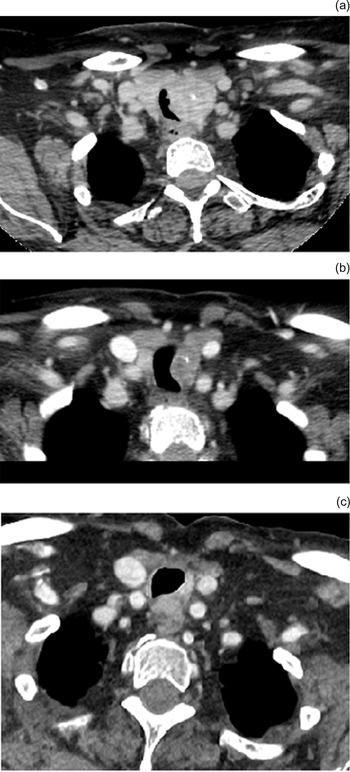
After 14 months of tyrosine kinase inhibitor treatment, computed tomography (CT) scan showed a marked reduction in tumour volume of 82 per cent. In particular, the extent of tracheal invasion was significantly reduced with no evidence of intraluminal disease, which was confirmed on tracheoscopy. Following discussion with the multi-disciplinary team (MDT), total thyroidectomy and left central neck dissection was performed.
Final pathological (p) staging was pT4a N1a M0 R1. The tumour was a papillary thyroid carcinoma measuring 14 mm with extra-thyroidal extension into surrounding fat, vascular invasion and perineural infiltration of large nerves. One lymph node was directly involved by primary tumour. Areas of necrosis, scarring and inflammatory change found on histological examination were thought to represent tumour regression in response to tyrosine kinase inhibitor therapy.
The patient received 3700 MBq of radioactive iodine subsequently, and the post-treatment scan showed uptake in the thyroid bed. Levothyroxine suppression was commenced, aiming for a target thyroid stimulating hormone of less than 0.1 mlU/l. A left vocal fold medialisation procedure was performed 4 months post-resection with no evidence of intraluminal tracheal disease seen.
The patient is currently asymptomatic at 75 months. Post-operative imaging confirmed an asymmetry of the trachea at the site of initial disease (Fig. 1). However, on serial scans this remains unchanged to date. In contrast, the previously observed pulmonary nodules have enlarged, suggesting these could be malignant. In light of this, an additional dose of radioactive iodine (6000 MBq) was delivered, with uptake in the central neck and without uptake in the lung. Her latest thyroglobulin Roche level was 0.08 ug/l.
Case 1. Computed tomography showing: (a) axial plane image of left thyroid mass at presentation, (b) axial plane image showing 82 per cent tumour volume reduction after 14 months of tyrosine kinase inhibitors and (c) axial plane image of stable mass appearance after 3 years.

Case 2
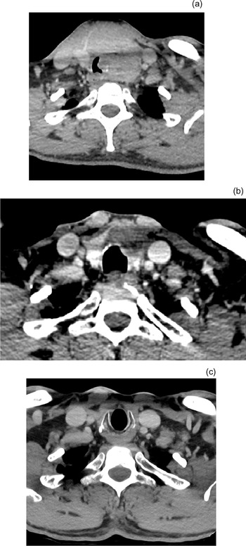
The second case was a 60-year-old male who presented with haemoptysis, increasing shortness of breath on exertion and a palpable central neck mass. Normal vocal fold movements were noted on direct visualisation. A CT scan showed a left lobe of thyroid mass eroding through the trachea at the subglottic level and reducing airway calibre (Fig. 2). On two serial scans six weeks apart, small but size-progressive nodes were reported as suspicious for involvement in the left level II and right level VI cervical lymph node stations. There was no evidence of distant metastases. On direct visualisation, there was an invasive lesion 1 cm below the cricoid, extending 2.5 cm distally. Biopsies were taken and features of follicular cell carcinoma of Hurthle cell type were seen, staged as cT4a N1b M0.
Case 2. Computed tomography showing: (a) axial plane image of left thyroid mass at presentation, (b) axial plane image showing 87 per cent tumour volume reduction after 17 months of tyrosine kinase inhibitors and (c) axial plane image with no evidence of local disease after 3 years.

While awaiting further treatment, the patient developed new biphasic stridor because of tumour enlargement, and this was debulked using a laryngeal Coblation device. Although the central neck disease was deemed operable, total laryngectomy was required for surgical clearance, but the patient refused consent. As an alternative to surgery, tyrosine kinase inhibitor therapy was suggested by the MDT. He was commenced on lenvatinib at 24 mg daily for 12 months. Because of QT corrected for heart rate prolongation, gastrointestinal and cutaneous side effects, the lenvatinib dose was reduced to 20 mg daily. Interval CT imaging at eight months of tyrosine kinase inhibitor therapy showed significant disease regression but also the formation of a small sinus from the trachea into the thyroid (Fig. 3). After 17 months of tyrosine kinase inhibitor therapy, there was a reduction in primary tumour volume of 87 per cent, and all nodes had returned to normal size and morphology.
Case 2. Axial plane computed tomography scan showing sinus developed from trachea into thyroid following eight months of tyrosine kinase inhibitor therapy which self-resolved.

Tracheoscopy was carried out immediately after the end of lenvatinib treatment, which found no frank tumour within the tracheal lumen. Imaging suggested all lateral neck nodal disease had responded. Therefore, a total thyroidectomy and left level 6 neck dissection with tracheal resection and anastomosis was carried out. A well differentiated 30 × 20 × 35 mm Hurthle cell tumour was found on histology. Extensive fibrosis and necrosis seen within the resection specimen was thought to be a result of tyrosine kinase inhibitor therapy. There were three metastatic level 6 lymph nodes found with no extracapsular spread. Final staging was pT4a N1a M0 R1.
Subsequently, the patient received 3700 MBq of radioactive iodine. Post-treatment iodine uptake scan showed one abnormal area of uptake at the site of the tracheal resection. Levothyroxine suppression was commenced. Interval nodal enlargement in the left lateral and right central neck was found on follow-up imaging eight months post-operatively, with further tumour size progression three months later and a rising thyroglobulin. Therefore, tracheoscopy with a left lateral and right central neck dissection was carried out, and there was no evidence of recurrent disease at the tracheal anastomosis. Nodal disease was confirmed in both central and lateral compartments. In view of this, an additional dose of radioactive iodine (6000 MBq) was delivered. His thyroglobulin Roche levels decreased from 5.17 ug/l to 1.96 ug/l. Other than a hoarse voice 4 months post-operatively, the patient remains asymptomatic 44 months from presentation.
Case 3
The third case was a 39-year-old female who presented with haemoptysis and longstanding chronic cough. Examination confirmed a vocal fold palsy. Contrast-enhanced CT showed a poorly defined mass arising from the right thyroid lobule with invasion of trachea, right level IV cervical lymphadenopathy and multiple pulmonary nodules suspicious for metastases. Rigid laryngoscopy confirmed the presence of an invasive tumour 1.5 cm inferior to the cricoid and extending 3–4 cm distally. Endoscopic debulking with a Coblation device was carried out, and initial staging was cT4a N1b M1 papillary thyroid cancer with Braf V600E mutation.
Given the presence of distant metastatic disease and the need for laryngectomy to excise locally advanced disease, the patient did not consent to radical surgery. Following discussion in the MDT, tyrosine kinase inhibitor treatment was suggested with the option to discuss surgical resection later if the disease was responsive to tyrosine kinase inhibitors. She commenced on 24 mg daily of lenvatinib for 4 months before a reduction in dose to 20 mg daily because of gastrointestinal side effects. She tolerated this dose for 3 months before a further reduction to 14 mg daily (because of QT corrected for heart rate prolongation on electrocardiogram). Tyrosine kinase inhibitor therapy was tolerated for a further 7 months (total 14 months).
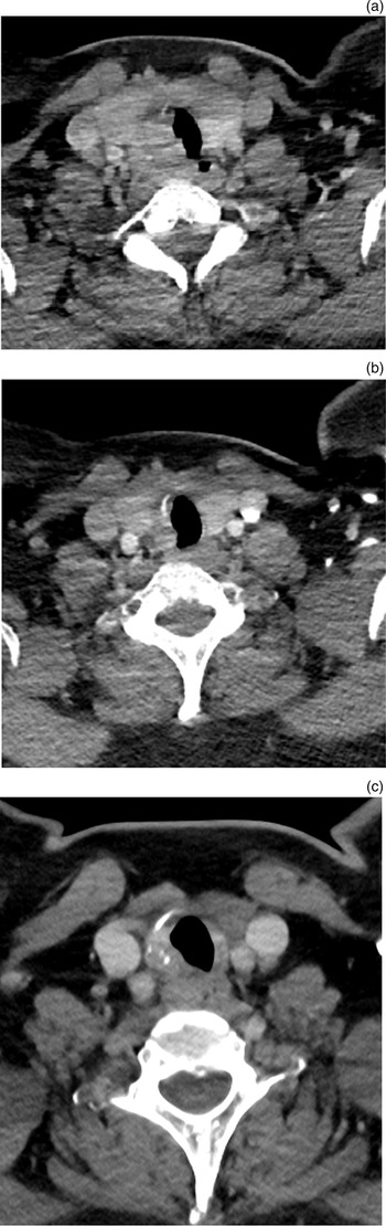
Interval imaging showed size reduction of the primary tumour with nodal disease regression after tyrosine kinase inhibitor treatment (Fig. 4). A total thyroidectomy was carried out following neoadjuvant tyrosine kinase inhibitor treatment to facilitate adjuvant radioactive iodine treatment of 3700 MBq. Fluoro-deoxy-glucose positron emission tomography following radioactive iodine treatment showed avid disease in the neck with multiple metastases suggesting radio-iodine resistant disease. Final staging was pT4a N1b M1 R2. She was recommenced on 14 mg of lenvatinib. Her latest thyroglobulin Roche levels were 4.05 ug/l.
Case 3. Computed tomography showing: (a) axial plane image of right thyroid mass at presentation, (b) axial plane image showing tumour regression after 14 months of tyrosine kinase inhibitors and (c) axial plane image of soft stable tissue mass in the right tracheo-oesophageal groove after 4 years.

She is currently alive at 53 months and has been on palliative lenvatinib for 32 months with stable disease and active monitoring.
Case 4
Our fourth case was a 70-year-old healthy female who presented with haemoptysis. On examination, there was a solid midline neck lump that moved on swallowing, and there was narrowing of the subglottis. Vocal fold function was normal. Contrast-enhanced CT demonstrated a poorly marginated mass arising in the right thyroid lobe, extending into the isthmus and with tracheal invasion. The mass measured 27 × 23 × 26 mm with ipsilateral cervical lymphadenopathy. Fine needle aspiration diagnosed papillary thyroid cancer with Braf V600E mutation, which was staged as cT4a N1a M0.
Because of concerns about open airway surgery at the height of the coronavirus disease 2019 pandemic, the patient was commenced on tyrosine kinase inhibitor therapy as an alternative. Lenvatinib was discontinued within a week because of migraines. Alternative treatment with sorafenib was also discontinued within a week because of myalgia and Steven–Johnson syndrome.
Therefore, the decision was made to proceed to surgery. The patient underwent a total thyroidectomy and central and right neck dissection with a tracheal resection and anastomosis. The tumour was 27 mm in diameter and was a well-differentiated papillary thyroid carcinoma that was partially encapsulated and partially infiltrative. Eight out of 10 lymph nodes recovered were positive; the largest was 7 mm with extra nodal extension. Final staging was pT4a N1a M0 R1. The patient was treated with adjuvant 3700 MBq of radioactive iodine and had thyroid hormone suppressive levothyroxine therapy.
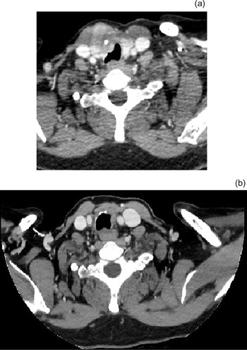
Interval imaging showed irregularity on the inner wall of the trachea with a small right superior mediastinal node that remained static in size and was later thought to be benign (Fig. 5). Mucosal hyperaemia was seen at the level of the anastomosis with no intraluminal disease on tracheoscopy. Her overall survival is 29 months, and she is currently being monitored with yearly follow-up imaging.
Case 4. Computed tomography showing: (a) axial plane right thyroid mass at presentation, and (b) axial plane image two years post-operatively showing minor irregularity along right anterior trachea which is stable.

Table 1 summarises the patient demographic information, disease, treatment and outcome.
Patient demographic data

TNM = tumour–node–metastasis; c = clinically; TKI = tyrosine kinase inhibitor; QTc = QT corrected for heart rate; p = pathologically; RAI = radioactive iodine; PET = positron emission tomography
Discussion
Curative treatment for patients with locally advanced differentiated thyroid cancer usually requires surgical resection of disease with a clear margin to allow adjuvant radioactive iodine treatment and optimise oncological outcome. However, not all cases present with surgically curable disease. Some patients have unresectable cancers, and others cannot accept the significant morbidity of airway and laryngeal resection, particularly in the palliative (M1) setting. In our experience, three quarters (75 per cent) of cases tolerated tyrosine kinase inhibitors with significant primary disease volume reduction that facilitated surgery that was significantly less morbid.
Tyrosine kinase inhibitor therapy has traditionally been reserved for patients with progressive, unresectable recurrent disease. However, in this small series of patients who presented with unresectable primary disease we used tyrosine kinase inhibitors in the ‘neoadjuvant’ setting. In 3 out of 4 cases, the aim was to gain control of local disease in order to allow later surgery without the need for airway resection, therefore avoiding laryngectomy. In the remaining patient with M1 disease at presentation, the aim was to control local symptoms, optimise local control and avoid laryngectomy, accepting that the disease was incurable but with the hope that adjuvant radioactive iodine would also benefit her distant disease.
Most patients tolerated the regimen of tyrosine kinase inhibitor treatment, with appropriate dose adjustment, for a suitable time (median, 15 months) to observe considerable disease regression on interval scanning. These patients went on to have limited surgical resection followed by adjuvant radioactive iodine. To date, despite suspicion of persistent local disease on imaging, all patients remain free of local disease with one patient requiring additional surgery because of interval nodal disease. The one patient who failed to tolerate tyrosine kinase inhibitor therapy underwent an airway resection and has had a good outcome to date. These cases were all deemed to have pathologically differentiated disease, but it is acknowledged that the degree of differentiation is a spectrum.
Fistula occurrence has been reported with tyrosine kinase inhibitors, and this risk should be discussed with the patient, particularly in cases with involvement of the oesophagus and great vessels. This complication has been reported in the recurrent disease setting but may also be pertinent in the ‘neoadjuvant’ setting as a relative contraindication. In our series, we experienced one sinus between the airway and thyroid, which was of no clinical consequence. However, two patients had anatomically anterior disease, and no patients had involvement of the oesophagus or great vessels. Careful assessment with endoscopy and imaging is essential to identify those at greatest risk of this potentially devastating complication.
Our approach in this limited series of patients has been to commence tyrosine kinase inhibitor therapy with three-monthly interval CT imaging. Once disease response has been confirmed, patients are followed for 18 months and then assessed for surgery. Treatment is withdrawn two weeks prior to surgery because of its anti-angiogenic effect. The approach to surgery was not to achieve a wide resection with negative pathological margins but rather to remove central neck disease and prepare the patient for radioactive iodine, aiming for a macroscopic clearance of evident loco-regional disease. Although external beam radiotherapy has been described in the post-operative setting, the outcomes have been poor.Reference Megwalu, Orloff and Ma8 To date, we have favoured adjuvant radioactive iodine with post-treatment monitoring. In one patient, pre-treatment lateral nodal disease responded to tyrosine kinase inhibitor therapy. Therefore, this was not included in the initial surgical procedure; however, later progression required neck dissection. Observation of low volume regional metastatic disease is an approach supported by international guidelines.Reference Tufano, Clayman, Heller, Inabnet, Kebebew and Shaha9
To date, we have not used external beam radiotherapy in this group of patients. There are a number of reasons for this. First, there is the concept that kinase inhibitor treatment may increase radioactive iodine avidity in a select group of patients.Reference Oh, Kalimuthu, Gangadaran, Baek, Zhu and Lee10 Second is the utility of radioactive iodine in determining potential disease trajectory. This is particularly useful for identifying those patients who have occult distant disease or those with radio-iodine refractory distant disease as both situations may impact on the long-term prospects of disease control. This must be balanced against the fact that, in the face of locally progressive recurrence and after less invasive primary surgery, additional surgical options may be available with the option of using external beam radiation at that point. To date, we have not had a patient with uncontrolled central neck recurrence; however, given the locally aggressive nature of the disease, we have treated it as likely that we will encounter this situation during follow up.
• Although the majority of patients with differentiated thyroid cancer have a good prognosis, those with locally advanced disease face significant disease and treatment-related morbidity and mortality
• Advanced disease, unresectable disease and radioactive iodine refractory disease are all poor prognostic factors
• Primary surgical resection, with wide local margins, is the mainstay of management with curative intent
• Tyrosine kinase inhibitors have been approved for use in radioactive iodine refractory, recurrent, unresectable differentiated thyroid cancer in the UK
• Neoadjuvant tyrosine kinase inhibitors can be effective in reducing primary tumour extent to potentially facilitate limited surgical resection for local disease control
• The tyrosine kinase inhibition approach was favoured for patients who have unresectable disease on presentation or patients who were unwilling to undergo a laryngectomy for complete resection of disease
Determination of unresectability in this cohort is complex. Accepting that almost all disease can be resected, a decision upon the suitability of the case for major ablative surgery or tyrosine kinase inhibitor therapy is based on the extent of tracheal and oesophageal involvement, involvement of great vessels (which may increase the potential for catastrophic fistulation), the fitness of the patient for both surgery and systemic therapy, the presence of regional and in particular distant disease (which confirms the palliative nature of any surgical intervention), and the opinion of the patient. By balancing all of these factors, the MDT in partnership with the patient determines the best course of treatment. During the time these 4 patients were treated, a total of 276 patients were managed by the MDT who treat a population of around 1.1 million. Of these, 12 patients were staged as T4. Therefore, this approach is suitable only for a small percentage of the patients presenting to an MDT.
The limitations of this study include that this was a small single-centre series of four cases. All of these cases would have died of airway obstruction had the progressive local disease not been managed. However, there remains uncertainty about the long-term outcome following neoadjuvant tyrosine kinase inhibitor therapy. Whether long-term outcomes will be compromised by this decision remains to be seen. In addition, treatment selection was not based on genetic profiling of the tumour, which may allow more targeted therapies in the future.
These limitations notwithstanding, our small case series suggests that neoadjuvant tyrosine kinase inhibitor therapy should be considered in highly selected patients with locally advanced differentiated thyroid cancer. Careful assessment of pre-operative imaging allows the identification of areas of potential inoperability. In patients who are fit for systemic therapy and in who there is a prediction of an ‘R2 interface’, pre-treatment with kinase inhibition may be considered. In particular, this approach seems favourable in older patients where the negative quality of life impact of major airway surgery can be avoided, potentially without impacting on overall survival. In younger patients, however, where oncological outcomes are likely to be optimised by radical surgery and radioactive iodine, a more cautious approach to the suggestion of tyrosine kinase inhibitors should be adopted.
Conclusion
Neoadjuvant tyrosine kinase inhibitors in locally advanced differentiated thyroid cancer can be effective in reducing primary tumour extent to potentially facilitate limited surgical resection for local disease control. However, this requires a highly selected patient group and requires further assessment with a larger case series.
Competing interests
None declared.