Introduction
The homogeneity of strong genetic risk factors within canine breeds makes them unique models for studying human genetic diseases. Importantly, development of disease model through a transgenesis approach provides entirely new avenues for identifying pathways involved in human complex diseases (Sutter & Ostrander, Reference Sutter and Ostrander2004; Chastant-Maillard et al., Reference Chastant-Maillard, Chebrout, Thoumire, Saint-Dizier, Chodkiewicz and Reynaud2010; Wilbe et al., Reference Wilbe, Jokinen, Truvé, Seppala, Karlsson, Biagi, Hughes, Bannasch, Andersson, Hansson-Hamlin, Lohi and Lindblad-Toh2010; Hasiwa et al., Reference Hasiwa, Bailey, Clausing, Daneshian, Eileraas, Farkas, Gyertyán, Hubrecht, Kobel, Krummenacher, Leist, Lohi, Miklósi, Ohl, Olejniczak, Schmitt, Sinnett-Smith, Smith, Wagner, Yager, Zurlo and Hartung2011). In this sense, the first cloned and then transgenic dogs were produced in 2005 (Lee et al., Reference Lee, Kim, Jang, Oh, Yuda, Kim, Hossein, Kim, Kang, Schatten and Hwang2005), and 2009 (Hong et al., Reference Hong, Kim, Jang, Oh, Park, Kang, Koo, Kim, Kwon, Koo, Ra, Kim, Ko and Lee2009), respectively, both in South Korea. However, the restricted supply of canine oocytes limits the worldwide application of SCNT in this valuable species.
Emerging evidence indicates that the central ooplasmic factors governing cellular reprogramming may be conserved among mammalian species (Dominko et al., Reference Dominko, Mitalipova, Haley, Beyhan, Memili, McKusick and First1999; Tecirlioglu et al., Reference Tecirlioglu, Guo and Trounson2006; Vallée et al., Reference Vallée, Aiba, Piao, Palin, Ko and Sirard2008). This has resulted in a parallel increased interest in the use of iSCNT to address basic questions of developmental biology and to improve the feasibility of cloning in species whose oocytes are not accessible or abundant (Loi et al., Reference Loi, Modlinski and Ptak2011; Hosseini et al., Reference Hosseini, Hajian, Forouzanfar, Moulavi, Abedi, Asgari, Tanhaei, Abbasi, Jafarpour, Ostadhosseini, Karamali, Karbaliaie, Baharvand and Nasr-Esfahani2012). Among mammalian species, bovine oocytes have been used for iSCNT in many species (for review see Tecirlioglu et al., Reference Tecirlioglu, Guo and Trounson2006). Importantly, Murakami et al. (Reference Murakami, Otoi, Wongsrikeao, Agung, Sambuu and Suzuki2005) used bovine oocytes for reprogramming of dog somatic cells, but only one embryo developed to the blastocyst stage from 250 reconstituted embryos and the reconstructed non-transgenic and transgenic dog-bovine iSCNT embryos in the study of Hong et al. (Reference Hong, Oh, Park, Kim, Kim, Koo, Jang and Lee2012) could only develop to the 8–16-cell stage before permanent arrest. This stark difference between cloning efficiency of intra- versus interspecies cloning indicates that the heterogeneity between the donor and recipient may hinder appropriate reprogramming of donor cell nucleus within the exogenous cytoplasm (Tecirlioglu et al., Reference Tecirlioglu, Guo and Trounson2006; Loi et al., Reference Loi, Modlinski and Ptak2011).
Irrespective of the heterogeneity between the donor and the recipient of the nucleus, the specific epigenetic status of differentiated donor chromatin, which is contributed by DNA hypermethylation and histone hypoacetylation/hypermethylation, also makes chromatin structure inaccessible or hard to access by the oocyte factors (Iager et al., Reference Iager, Ragina, Ross, Beyhan, Cunniff, Rodriguez and Cibelli2008; Tian et al., Reference Tian, Park, Bruno, French, Jiang and Prather2009). Recent studies on epigenetic modification of donor cells and cloned embryos showed well that chemically assisted relaxation of donor chromatin structure using the inhibitors of histone-deacetylase (HDAC) and DNA methyl-transferase (DNMT) leads to remarkable changes in the transcription status and developmental competence of the resultant bovine SCNT embryos (Jafarpour et al., Reference Jafarpour, Hosseini, Hajian, Forouzanfar, Ostadhosseini, Abedi, Gholami, Ghaedi, Gourabi, Shahverdi, Vosough and Nasr-Esfahani2011a; Jafari et al., Reference Jafari, Hosseini, Hajian, Forouzanfar, Jafarpour, Abedi, Ostadhosseini, Abbasi, Gourabi, Shahverdi, Dizaj, Anjomshoaa, Haron, Noorshariza, Yakub and Nasr-Esfahani2011a, Reference Jafari, Hosseini, Hajian, Forouzanfar, Jafarpour, Abedi, Ostadhosseini, Abbasi, Gourabi, Shahverdi, Vosough, Anjomshoaa, Haron, Nordin, Yaakub and Nasr-Esfahanib). Therefore, it is interesting to know if treatment of donor chromatin with these epigenetic modifiers can improve the efficiency of iSCNT in canine. More importantly, considering the genome-wide epigenetic effects of HDAC and DNMT inhibitors (Tian et al., Reference Tian, Park, Bruno, French, Jiang and Prather2009), one may argue that the relaxation of chromosome at loci that are linked to pluripotency may provide a chromatin conformation that is acquiescent to transcriptional activation. Therefore, it is important to investigate if HDAC and DNMT inhibitors have any effect on the capacity and time-window of pluripotency gene expression in iSCNT embryos.
POU5F1 is a well known key regulator of pluripotency and cell differentiation and differentiated cells transfected with POU5F1 have provided a good model for such important investigations (Sylvestre et al., Reference Sylvestre, Pennetier, Bureau, Robert and Sirard2010; Jafari et al., Reference Jafari, Hosseini, Hajian, Forouzanfar, Jafarpour, Abedi, Ostadhosseini, Abbasi, Gourabi, Shahverdi, Dizaj, Anjomshoaa, Haron, Noorshariza, Yakub and Nasr-Esfahani2011a). In this study, canine fibroblasts were first stably transfected with two reporter genes: (1) CMV promoter-driven enhanced green fluorescent protein (EGFP) as a simple reporter of presence of canine genome in the iSCNT embryos; and (2) POU5F1 promoter-driven EGFP to provide a visible marker of time and capacity of pluripotency gene expression in iSCNT embryos. The POU5F1–EGFP positive fibroblasts were used to investigate the effect of pre-treatment of donor cells with trichostatin A (TSA), a synthetic anti-neoplastic drug which inhibits HDAC, and S-adenosylhomocysteine (SAH), a natural metabolite of DNMT pathway that reversibly inhibits DNMTs, on epigenetic status and gene expression of donor cells and on remodeling, reprogramming, and embryonic development of the reconstituted embryos.
Materials and methods
All chemicals were from Sigma Aldrich Chemicals (St. Louis, MO, USA) and all media were purchased from Gibco, Invitrogen Corporation (Grand Island, NY, USA) unless otherwise indicated.
Establishment and transfection of fibroblasts
Adult fibroblasts were obtained from an ear skin biopsy taken from a 2-year-old male dog (Labrador breed) according to Jafari et al. (Reference Jafari, Hosseini, Hajian, Forouzanfar, Jafarpour, Abedi, Ostadhosseini, Abbasi, Gourabi, Shahverdi, Dizaj, Anjomshoaa, Haron, Noorshariza, Yakub and Nasr-Esfahani2011a). After through washing with Ca2+- and Mg2+-free phosphate-buffered saline (PBS−) containing antibiotic (penicillin 100 IU/ml, streptomycin 100 mg/ml, and amphotericin-B 0.025 μg/ml), the tissue was cut into small pieces (2–3 mm2), and the explants were cultured in 5 ml Dulbecco's modified Eagle's medium F12 (DMEM/F-12) supplemented with 10% (v/v) fetal calf serum (FCS) and 10 μg/ml penicillin/streptomycin solution at 37°C in a humidified atmosphere of 5% CO2 in air. After 6–7 days, when monolayer cells were observed around the explants’ edges, the explants were removed and attached cells were cultured to reach 80% confluence. The cells were then trypsinized, and sub-cultured to prepare adequate numbers of cells for subsequent experiments. To confirm fibroblastic phenotype, cells at passage 2 were used for immunostaining against anti-vimentin (for fibroblasts) and anti-pancytokeratin (for epithelial cells), as described previously (Jafarpour et al., Reference Jafarpour, Hosseini, Hajian, Forouzanfar, Abedi, Hosseini, Ostadhosseini, Gholami and Nasr-Esfhani2011b).
For transfection, established canine ear fibroblast cells (CEF) in density of 2 × 10⁵ were cultured in wells of 6-well plates (BD Falcon™) for 4–5 days until 80% confluent. The transfection reagent was comprised of solution: (1) 4 μg plasmid (either POU5F1–EGFP—neomycin cDNA or CMV–EGFP–neomycin cDNA) (Ghorbani et al., Reference Ghorbani, Emamzadeh, Khazaie, Dormiani, Ghaedi, Rabbani, Foruzanfar, Karbalaie, Karamali, Lachinani, Kiani-Esfahani, Nematollahi and Nasr-Esfahani2012) (Fig. 1A, B) in 250 μl of Opti-MEM®; and solution (2) 10 μl Lipofectamine 2000™ in 250 μl Opti-MEM®. After incubation for 5 min at room temperature (RT), solutions 1 and 2 were mixed gently and incubated for 20 min at RT. Then, 500 μl Opti-MEM® was added to the transfection mixture and the resultant solution was added to the wells containing 1000 μl Opti-MEM® without FCS. Four hours after incubation at 37°C, and 5% CO2, the transfection medium was removed and cells were seeded in DMEM/F-12 supplemented with 10% (v/v) FCS for 3 days. Next, cells were cultured in dishes containing selection medium (DMEM/F-12 plus 400 μg/ml neomycin). Selection medium was refreshed at 2-day intervals for 10–12 days, and G418-resistant colonies isolated, passaged and cryopreserved by standard procedure. A portion of each expanded colony was analyzed for transgenes using polymerase chain reaction (PCR) (Fig. 1C) and among confirmed transgenic colonies of each transgene, one colony with reasonable growth rate and cell morphology was selected for subsequent studies (Fig. 2A).
(A) Schematic map of the POU5F1 promoter/EGFP cassette in the pDB2 vector (kindly gifted by Dr K. Ghaedi). (B) Schematic map of the CMV promoter/EGFP (obtained from BD Bioscience Clontech: www.clontech.com). (C) PCR amplification of EGFP indicating the presence of the EGFP gene in EGFP–CMV and EGFP–POU5F1 transfected cell lines. From left to the right: positive control (pEGFP-C1 vector, canine ear fibroblasts (CEF) transfected with EGFP–CMV, positive control (pDB2–POU5F1 vector), CEF transfected with EGFP–POU5F1, negative control, ladder M100.

(A) Bright-field and fluorescent images of CMV–EGFP transfected canine ear fibroblasts along with canine–bovine iSCNT embryos in different developmental stages derived from these cells. (B) EGFP fluorescence was not detected in any stages, even in the morula stage, of iSCNT embryos derive from POU5F1–EGFP transfected canine fibroblast. (C) Well developed bovine cloned blastocyst derived from the POU5F1–EGFP fibroblast. As seen, EGFP signal was not restricted to the inner cell mass (ICM) or to the trophectoderm (TE), although the signal intensity in the ICM was higher than in the TE. Bars represent 50 μm.
In this study, bovine fibroblasts stably transfected with POU5F1–EGFP in a recent study (Jafari et al., Reference Jafari, Hosseini, Hajian, Forouzanfar, Jafarpour, Abedi, Ostadhosseini, Abbasi, Gourabi, Shahverdi, Dizaj, Anjomshoaa, Haron, Noorshariza, Yakub and Nasr-Esfahani2011a) were used for control bovine SCNT experiments.
Assisted epigenetic modification and immunostaining of donor cells
To our knowledge, there was no report on optimum TSA and SAH concentrations/durations for SCNT in dog. Therefore, during a pilot study we tested different doses/concentrations of TSA and SAH (TSA: 0.1, 0.5, 1.5, 2, or 3 μM for 1 or 2 days; SAH: 0.1, 0.5, 1.5, 2, or 3 mM for 2, 4, 6, 8 or 10 days) based on available infield studies (Iager et al., Reference Iager, Ragina, Ross, Beyhan, Cunniff, Rodriguez and Cibelli2008; Jafarpour et al., Reference Jafarpour, Hosseini, Hajian, Forouzanfar, Ostadhosseini, Abedi, Gholami, Ghaedi, Gourabi, Shahverdi, Vosough and Nasr-Esfahani2011a; Jafari et al., Reference Jafari, Hosseini, Hajian, Forouzanfar, Jafarpour, Abedi, Ostadhosseini, Abbasi, Gourabi, Shahverdi, Vosough, Anjomshoaa, Haron, Nordin, Yaakub and Nasr-Esfahani2011b). Treated cells along with their corresponding controls were used for the morphology assay, TUNEL assessment of DNA fragmentation, viable staining using propidium iodide, and SCNT as described elsewhere (Jafarpour et al., Reference Jafarpour, Hosseini, Hajian, Forouzanfar, Ostadhosseini, Abedi, Gholami, Ghaedi, Gourabi, Shahverdi, Vosough and Nasr-Esfahani2011a). We observed that at higher concentrations and durations of both drugs, cells were totally arrested with abnormal morphologies (extremely pyknotic nuclei, enlarged in size) and were floated in the culture medium. SCNT using these cells was not possible because they ruptured upon electrofusion. Conversely, although concentrations/durations lower than 1 μM TSA were not toxic and had the same viability parameters compared with 1 μM TSA and 1 mM SAH, their DNA methylation statuses were almost unchanged compared with the control. Therefore, we were convinced to use 1 μM TSA for 24 h and 1 mM SAH for 144 h as the most safe/effective treatment protocols for SCNT experiments (details not shown).
Transfected and non-transfected CEF cells were added in equal densities to 6 mm culture dishes containing DMEM/F-12, 10% FCS. The cells were further supplemented with either 1 μM TSA or 1 mM SAH and cultured for 24 h or 144 h, respectively. Concentrations and durations of TSA and SAH were selected based on the available reports (Shi et al., Reference Shi, Hoeflich, Flaswinkel, Stojkovic, Wolf and Zakhartchenko2003; Enright et al., Reference Enright, Kubota, Yang and Tian2003), our recent study (Jafarpour et al., Reference Jafarpour, Hosseini, Hajian, Forouzanfar, Ostadhosseini, Abedi, Gholami, Ghaedi, Gourabi, Shahverdi, Vosough and Nasr-Esfahani2011a) and preliminary studies on dog somatic cells (data not shown). With regards to the stability and half-life of SAH, the culture medium was refreshed every 24 h (Jafari et al., Reference Jafari, Hosseini, Hajian, Forouzanfar, Jafarpour, Abedi, Ostadhosseini, Abbasi, Gourabi, Shahverdi, Dizaj, Anjomshoaa, Haron, Noorshariza, Yakub and Nasr-Esfahani2011a). Treated and non-treated cells were then used for the assessment of histone H3K9-acetylation and DNA methylation using FACS as described previously (Jafarpour et al., Reference Jafarpour, Hosseini, Hajian, Forouzanfar, Ostadhosseini, Abedi, Gholami, Ghaedi, Gourabi, Shahverdi, Vosough and Nasr-Esfahani2011a). In brief, cells were first trypsinized, washed with PBS, and the pellet cells were resuspended and fixed with cooled (4°C) ethanol 75% for 1 h. Fixed cells were then incubated in PBS containing 0.5% Triton X-100 for 20 min at RT for cell permeabilization. Quantitative assessment of DNA methylation and histone H3K9 acetylation was carried out by incubating the treated cells with 1:400 and 1:200 dilutions of mouse anti-5-methyl cytosine and anti-H3K9 monoclonal antibodies, respectively. Fluorescein isothiocyanate-conjugated goat anti-mouse immunoglobin was used as the secondary antibody at 1:50 dilution. Corresponding controls for each experiment were conducted. Cells were filtered through a 40-μm nylon mesh, in order to exclude aggregation. Ten thousand cells were collected with the FACS-Caliber and analyzed using CELL QUEST® 3.1 software (Becton Dickinson, USA). Each treatment was repeated three times and, in each replicate, appropriate controls for autofluorescence and non-specific binding by the secondary antibody were included. Moreover, to assess the quality of immunostaining, samples of treated/non-treated CEF cells were cultured on 22-mm2 glass coverslips in 12-well plates, immunostained as described above, and were used for observation under an epifluorescence microscope (excitation: 557 nm and emission: 576 nm).
Recipient cytoplast preparation
The process of oocyte in vitro maturation (IVM) was as described previously (Moulavi et al., Reference Moulavi, Hosseini, Ashtiani, Shahverdi and Nasr-Esfahani2006). Briefly, bovine ovaries were obtained from a local slaughterhouse and transported at 25–30°C in saline solution to the laboratory within 2–3 h. Immature cumulus–oocyte complexes (COCs) were aspirated from antral follicles (2–8 mm diameter) using a disposable 18-gauge needle and COCs having compact multilayer cumulus cells and homogeneous cytoplasm were collected in HEPES-buffered tissue culture medium-199 (HTCM199). COCs were cultured in maturation medium (TCM199) containing 10% (v/v) FCS, follicle-stimulating hormone (FSH) (10 μg/ml), luteinising hormone (LH) (10 μg/ml) and 17-β-estradiol (1 μg/ml) at 38.5°C, and a humidified atmosphere of 5% CO2 in air. At 18–20 h post IVM, COCs were mechanically denuded from cumulus cells by vortexing in HTCM199 containing 10% FCS and 300 IU/ml hyaluronidase for 4 min. Denuded oocytes with normal cytoplasm and first polar extruded were selected for the experiments.
SCNT procedure
The zona-free method for SCNT described by Oback et al. (Reference Oback, Wiersema, Gaynor, Laible, Tucker, Oliver, Miller, Troskie, Wilson, Forsyth, Berg, Cockrem, McMillan, Tervit and Wells2003) was used in this study with minor modifications (Nasr-Esfahani et al., Reference Nasr-Esfahani, Hosseini, Hajian, Forouzanfar, Ostadhosseini, Abedi, Khazaie, Dormiani, Ghaedi, Forozanfar, Gourabi, Shahverdi, Vosough and Vojgani2011). In brief, the zona pellucida was removed by incubation in 5% proteinase dissolved in HTCM199 plus 10% FCS for up to 3 min before being incubated in TCM199 + 20% FCS to neutralize the enzyme. Oocytes were then incubated with H33342 (5 μg/ml for 5 min), transferred on the microscope stage (Olympus; IX71) equipped with Narishige micromanipulators (Olympus). Zona-free oocytes were enucleated at ×100 magnification under UV exposure, using a blunt (10–15 μm inner diameter) micropipette. For nuclear transfer, oocytes were adhered to individual CEF cells in medium containing 10 mg/ml phytohemagglutinin. The couplets were then electrofused [a sinusoidal electric current (7 V/cm) for 10 s followed by two direct currents (1.75 kV/cm for 30 μs and a 1 s delay)] in 290 mOsm fusion buffer free of Ca2+ and Mg2+ and the reconstructed oocytes were activated using ionomycin (5 μM, 5 min) followed by incubation with 2 mM 6-DMAP for 4 h (Hosseini et al., Reference Hosseini, Hajian, Moulavi, Shahverdi and Nasr-Esfahani2006). Five to seven activated reconstructed iSCNT embryos were cultured for up to 8 days in the well-of-the-well system (Vajta et al., Reference Vajta, Peura, Holm, Paldi, Greve, Trounson and Callesen2000) in 20 μl of a modified synthetic oviductal fluid (mSOF) (Hosseini et al., Reference Hosseini, Moulavi, Hajian and Abedi2008), under mineral oil at 38.5°C, in 5% CO2, 5% O2 and humidified air.
Nuclear remodeling
Stepwise assessment of nuclear remodeling, pronuclear formation and mitotic division (of the early embryo) were evaluated at 0.5–6 h post oocyte reconstitution (hpr), and 5–8 and 12–16 h post activation (hpa), respectively, as described previously (Asgari et al., Reference Asgari, Hosseini, Ostadhosseini, Hajian, Azhdari, Mosaie and Nasr-Esfahani2012). In brief, reconstituted oocytes were fixed in 4% paraformaldehyde for 15 min. Spindle microtubules of samples were then immunostained with anti-β-tubulin monoclonal primary antibody (1:100), and FITC-labeled anti-mouse-IgG secondary antibody (1:100). The chromosomes were counterstained with 2 μg/ml H33342, and then, samples were washed and mounted on glass slides in glycerol droplets to be observed using epifluorescence microscope (Olympus, BX51), at ×400 magnification. Upon exposure, a digital image of each sample was taken with a high sensitive camera (DP-72, Olympus, Japan), operated on DP2-BSW software.
Fluorescence imaging
Interspecies SCNT embryos retrieved from either POU5F1–EGFP or CMV–EGFP cell lines were checked for EGFP fluorescence as described previously (Jafari et al., Reference Jafari, Hosseini, Hajian, Forouzanfar, Jafarpour, Abedi, Ostadhosseini, Abbasi, Gourabi, Shahverdi, Dizaj, Anjomshoaa, Haron, Noorshariza, Yakub and Nasr-Esfahani2011a). In brief, blastocysts were first checked for their EGFP fluorescence using an inverted microscope (Olympus, IX71) equipped with fluorescence system (excitation: 450–490 nm; emission: 515–569 nm) for any sign of green light emission. Immediately after exposure, a digital image of each iSCNT embryo was taken with a high sensitivity camera (DP-72, Olympus, Japan) operated on DP2-BSW software. As a control to verify the activity of POU5F1–EGFP vector, bovine cloned embryos derived from POU5F1–EGFP bovine fibroblasts were used in parallel to iSCNT embryos.
Quantitative RT-PCR analysis of apoptosis
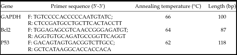
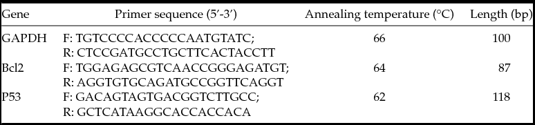
In order to assess if cell transfection and epigenetic modification have affected cell survival, comparative quantitative RT-PCR (qRT-PCR) analysis of two apoptotic genes (P53 and BCL2) (Table 1) was carried out for treated and non-treated cells. In brief, total RNA was extracted from CEF cells using RNeasy mini kit (Qiagen®, Germany) according to the manufacturer's instructions. Samples were treated with DNase I (Fermenas; EN0521) and cDNA was reverse transcribed from 1 μg total RNA, using RevertAid™ First Strand cDNA Synthesis kit (Fermentas; K1622, Germany) in total volume of 20 μl containing 1 μl random hexamers, 4 μl 5× reaction buffer, 2 μl dNTP Mix (10 mM), 1 μl Ribolock™ RNase inhibitor (20 U/μl) and 1 μl RevertAid M-MuLV reverse transcriptase (200 U/μl) and adjusted to a volume of 20 μl using DEPC-treated water. qPCR was performed with the Quantitect SYBR Green PCR kit (Takara, Japan) using 20 μl of reaction solution containing 10 μl of 2× Quantitect SYBR Green PCR master mix, 25 ng of cDNA, 10 pmol primers and reached to a final volume of 20 μl by addition of sterile distilled water. Gene expression levels were calculated using determined of cycle threshold (CT) value, then expression of either gene was calculated comparing the Ct of genes with the house-keeping gene GAPDH (Table 1). Relative gene expression for each gene was calculated as a ratio of target gene expression to that of the reference by ΔΔCT analysis. For each cDNA sample, three replicates of PCR were carried out.
Primers used for real-time PCR experiment
Statistical analysis
All experiments were replicated at least three times in this study. Percentage data of iSCNT embryo development were modeled to the binomial model of parameters by ArcSin transformation. The transformed data, along with crude data of the cellular characteristics, real-time PCR expression of BCL2, and P53 were analyzed by the SPSS-17 one-way analysis of variance (ANOVA) model. Differences were compared by Tukey multiple comparison post-hoc-test. All data were presented as means ± standard error of the mean (SEM). Differences between experimental groups were considered significant at P < 0.05.
Results
Stable transfection of CEFs with the transgenes
Figure 1C shows the presence of both transgenes in CEFs that were confirmed by PCR. Figure 2A shows the presence of green fluorescent protein in CEF cells transfected with CMV–EGFP that were selected after treatment with G418 neomycin for about 12–14 days. Importantly, iSCNT embryos derived from fibroblasts transfected with CMV–EGFP expressed EGFP at all developmental preimplantation stages (2-cell, 4-cell, 8–16-cell and morula) without mosaicism (Fig. 2A).
Epigenetic states of donor cells
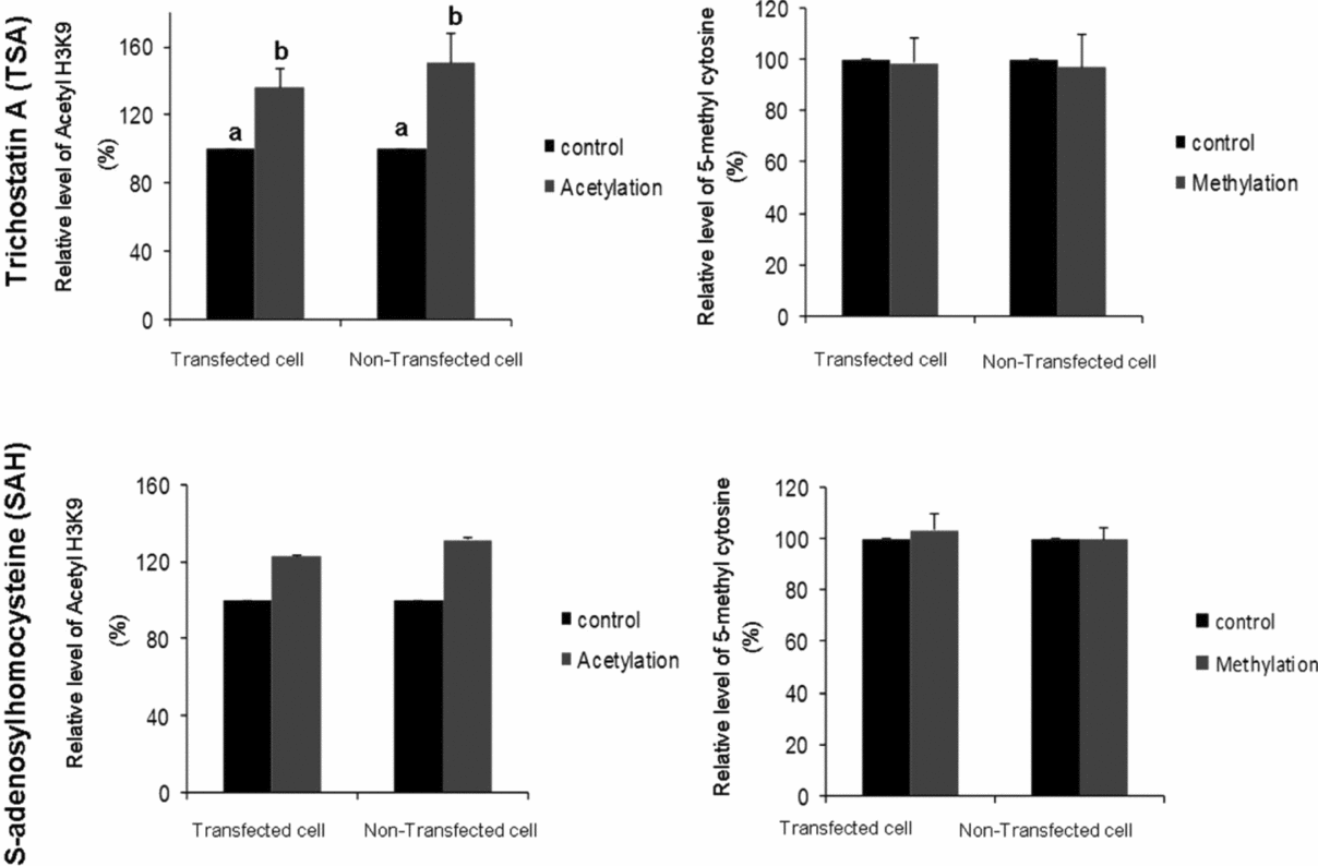
Figure 3 shows the effect of epigenetic treatment on epigenetic statuses of transgenic and non-transgenic CEF cells. As depicted, FACS results indicated that irrespective of transgenesis, TSA treatment of CEF cells significantly increased acetylation levels of the cells compared with untreated cells. DNA methylation of treated cells, however, was not significantly different between the groups. This effect was irrespective of donor cell transgenesis. As shown Fig. 3, SAH treatment could not change the levels of H3K9-acetylation and DNA methylation in CEF cells compared with untreated CEF cells. These facts were again irrespective of cell transgenesis.
Relative levels of histone H3K9 acetylation and DNA methylation in transfected and non-transfected canine fibroblasts treated with trichostatin A (TSA) and S-adenosylhomocysteine (SAH). a,bDifferent labels indicate significant difference between the groups (P < 0.05).
Apoptotic genes expression
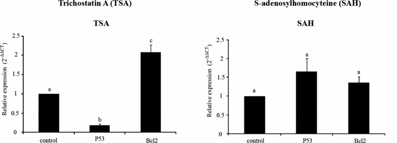
As shown in Fig. 4, the expression level of Bcl2 was significantly increased in CEF cells treated with TSA compared with non-treated cells (~2.1-fold). Further, the expression level of P53 significantly decreased in these cells when compared with non-TSA-treated cells. These facts were irrespective of cell transgenesis (data not shown). In contrast, SAH treatment of CEF cells had no significant effect on the expression levels of Bcl2 and P53 compared with control cells. These effects were also irrespective of cell transgenesis (data not shown).
Relative gene expression of Bcl2 and P53 in canine fibroblasts treated with trichostatin A (TSA) and S-adenosylhomocysteine (SAH). a,b,cDifferent labels indicate significant difference between the groups (P < 0.05).
Nuclear remodeling
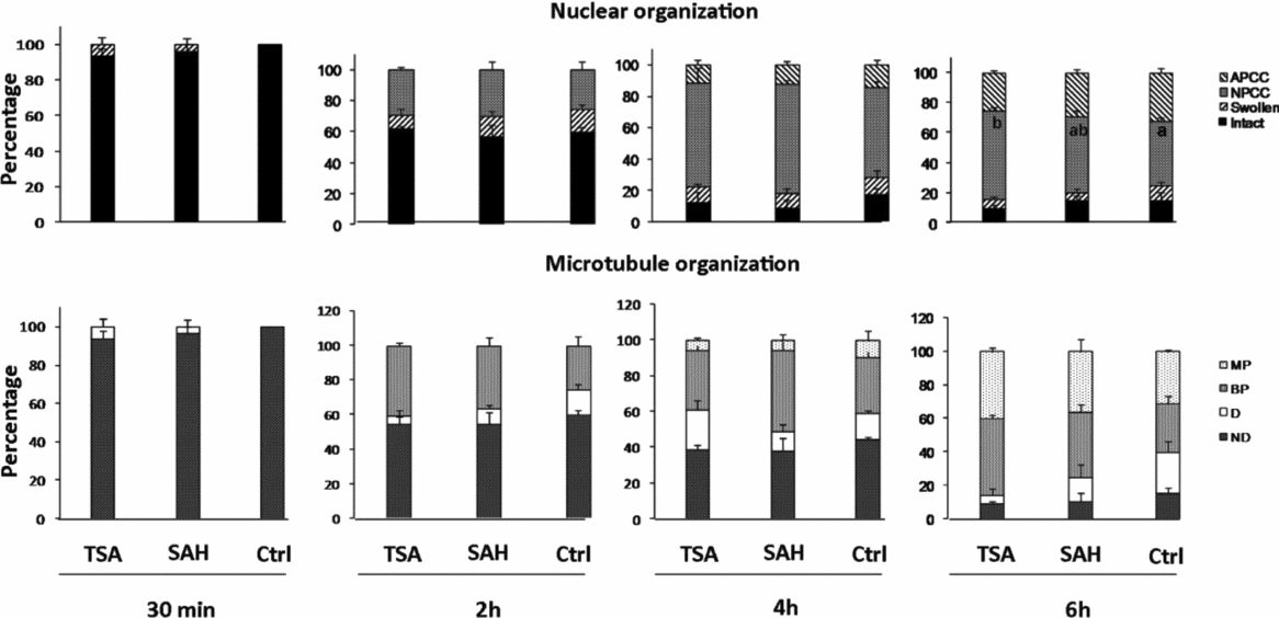
Figure 5 shows a stepwise pattern of nuclear remodeling and microtubule organization following dog-bovine iSCNT using non-transgenic CEF cells. Figure 6 compares the effect of donor cells treatment using TSA or SAH on the pattern of chromosome and spindle organization of iSCNT embryos. As shown, there was a time-dependent effect of post-fusion interval on nuclear remodeling. Accordingly, at 0.5 h post reconstitution (hpr), most nuclei were intact with no sign of spindle formation. At 2 hpr, less than half of the reconstituted embryos had partially remodeled nuclei with either swollen or prematurely condensed nuclei and with developed bipolar spindles. Donor cell pre-treatment had no effect on the pattern of nuclear remodeling in iSCNT embryos. At 4 hpr, the majority of reconstituted embryos had premature chromosome condensation (PCC). Developed bipolar spindle was also observed in half of the reconstituted embryos. At this stage also there was no effect of pre-iSCNT epigenetic modification of donor cells. At 6 hpr, while the overall percentages of remodeled nuclei were not different with those observed 4 hpr, the frequencies of abnormal PCC and abnormal multipolar spindles were increased. Importantly, donor cell pre-treatment with TSA could reverse this adverse effect as the percentage of normal PCC in this group was significantly higher than in the control group. Single pronuclear formation was observed from 6 h post activation (hpa) and approached maximum at 12 hpa. Moreover, mitotic nuclei were observed from 15 hpa and reached maximum at 21 hpa (data not shown).
Representative images describe different organizations of microtubules (A–F), chromosomes (A′–F′) and the merged images showing the organizations of both microtubules and chromosomes (A″–F″). (A, A′) No sign of nuclear swelling or spindle formation. (B, B′) Early PCC with developing spindle. (C, C′) Mid-stage PCC and spindle arrangement. (D, D′) Complete chromosome and bipolar spindle organization. (E, E′, F, F′) Two different patterns of pulverized spindle and chromosome organization. Bar represents 100 μm.
Time-dependent effect of SCNT technique on remodeling events of the reconstituted oocytes. APCC: abnormal prematurely condensed chromosomes; BP: bipolar; D: developing/developed; MP: multipolar; ND: non-developed; NPCC: normal prematurely condensed chromosomes; PCC: prematurely condensed chromosomes. a,bBars with different letters are significantly different at P < 0.05.
Preimplantation development of iSCNT and SCNT embryos
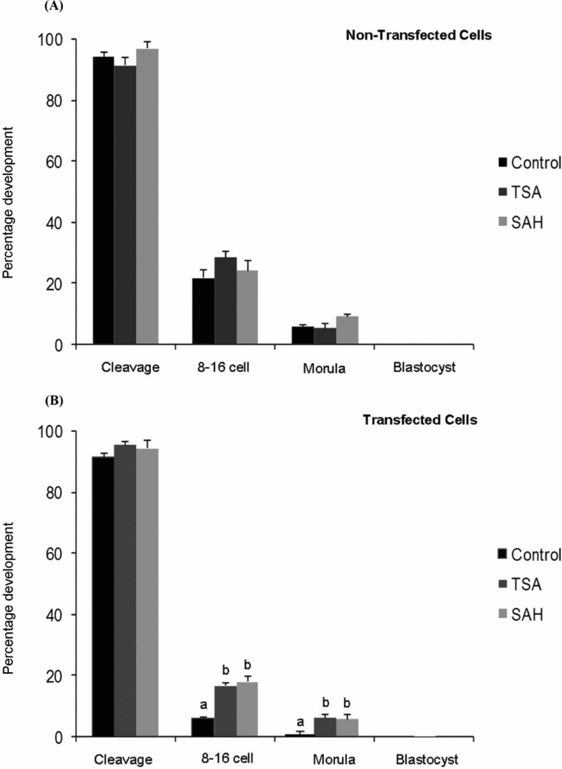
As shown in Fig. 7, non-transgenic iSCNT reconstructed embryos had overall similar rates of cleavage [control: 94.2% (599/636); TSA: 97.2% (106/109); and SAH: 91.6% (174/190)], development to the 8–16-cell stage [control: 21.7% (130/599); TSA: 28.3% (30/106); and SAH: 24.14% (42/174)] and morula [control: 5.51% (33/599); TSA: 4.72% (5/106); and SAH: 9.19% (16/174)] stages in control, TSA and SAH groups, and there was no significant difference between the groups. Donor cell pre-treatment had no significant effect on the efficiency of iSCNT embryo development. For transgenic iSCNT embryos, TSA and SAH treatment could significantly improve the development to the 8–16-cell stage [16.2% (23/142), 17.82% (18/101) respectively] and morula stages [6.33% (9/142), 5.94% (6/101) respectively)] compared with control [8–16-cell stage: 6.54% (7/107); morula: 0% (0/107)]. Despite these improvements, all the embryos arrested at morula stage and did not progress to the blastocyst stage in any group. Noteworthy, using bovine POU5F1–EGFP fibroblasts, the efficiency of cloned embryo development was quite comparable with the previous study by the same group (Jafari et al., Reference Jafari, Hosseini, Hajian, Forouzanfar, Jafarpour, Abedi, Ostadhosseini, Abbasi, Gourabi, Shahverdi, Dizaj, Anjomshoaa, Haron, Noorshariza, Yakub and Nasr-Esfahani2011a). Of 128 oocytes reconstituted by bovine transgenic fibroblasts, 118 (92.2%) cleaved and of these cleaved embryos, 37 (31.35%) developed to the blastocyst stage.
Effect of epigenetic modification of canine transfected (A) and non-transfected (B) somatic cells with trichostatin A (TSA) and S-adenosylhomocysteine (SAH) on in vitro embryo development of interspecies canine–bovine reconstructs. a,bWithin each stage of embryo development, values with different superscripts are significantly different (P < 0.05).
EGFP fluorescence
Interspecies SCNT embryos derived from fibroblasts transfected with CMV–EGFP expressed green fluorescent protein (GFP) at all preimplantation developmental stages (2-cell, 4-cell, and 8–16-cell stages and morula) without mosaicism (Fig. 2A). However, for iSCNT embryos derived from POU5F1–EGFP transfected fibroblasts, EGFP fluorescence was not detected in none of iSCNT embryos at any stage and this observation was irrespective of epigenetic modification and cell transfection (Fig. 2B). In the same way, stepwise assessment of POU5F1–EGFP expression in cloned bovine embryos revealed no EGFP expression until the 8–16-cell stage. Beyond this stage, the EGFP signal was observable in developing embryos and, importantly, the peak intensity of EGFP was observed when the embryos reached to the blastocyst stage. For the blastocysts, the EGFP signal was not restricted to the inner cell mass (ICM) or trophectoderm (TE), although the signal intensity in the ICM was higher than in the TE (Fig. 2C).
Discussion
Despite highlighted potential applications of dogs as a transgenic model of human diseases (Sutter et al., Reference Sutter and Ostrander2004; Chastant-Maillard et al., Reference Chastant-Maillard, Chebrout, Thoumire, Saint-Dizier, Chodkiewicz and Reynaud2010; Wilbe et al., Reference Wilbe, Jokinen, Truvé, Seppala, Karlsson, Biagi, Hughes, Bannasch, Andersson, Hansson-Hamlin, Lohi and Lindblad-Toh2010; Hasiwa et al., Reference Hasiwa, Bailey, Clausing, Daneshian, Eileraas, Farkas, Gyertyán, Hubrecht, Kobel, Krummenacher, Leist, Lohi, Miklósi, Ohl, Olejniczak, Schmitt, Sinnett-Smith, Smith, Wagner, Yager, Zurlo and Hartung2011), canine SCNT studies are very limited. Therefore, this study was undertaken to investigate if bovine in vitro matured oocytes, with concomitant application of epigenetic modifiers, can be efficiently used for canine iSCNT.
One first point highlighted here was that canine fibroblasts possibly are not as responsive as bovine fibroblasts to the assisted epigenetic medications induced by TSA and SAH. For example, bovine fibroblasts have shown direct dose-dependent changes in the levels of DNA methylation and histone acetylation in response to TSA treatments (Iager et al., Reference Iager, Ragina, Ross, Beyhan, Cunniff, Rodriguez and Cibelli2008; Jafarpour et al., Reference Jafarpour, Hosseini, Hajian, Forouzanfar, Ostadhosseini, Abedi, Gholami, Ghaedi, Gourabi, Shahverdi, Vosough and Nasr-Esfahani2011a; Jafari et al., Reference Jafari, Hosseini, Hajian, Forouzanfar, Jafarpour, Abedi, Ostadhosseini, Abbasi, Gourabi, Shahverdi, Vosough, Anjomshoaa, Haron, Nordin, Yaakub and Nasr-Esfahani2011b). However, we observed no change in the methylation state of canine fibroblasts in response to TSA and only their acetylation levels were increased. Moreover, while SAH treatment could dramatically change epigenetic state of bovine fibroblasts to a more relaxed state based on DNA methylation and H3K9 acetylation (Jeon et al., Reference Jeon, Coppola, Perrault, Rho, Betts and King2008; Jafari et al., Reference Jafari, Hosseini, Hajian, Forouzanfar, Jafarpour, Abedi, Ostadhosseini, Abbasi, Gourabi, Shahverdi, Dizaj, Anjomshoaa, Haron, Noorshariza, Yakub and Nasr-Esfahani2011a), it had no effect on DNA methylation of canine fibroblasts concomitant with a marginal increase in H3K9 acetylation. This may simply explain species–species differences in initial epigenetic status, or responsiveness to these drugs. Also, one may consider that canine fibroblasts may have a robust chromatin conformation which either does not respond to SAH or needs alternative epigenetic modifiers. It also may explain donor cell-type differences in epigenetic regulation as Kishigami et al. (Reference Kishigami, Mizutani, Ohta, Hikichi, Thuan, Wakayama, Bui and Wakayama2005) demonstrated that TSA had variable donor cell-dependent effects (tail tip cells, spleen cells, neural stem cells, and cumulus cells) on cloning in mice. Further studies are needed to clarify these issues.
During the first hours following SCNT, the transferred nucleus undergoes dramatic structural changes including nuclear envelope breakdown (NEBD), PCC, nuclear swelling, and microtubule organization (Moulavi et al., Reference Moulavi, Hosseini, Hajian, Forouznfar, Asgari and Nasr-Esfahani2013). Given the regulatory role of sub-nuclear compartmentalization on gene expression, nuclear remodeling significantly impacts the transcriptional status of the SCNT embryos (Hosseini et al., Reference Hosseini, Hajian, Forouzanfar, Moulavi, Abedi, Asgari, Tanhaei, Abbasi, Jafarpour, Ostadhosseini, Karamali, Karbaliaie, Baharvand and Nasr-Esfahani2012). Indeed, it has been demonstrated that the capability of oocyte to induce nuclear remodeling is instrumental in efficient reprogramming. Accordingly, the results of this study demonstrated that in vitro matured bovine oocytes were able to remodel canine somatic cells. This remodeling capability of oocyte has been associated with high MPF activity of MII oocytes. Mechanistically, active MPF induces NEBD through phosphorylation of nucleoporins, lamins, and inner nuclear membrane proteins which weaken protein–protein interactions, thus compromising the integrity of higher-order structures associated with the nuclear envelope (Prunuske et al., Reference Prunuske, Liu, Elgort, Joseph, Dasso and Ullman2006). Importantly, this study showed that prolonged exposure of chromatin to MII ooplasm (beyond 4 h) adversely affected chromosomes and the spindle organization of the reconstituted iSCNT embryos, an observation that is similar to our previous studies in sheep SCNT (Moulavi et al., Reference Moulavi, Hosseini, Hajian, Forouznfar, Asgari and Nasr-Esfahani2013). Therefore, if we consider that canine somatic cells are resilient to epigenetic drugs, bovine in vitro matured oocytes may support the partial remodeling of canine somatic cells, and this remodeling may make the way for subsequent reprogramming of donor chromatin.
In this study, we used CMV–EGFP and POU5F1–EGFP transgenic cell lines as proof of iSCNT embryo development to investigate if transgenesis has any effect on the epigenetic characteristics or iSCNT efficiency. As expected, transgenesis had no significant effect on none of the mentioned criteria. Then, we used the POU5F1–EGFP transgenic cell line to assess firstly the time of pluripotency gene activation and secondly the possible effect of epigenetic modifiers on this important phenomenon which is linked to another important criterion, embryonic genome activation (EGA) (Sirard, Reference Sirard2010). For the first time in dog, Chastant-Maillard et al. (Reference Chastant-Maillard, Viaris de Lesegno, Thoumire, Chebrout and Reynaud2011) demonstrated that minor activation of EGA occurs in the canine embryo as early as the two pronuclei stage, with major activation taking place during the 8-cell stage. However, we could not observe any sign of POU5F1 gene activation during development of iSCNT embryos before being arrested at the morula stage, and epigenetic modification did not favor this phenomenon. In contrast, bovine SCNT embryos here (Fig. 2C) and in a previous study showed early signs of POU5F1 gene expression at the 8–16-cell stage, corresponded with EGA in bovine, and reached its maximum at the blastocyst stage (Jafari et al., Reference Jafari, Hosseini, Hajian, Forouzanfar, Jafarpour, Abedi, Ostadhosseini, Abbasi, Gourabi, Shahverdi, Vosough, Anjomshoaa, Haron, Nordin, Yaakub and Nasr-Esfahani2011b).
The ultimate goal of animal SCNT in vitro is to produce a cloned blastocyst with appropriate quality. In this sense, we observed that bovine oocytes could support early cleavage rates comparable with bovine–bovine SCNT. However, all of the iSCNT embryos were arrested before the morula stage, and could not produce blastocysts. Importantly, the 8–16-cell stage in bovine is the stage of EGA and therefore, one may consider that successful progression of canine–bovine embryos until 8–16-cell stage may be more a contribution of oocyte-inherited mRNA and regulatory molecules rather than proper gene expression of the partially remodel canine chromatin. In agreement with our results, using EGFP transgenic cell line, Hong et al. (Reference Hong, Oh, Park, Kim, Kim, Koo, Jang and Lee2012) observed no difference between transgenic iSCNT versus non-transgenic iSCNT embryos in terms of fusion, cleavage and development to the 8–16-cell stage. And they also observed no embryos progressed beyond 8–16-cell stage. It is of note that almost all studies on canine SCNT tend to transfer clone embryos at early stages without waiting for blastocyst development (Lee et al., Reference Lee, Kim, Jang, Oh, Yuda, Kim, Hossein, Kim, Kang, Schatten and Hwang2005; Hong et al., Reference Hong, Kim, Jang, Oh, Park, Kang, Koo, Kim, Kwon, Koo, Ra, Kim, Ko and Lee2009), possibly to avoid poor efficiency of in vitro embryo culture systems, and there is limited information regarding the exact reprogramming ability of canine oocytes for better comparison with the canine–bovine model of SCNT.
Undoubtedly, EGA and pluripotency gene activation are precisely regulated phenomena and require proper remodeling and reprogramming of chromatin structure (Sirard, Reference Sirard2010). This further indicates that bovine in vitro matured oocytes could not properly reprogram the chromatin of canine donor cells. As the EGA stage in bovine embryos commences around the 8–16-cell stage, which is 2–3-cell cycles later than in canine embryos, one may argue that bovine oocytes may need more time to properly remodel and reprogram canine somatic cells before acquisition of EGA. This issue comes from other successful interspecies studies on human–sheep (Hosseini et al., Reference Hosseini, Hajian, Forouzanfar, Moulavi, Abedi, Asgari, Tanhaei, Abbasi, Jafarpour, Ostadhosseini, Karamali, Karbaliaie, Baharvand and Nasr-Esfahani2012), muflon–sheep cross-species (Loi et al., Reference Loi, Ptak, Barboni, Fulka, Cappai and Clinton2001), sheep and pig–bovine (Dominko et al., Reference Dominko, Mitalipova, Haley, Beyhan, Memili, McKusick and First1999) and gaur–bovine (Srirattana et al., Reference Srirattana, Imsoonthornruksa, Laowtammathron, Sangmalee, Tunwattana, Thongprapai, Chaimongkol, Ketudat-Cairns and Parnpai2012) studies in which the species-specific stage of EGA in the donor and recipient of nucleus is the same or close. One other important issue to consider is the recent study of Noggle et al. (Reference Noggle, Fung, Gore, Martinez, Satriani, Prosser, Oum, Paull, Druckenmiller, Freeby, Greenberg, Zhang, Goland, Sauer, Leibel and Egli2011) who demonstrated the feasibility of reprogramming human somatic cells using non-enucleated oocytes. In this study they showed that removal of the oocyte genome may be considered as the primary cause of developmental failure after nuclear transfer. In this sense, an important question would be to know whether the non-enucleated bovine oocyte could fully remodel and reprogram dog somatic cells back to the embryonic stage. The answer to this question can provide insights as to whether reprogramming factors are conserved among species or not.
In this study we observed no beneficial effect of treatment with either TSA or SAH on in vitro developmental competence of non-transgenic iSCNT embryos (Fig. 7A). For transgenic iSCNT embryos, however, treatment with either TSA or SAH improved in vitro embryo development compared with the control group (Fig. 7B). It has been reported that transgenesis may induce different degrees of apoptosis and this may affect the developmental competence of SCNT embryos (Liu et al., Reference Liu, Jan, Chou, Chen and Ke1999). In this study we showed that, in the transfected TSA group, P53 expression was significantly reduced and Bcl2 expression was significantly increased, while SAH had no effect on relative expression of both genes. This anti-apoptotic effect of TSA has been previously reported in bovine SCNT (Cui et al., Reference Cui, Xu, Shen, Zhang, Zhang and Kim2011). Considering the fact that the rates of developmental competency of iSCNT embryos were improved both for TSA- and SAH-treated canine transfected fibroblast cells compared with control and that this effect was not observed in the non-transfected cells, other reason(s) may account for the difference, for which we could not provide an explanation.
In summary, our results demonstrated that following the successful isolation of canine transgenic fibroblasts, iSCNT embryos developed to early preimplantation stages in vitro, showing stable EGFP expression. These canine–bovine iSCNT embryos can be used for further in vitro analysis of canine transgenic cells and will contribute to the production of various transgenic dogs for use as specific human disease models. Our study also demonstrates that canine somatic cells respond to TSA but are resistant to SAH treatment. Further studies are needed to establish this issue and to find alternative chemicals for in vitro epigenetic modification in canine somatic cells.
Acknowledgements
This study was funded by a grant from the Royan Institute of the IRI. Author roles: HSM and NMH conceived and designed the manuscript. MH, HSM, MM, JF, FM, and AV performed the experiments. JF and MM analyzed data. HSM and NMH wrote the paper.
Conflict of interest statement
The authors declare that there is no conflict of interest in this study.