Introduction
Liu (Reference Liu2002) first reported information about adult worms of Lasiotocus lizae from the intestines of Liza carinata (Valenciennes) from the Taiwan Strait. Type species of the genus Paucivitellosus, P. fragilis Coil, Reid & Kuntz, Reference Coil, Reid and Kuntz1965 (Bivesiculidae), have been found in Chelon troscheli Bleeker (now C. macrolepis) from the same area (Coil et al., Reference Coil, Reid and Kuntz1965). Peng et al. (Reference Peng, Wang, Yu and Liu2004) presented morphometrics and illustrations for P. fragilis from L. carinata of the Taiwan Strait. Pearson (Reference Pearson1968) studied the life cycle and morphology of various developmental stages of this species. Mani (Reference Mani1989) included a new species, P. hanumanthai, in the genus Paucivitellosus from the Indian Mugil cephalus, and provided data on the life cycle and morphology of various developmental stages of the described trematode species. Subsequently, Cribb et al. (Reference Cribb, Bray and Barker1994) analysed numerous specimens of the genus Paucivitellosus from various species of Mugilidae and Blenniidae, and concluded that P. hanumanthai is a synonym of P. fragilis.
We have identified adults of L. lizae and Paucivitellosus spp. in mullet fish from the coastal waters of Cat Ba Island, Tonkin Bay, Vietnam and present the morphometric and molecular data for these species.
Materials and methods
Collection of trematodes
Adult worms, including 49 specimens of L. lizae from the intestines of Liza longimanus and a number of specimens (n) of Paucivitellosus spp. from Liza subviridis (n = 14), Liza engeli (n = 2) and Valamugil seheli (n = 2) were collected in the coastal waters near Cat Ba Island Tonkin Bay, Vietnam. Trematode specimens were rinsed in saline and then identified under a light microscope. For DNA analysis, worms were killed in hot distilled water and preserved in 70% ethanol. Following fixation, flukes were transferred to 96% ethanol and whole mounts were stained in alum carmine, dehydrated in an ethanol series, cleared in xylene and mounted in Canada balsam.
We were unable to perform detailed morphological analysis of Paucivitellosus spp. from L. engeli and V. seheli due to the small number of specimens collected. These worms were analysed with molecular methods only.
All measurements are given in millimetres (mm).
DNA extraction, amplification and sequencing
Five adult L. lizae specimens from L. longimanus and six trematode specimens of the genus Paucivitellosus from L. subviridis (n = 2), L. engeli (n = 2) and V. seheli (n = 2) were used for molecular phylogenetic analysis. Total DNA was extracted from whole worms using the ‘hot shot’ technique (Truett, Reference Truett and Kieleczawa2006).
Nuclear 28S rDNA was amplified by polymerase chain reaction (PCR) using the following primers: forward DIG12 (5′-AAG CAT ATC ACT AAG CGG-3′), forward LSU5 (5′-TAG GTC GAC CCG CTG AAY TAA AGC-3′) and reverse 1500R (5′-GCT ATC CTG AGG GAA ACT TCG-3′) (Tkach et al., Reference Tkach, Littlewood, Olson, Kinsella and Swiderski2003). Nuclear 18S rDNA was amplified using the primers 18S-E and 18S-F (Littlewood & Olson, Reference Littlewood, Olson, Littlewood and Bray2001). Initial PCR reactions were carried out using Q5 High Fidelity polymerase (New England Biolabs, Hitchin, UK). Amplification was performed in a GeneAmp 9700 thermo cycler (Applied Biosystems, Waltham, Massachusetts, USA) with annealing temperatures of 55°C for the partial 28S rRNA gene, 58°C for the complete 18S rRNA gene and 54°C for the complete internal transcribed spacer (ITS)1–5.8S–ITS2 rDNA fragment. Negative and positive controls were used. The 28S rDNA PCR products were directly sequenced using an ABI Big Dye Terminator v.3.1 Cycle Sequencing Kit (Applied Biosystems), as recommended by the manufacturer, with the internal sequencing primers described in Tkach et al. (Reference Tkach, Littlewood, Olson, Kinsella and Swiderski2003). Amplified 18S rDNA fragments were sequenced with the primers for amplification and additional internal primers, as described in Littlewood & Olson (Reference Littlewood, Olson, Littlewood and Bray2001). The PCR products were analysed using an ABI 3130 genetic analyser (Applied Biosystems) at the Institute of Biology and Soil Science. The sequences were submitted to the European Nucleotide Archive (ENA) (see table 1).
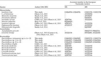
List of taxa incorporated in the molecular analysis of different trematode families, with the number of DNA sequences given in parentheses.

Alignments and phylogenetic analysis
The ribosomal DNA sequences were assembled with SeqScape (v. 2.6) software (Applied Biosystems) and aligned using the MEGA (v. 6.0) alignment explorer (Tamura et al., Reference Tamura, Stecher, Peterson, Filipski and Kumar2013) with default options. Estimation of a number of variable and parsimony-informative sites was performed using MEGA 6.0. Phylogenetic analysis of the nucleotide sequences was performed using Bayesian algorithms with MrBayes (v. 3.1.2) software (Huelsenbeck et al., Reference Huelsenbeck, Ronquist, Nielsen and Bollback2001). Phylogenetic algorithms were used with the TPM3uf + I model (Darriba et al., Reference Curran, Tkach and Overstreet2012) for 28S data of trematodes of Paucivitellosus; general time reversible model, including gamma-distributions (GTR + G) (Tavare, Reference Tavare1986) for 28S data of L. lizae; K80 + G model (Kimura, Reference Kimura1980) for 18S data of trematodes of Paucivitellosus; and TPM3 + G model (Darriba et al., Reference Curran, Tkach and Overstreet2012) for 18S data of L. lizae. These models showed the best fit to the data using Bayesian information criterion (BIC) in jModeltest (v. 3.07) software (Darriba et al., Reference Darriba, Taboada, Doallo and Posada2012). Bayesian analysis was performed using 10,000,000 generations and with four independent runs. The first 1,000,000–2,500,000 generations were burned, depending on the DNA fragment and species investigated. These values were established with the Tracer software (Rambaut & Drummond, Reference Rambaut and Drummond2009). The significance of the phylogenetic relationship was estimated by posterior probabilities (Huelsenbeck et al., Reference Huelsenbeck, Ronquist, Nielsen and Bollback2001).
The phylogenetic relationships of L. lizae and Paucivitellosus species were inferred from our data and the 18S rDNA and 28S rDNA nucleotide sequences of other trematode specimens obtained from GenBank (National Center for Biotechnology Information (NCBI)) (Cribb et al., Reference Cribb, Bray, Littlewood, Pichelin, Herniou, Littlewood and Bray2001; Tkach et al., Reference Tkach, Pawlowski, Mariaux, Swiderski, Littlewood and Bray2001; Olson et al., Reference Olson, Cribb, Tkach, Bray and Littlewood2003; Bray et al., Reference Bray, Webster, Bartoli and Littlewood2005; Besprozvannykh et al., Reference Besprozvannykh, Ermolenko and Atopkin2012) (table 1).
Results
Lasiotocus lizae Liu, Reference Liu2002
Taxonomic summary
Host
Liza longimanus – four fish examined, one infected with 49 worms.
Locality
Coastal waters off Cat Ba Island, Tonkin Bay, northern Vietnam (20°84′N, 106°59′E).
Site
Intestine.
Deposited
Slides (numbers 68–72Tr.) were placed in the collection of the Zoological Museum (Institute of Biology and Soil Sciences, Far East Branch of the Russian Academy of Sciences, Vladivostok, Russia); E-mail: petrova@ibss.dvo.ru. Deposited 25 September 2015.
Description
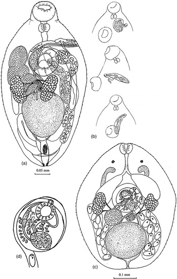
Based on seven specimens (fig. 1a, b; table 2). Body oval, spined from anterior to posterior end. Oral sucker subterminal, prepharynx short or absent, oesophagus present, oesophageal bifurcation before ventral sucker or on level of its anterior margin. Caeca terminated at level of middle of testis. Ventral sucker on border of anterior and middle third of body, larger than oral one. The ratio of sucker's length was 1:1.12–1.57 and of sucker's width was 1:0.96–1.26. Testis single, round or oval, in posterior half of body on median line. Two seminal ducts, which merge before cirrus-sac, moving away from testis. Anterior part of cirrus-sac before ventral sucker. Distal part of cirrus-sac with sac-shaped seminal vesicle.
(a, b) Lasiotocus lizae Liu, Reference Liu2002: (a) adult worm; (b) variants of location of the cirrus-sac. (c, d) Paucivitellosus vietnamensis sp. n.: (c) adult worm; (d) cirrus-sac.

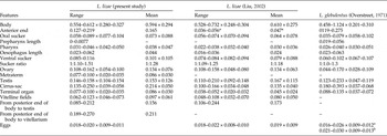
A comparison of the morphometrics (in mm) of adult Lasiotocus lizae with L. glebulentus.

* Probably, Liu (Reference Liu2002) made a mistake. Length of anterior end of the body, described by him, is not identical to the percentage of body length. According to Liu, the anterior end of the body is 28–33% (mean 31%) of the body length, which corresponds to a mean length of 0.189.
aMounted specimens; bliving ones.
Posterior part of cirrus-sac right from median line, for most part covered by ventral sucker, never crosses the posterior margin. Pars prostatica tubular, surrounded by small number of prostatic cells. Cirrus spined. Genital pore lateral or sublateral, left from ventral sucker, on level of oesophageal bifurcation. Genital atrium non-armed. Ovary large, three-lobed, to the right of median line of body, a short distance before or adjacent to the testis. Anterior margin of ovary on a level with anterior border-middle of ventral sucker. Seminal receptacle uterine. Mehlis' gland and Laurer's canal present. Uterine loops from posterior end of body to anterior margin of ventral sucker. Metraterm thin-walled, opening in bipartite terminal organ. Anterior part of terminal organ spined. Vitellarium from two compact masses, each from four large undivided follicles. Vitelline fields lateral in middle part of body, from posterior margin of ovary to anterior margin of testis. Eggs light yellow, operculated. Excretory bladder I-shaped, consists of one large or one large and 1–3 small concretions.
Molecular data
A 1345-bp fragment of the 18S rRNA gene was successfully sequenced and aligned for L. lizae. There were no variable sites between 18S sequences of L. lizae.
A 1068-bp fragment of the 28S rRNA gene sequence of L. lizae from Vietnam, obtained in our study, was aligned with 28S rDNA sequences of trematodes of the family Monorchidae from the GenBank database. There were three variable sites within L. lizae from Vietnam, which provide 0.28% of intraspecific genetic differentiation.
Remarks
The morphology and morphometrics of the present trematode species are similar to those of L. glebulentus Overstreet, Reference Overstreet1971 from M. cephalus of the estuarine waters of the Gulf of Mexico (Overstreet, Reference Overstreet1971) and L. lizae Liu, Reference Liu2002 from L. carinata of Taiwan Strait, China (Liu, Reference Liu2002). The main differences relate to the cirrus-sac, terminal organ, ovary and excretory bladder (table 2). The cirrus-sac lies between the anterior margin of the testis and the oesophageal bifurcation, and its distal part reaches the level of the vitellarium, vs. cirrus-sac is placed between the anterior margin of the ventral sucker and the pharynx, and its distal part is partially covered by the ventral sucker. The proximal part of terminal organ is spined or non-spined vs. the proximal part of terminal organ is non-spined. The ovary is non-lobed vs. the ovary is three-lobed. The excretory bladder contains 5–13 concretions vs. an excretory bladder with 1–4 concretions. Based on these features, the worms from our study are most similar to L. lizae, differing insignificantly from the latter only by the position of the cirrus-sac. We expect that adult worms collected from mullet fish from Tonkin Bay belong to L. lizae.
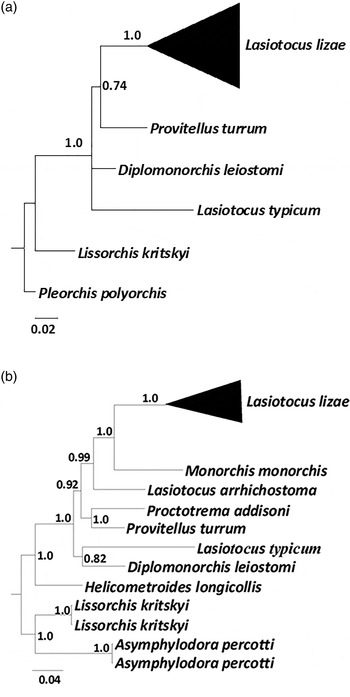
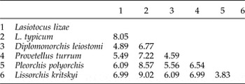
The type species of Lasiotocus, L. mulli (Stossich, 1883), like the most numerous species of this genus, has a median or submedian genital pore before the ventral sucker. For the species L. odhneri (Srivastava, 1939), L. minutus (Manter, 1931), L. elongates (Manter, 1931) and L. mugilis Overstreet, 1969, the submedian genital pore is placed in the borders of projection of the ventral sucker. For the trematode specimens studied in the present work, the genital pore is sublaterally or laterally located. At the same time, the genital pore lies left from the ventral sucker in the species L. glebulentus and L. lizae. Unlike L. mulli, which has a vitellarium consisting of a group of separate follicles, these two species have vitellaria consisting of two compact masses of undivided follicles. Similar vitellarial structures are inherent for the type species of Pseudoproctotrema, P. parupenei Yamaguti, 1924. Ovaries of L. lizae are three-lobed and longitudinally extended, similar to the ovaries in the type species of Paraproctotrema, P. fusiforme Yamaguti, 1934. The presence of significant morphological differences between L. glebulentus and L. lizae and the type species of Lasiotocus, L. mulli, and the similarity of these species with other species of this genus by a number of diagnostic criteria suggest that L. glebulentus and L. lizae may belong to the genus of Monorchiinae Odhner, 1911. Accordingly, the molecular data demonstrate that L. lizae is highly differentiated from other Monorchiidae (tables 3 and 4). Genetic differentiation by 18S rDNA analyses of L. lizae with members of Monorchiidae and Lissorchiidae ranged from 4.9% (Diplomonorchis leiostomi) to 8% (Lasiotocus typicum). The Bayesian algorithm of phylogenetic reconstructions based on 18S rRNA gene sequences showed unresolved topology for Monorchiidae, and put L. lizae and Provitelus turrum (Monorchiidae) into the same cluster with high statistical support (fig. 2a). Analysis of genetic distances, calculated for 28S rDNA sequence data, showed that L. lizae has the same level of genetic differentiation (9.42–9.59%) as Monorchis monorchis, P. turrum and Lasiotocus arrhichostoma (table 4). However, Bayesian analysis of the phylogenetic reconstruction showed that L. lizae was closely related to M. monorchis (fig. 2b), and L. arrhichostoma appears as a sister species to the cluster L. lizae/M. monorchis.
Bayesian phylogenetic tree of species of Monorchioidea based on: (a) partial 18S rRNA gene sequences; (b) partial 28S rRNA gene sequences.

Genetic differentiation (%) of species of Monorchioidea by 18S rRNA gene sequences.

Genetic differentiation (%) of species of Monorchioidea by 28S rRNA gene sequences.

In spite of the non-compliance of morphological characteristics of L. glebulentus and L. lizae to diagnostic criteria for Lasiotocus, we consider the data inappropriate to establish a new genus or include these two species into any existing genera of Monorchiinae. Our data support the point of Searle et al. (Reference Searle, Cutmore and Cribb2014) about the necessity for revision of the genus Lasiotocus and the subfamily Monorchiinae, using molecular data for all type species.
Paucivitellosus vietnamensis sp. n.
Taxonomic summary
Type host
Liza subviridis.
Site
Intestine.
Intensity of infection
Ten fish examined, one infected with 14 worms.
Type locality
Coastal waters of Cat Ba Island, Tonkin Bay, Vietnam (20°84′N, 106°59′E).
Type deposited
Holotype (no. 60-Tr.) and paratypes (nos 61–64-Tr.) were placed in the collection of the Zoological Museum (Institute of Biology and Soil Sciences, Far East Branch of the Russian Academy of Sciences, Vladivostok, Russia); E-mail: petrova@ibss.dvo.ru. Deposited 25 September 2015.
Etymology: The specific name refers to Vietnam, the type locality.
Description
Based on six specimens (fig. 1c and d; table 5). Body trapezoidal, spined. Two eyespots in first third of body, on level of oesophagus. Oral sucker absent, pharynx opened in short oesophagus, oesophageal bifurcation before cirrus-sac. Caeca sac-shaped, short, terminated level of posterior margin of cirrus-sac or anterior margin of testis. Ventral sucker absent, testis single, round or cross-oval, in posterior half of body. Two seminal ducts, which merge before cirrus-sac, moving away from testis. Cirrus-sac large, round with oval seminal vesicle. Duct of seminal vesicle thin-walled, often with sperm, opens in C-shaped pars prostatica in middle part. Pars prostatica opens in genital atrium by thick-walled pore. Prostatic cells numerous. Ovary round, to the right of median line between cirrus-sac and testis. Uterine loops lie from posterior margin on both sides of testis to cirrus-sac. Genital pore opens in genital atrium on median line of body. Vitellarium two- to four-lobed, in two lateral fields, placed on both sides of median line from cirrus-sac to ovary. Eggs oval, operculated, with miracidiae. Miracidium with eyespot. V-shaped excretory bladder, branches reach level of oesophagus.
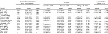
A comparison of the morphometrics (in mm) of adult Paucivitellosus vietnamensis sp. n. with P. fragilis.

Molecular data
Two sequences of the 18S rRNA gene fragment (268 bp) were successfully sequenced and aligned for P. vietnamensis sp. n. from L. subviridis from Vietnam. These two sequences were identical to each other.
Two 1081-bp sequences of the 28S rRNA gene fragment were obtained for P. vietnamensis sp. n. from L. subviridis from Vietnam. These sequences were also identical to each other.
Trematodes identified as Paucivitellosus sp. 1 and Paucivitellosus sp. 2 from L. engeli and V. seheli, respectively, have no intraspecific rRNA gene sequence variations.
Remarks
According to WoRMS Editorial Board (2014), this genus includes two species: P. fragilis Coil, Reid & Kuntz, Reference Coil, Reid and Kuntz1965 and P. hanumanthai (Mani, Reference Mani1989). Adult P. vietnamensis sp. n. from L. subviridis meets all morphological criteria (Cribb, Reference Cribb, Gibson, Jones and Bray2002) of the genus Paucivitellosus. On the other hand, P. vietnamensis sp. n. differs from P. fragilis and P. hanumanthai by a number of metric features (Coil et al., Reference Coil, Reid and Kuntz1965; Pearson, Reference Pearson1968; Mani, Reference Mani1989). In particular, P. vietnamensis sp. n. is characterized by larger body size, oesophagus, cirrus-sac and eggs in comparison with P. fragilis (table 5). The newly described species is closer to P. hanumanthai by most metric characteristics, with the exception of sizes of body width, testis, ovary and cirrus-sac length (table 5).
Cribb et al. (Reference Cribb, Bray and Barker1994) reported that Paucivitellosus includes the single type-species, P. fragilis. On the basis of analysis of numerous specimens of Paucivitellosus from various Australian, Indian and Chinese Mugilidae and Blenniidae, Cribb et al. (Reference Cribb, Bray and Barker1994) have come to the conclusion that P. hanumanthai is a synonym of P. fragilis. They reported that high variation in length (from 0.272 to 0.812 mm) and width (from 0.216 to 0.525 mm) of the body is typical for P. fragilis. However, cercariae of P. hanumanthai have sizes of 0.272–0.304 × 0.196–0.256 mm (Mani, Reference Mani1989). On the contrary, sizes of cercariae of P. fragilis are of size 0.150–0.170 × 0.110–0.130 mm (Pearson, Reference Pearson1968). According to Mani (Reference Mani1989), it is unlikely that adult worms from naturally infected hosts (Cribb et al., Reference Cribb, Bray and Barker1994) have smaller body sizes than cercariae. Mani (Reference Mani1989) also established that specimens of mature P. hanumanthai at 10 days of age have a minimum body length and width of 0.480 and 0.320 mm, respectively. Usually, small worms that can be attributed to specimens of the genus Paucivitellosus increase in size insignificantly after puberty. Based on the data of Crib et al. (Reference Cribb, Bray and Barker1994), we can note that specimens of P. fragilis almost tripled their body lengths after puberty. The largest specimens, identified as P. fragilis, were detected in L. carinata of the Taiwan Strait (Peng et al., Reference Peng, Wang, Yu and Liu2004) (table 5). These specimens are characterized by higher values of morphometric parameters in comparison with specimens of P. fragilis and P. hanumanthai reported by Coil et al. (Reference Coil, Reid and Kuntz1965), Pearson (Reference Pearson1968) and Mani (Reference Mani1989), respectively. Given that specimens of various species can be barely discernible morphologically, metric criteria, together with molecular data, can determine the main features for species differentiation.
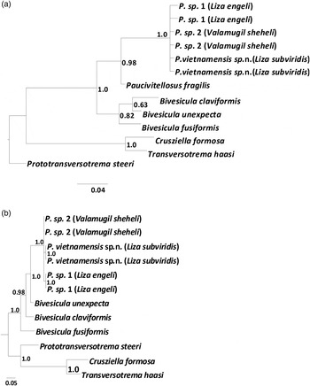
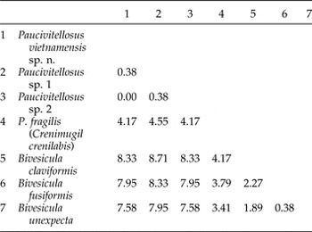
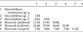
The results of genetic investigation showed that specimens of P. vietnamensis sp. n. from L. subviridis, and Paucivitellosus sp. 1 and Paucivitellosus sp. 2 from L. engeli and V. seheli, are highly differentiated from specimens of P. fragilis from the Australian Crenimugil crenilabis (Cribb et al., Reference Cribb, Bray, Littlewood, Pichelin, Herniou, Littlewood and Bray2001). Specimens of Paucivitellosus spp. were genetically distant from Australian P. fragilis by 4.17–4.55%, by analysis of 18S rRNA gene sequences (table 6). Our study also showed that genetic differentiation of P. vietnamensis sp. n. from Paucivitellosus sp. 1 from L. engeli was 0.38% by 18S rRNA gene sequences, and 1.58% by 28S rRNA gene sequences (tables 6 and 7). Nucleotide sequences of 18S rRNA of P. vietnamensis sp. n. and Paucivitellosus sp. 2 were identical. These worms differ from each other by a 28S rRNA gene fragment (0.62%). Moreover, specimens of Paucivitellosus from L. engeli and V. seheli show a slight genetic differentiation by analysis of the 268-bp 18S rRNA gene fragment (0.38%) and 1081-bp 28S rRNA gene fragment (0.95%) (tables 6 and 7). Bayesian phylogenetic analysis also showed high differentiation between trematode specimens of the genus Paucivitellosus from Vietnam and P. fragilis from C. crenilabis (fig. 3a) and indicated genetic heterogeneity of specimens of this genus from different mugilid species from Vietnam (fig. 3b). These observations do not preclude P. fragilis sensu Cribb et al. (Reference Cribb, Bray and Barker1994) from representing a set of cryptic species.
Bayesian phylogenetic tree of Bivesiculidae based on: (a) partial 18S rRNA gene sequences; (b) partial 28S rRNA gene sequences.

Genetic differentiation (%) of species of Bivesiculidae by 18S rRNA gene sequences.

Genetic differentiation (%) of species of Bivesiculidae by 28S rRNA gene sequences.

On the basis of the observed genetic differences between Paucivitellosus specimens from different Vietnamese species of mullet fish and the absence of morphological data of trematodes from L. engeli and V. seheli, we recognize a new trematode species, P. vietnamensis sp. n., from L. subviridis.
Financial support
This study was partially supported by grants RFBR No. 16-34-00222_mol_a and VAST.DA47.DA 12/15-18.
Conflict of interest
None.