Introduction
Between 1971 and 1993, Germany’s most notorious terrorist group, the Red Army Fraction (RAF), killed 33 people and injured more than 200 others, among them many representatives of the justice system, the political establishment, and the economy. The intellectual head of this left lunatic fringe group was Ulrike Meinhof. Before she entered the RAF in 1970, she was a well-known and recognized journalist who committed herself by peaceful means to promote her political ideas. Before she joined the RAF, she underwent neurosurgery in 1962 after developing neurological symptoms because of a vascular tumor (angioma) at the base of her brain next to the right medial temporal lobe. In the years after this surgery, she developed a change of personality that included increasingly aggressive traits. She later wrote the “The Concept of the Urban Guerilla,” by which she tried to adapt the strategies of South American guerilla groups to West German cities. After she was captured, she committed suicide in 1976 during the court proceedings against leading members of the RAF. An autopsy of her brain was performed, and the neuropathologist described small circumscribed damage to the cortical tissue and adjoining white matter in the right anterior medial temporal lobe, very close to the amygdala, as a result of the brain surgery she had in 1962, but no damage to other brain structures.Reference Bogerts 1 Her brain lesion was localized in a key limbic region involved in neuronal control of basic emotions, including aggressive and violent behaviors. The content of the radical political ideas she fought for can of course not be explained by the postsurgical limbic brain damage but are rather a result of the special political and social environment of her time. But the fact that she developed a personality change with increasing aggressiveness and violence has to be regarded as a result of the brain injury closely related to the amygdala. This regionally localized type of brain damage might not be representative for terroristic behavior in general, but we are not aware of any other postmortem or neuroimaging investigation of a terrorist’s brain.
To our knowledge, with two exceptions, there are also no autopsy findings in persons who ran amok. The best-documented historic case of an amok runner, at least in European psychiatry, belongs to the teacher Ernst Wagner, who in 1913 first killed his wife and 4 children, then burned down several buildings, and then shot and killed 9 male inhabitants and injured 11 others during one night in a village near the city of Stuttgart. Months before the mass killings, he began to train himself to shoot pistols. He was eventually overpowered and subdued during the course of his mass murder spree and then examined by a psychiatrist, who diagnosed him as suffering from paranoia, since for years he had felt persecuted and threatened by his victims. After he died years later in a forensic hospital, a brain autopsy revealed a small circumscribed lesion in the left anterior entorhinal cortex, next to the hippocampus and amygdala, while other parts of the brain looked inconspicuous.Reference Bogerts 1
The second case of a person who ran amok and underwent a brain autopsy was Charles Whitman, a 25-year-old college student at the University of Texas. In 1966, he first stabbed his mother and his wife, and he then shot and killed 17 people from his sniper’s next in the university tower, injuring 32 others. Months before the shootings, he sought psychiatric help because he was suffering from increasing personal stress and psychological isolation. He felt that something was going wrong in his head, and in his suicide note he requested that an autopsy be performed to determine if something had changed in his brain. After he was shot by security forces at the top of the tower, an autopsy was indeed performed. Aside from many of his brain parts being damaged by penetrating fragments of bone created by his gunshot wounds, a small tumor (a glioblastoma) was found beneath the thalamus, impinging upon the hypothalamus and compressing the amygdala. 2 , Reference Eagleman 3
Neuronal Correlates of Violence
The brain lesions in these three well-known cases have at least one thing in common: they were located in limbic structures of the anterior temporal lobe involved in the neuronal control of amygdala activities and, via the amygdala, also of the hypothalamic and lower brainstem areas, called by McLean the “reptilian brain.”Reference MacLean 4 In these phylogenetically very old brain structures, groups of neurons are located that play a central role in the brain networks responsible for aggressive behavior and thus can be regarded as some kind of neuronal “generators” of aggression. Already in 1932, the Swiss physiologist Hess could demonstrate in cats that electrical stimulation of hypothalamic and other regions of the “reptilian brain” immediately provoked aggressive behavior.Reference Hess 5 , Reference Wasman and Flynn 6 Years later, these experiments were extended by Ploog,Reference Ploog 7 who detected cell groups in the medial hypothalamus and medial amygdala related to the reactive/defensive rage type of aggression. The proactive/predatory attack type of aggression was elicited by stimulation of cell groups in the lateral hypothalamus and lateral amygdala. Under certain physiological conditions, these cell groups in the medial and lateral hypothalamus come into action if either threatening environmental conditions or aggression-inducing cue stimuli activate them via fiber tracts from the amygdala. Under pathological conditions, certain brain diseases can cause abnormal activation or a lack of inhibition of these cell groups by afferent fibers from the limbic or cortical association areasReference Bogerts and Möller-Leimkühler 8 or, as shown in the experiments of Hess and Ploog, if they are activated experimentally by direct electrical stimulation. Even in humans, electrical stimulation of the amygdala, performed for diagnostic purposes before stereotactic surgery in patients suffering from temporal lobe epilepsy, has caused severe impulsive aggressive outbursts.Reference Mark and Erwin 9 Already in 1937, Klüver and BucyReference Klüver and Bucy 10 showed that their quite aggressive monkeys after destruction of the anterior temporal lobe (including the amygdala) displayed a “psychic blindness” characterized by a lack of anxiety and by steadily tame behavior.
Aside from temporal lobe epilepsy, which is caused by abnormal neuronal discharges in temporolimbic groups of neurons and is sometimes associated with sudden violent acts, limbic structures in the medial temporal lobe also belong to the regions that exhibit more or less subtle structural or functional damage in a significant proportion of patients suffering from psychotic diseases (e.g., schizophrenia),Reference Bogerts 11 , Reference Bogerts 12 which might explain the increased frequency of aggressive attacks in such psychotic syndromes.Reference Fazel, Gulati, Linsell, Geddes and Grann 13
The neuronal generators of violence in the limbic system and hypothalamus did not arise incidentally: they are—aside from neuronal networks for prosocial attitudes—a product of a long evolutionary process that provided for the more aggressive male individuals or species an advantage in reproducing themselves while diminishing this chance for their victims. It has been shown that the primates (including homo sapiens), among all the vertebrata, have the highest rates of killing members of their own species, with about 2% dying as the result of an attack from their own group or from other human groups.Reference Gómez, Verdú, González-Megías and Méndez 14 Moreover, hostile and martial attitudes against other groups combined with parochial altruistic behavior for in-group members also provide a strong evolutionary advantage.Reference Choi and Bowles 15 Thus, neuronal substrates that are responsible for violence belong to the phylogenetically old equipment present in the brains of the human species in general.
Do Terrorists Have a Peculiar Neurobiological Predisposition?
There are several reasons to assume that at least a significant proportion of terrorist brains has a biological predisposition to violent behavior, so that one can conclude that the various ideologies claimed by them as justification for their terrorist acts are not the real reason for their behavior. Such adverse early life experiences as misuse in childhood, oppressive education, and lack of positive parental emotional attention, which are frequently characteristic of violent offenders, have plastic effects on such limbic brain regions as the amygdala and hippocampus. They cause not only enduring psychological scars but also long-lasting functional and even structural changes in the brain.Reference Jung, Herrenkohl, Lee, Klika and Skinner 16 A large body of evidence also demonstrates the considerable influence of genes for the emergence of aggressive personality traits, psychopathy, and even religious fundamentalism.Reference DiLalla, Carey, Gottesman and Bouchard 17 Adoption studies and investigations of identical twins have shown that genetic factors may explain up to 50% of the causative variance of criminal offences.Reference Joyal, Putkonen and Mancini-Marïe 18 , Reference Rhee and Waldman 19 Among the genetic components showing an affinity for aggressive behavior, the monoamine oxidase A (MAOA) gene received special attention.Reference Alia-Klein, Goldstein and Kriplani 20 , Reference Buckholtz and Meyer-Lindenberg 21 The low-expression variant of this gene has a particularly high affinity for violent behavior, especially if it occurs in combination with traumatic childhood experiences.Reference Caspi and Moffitt 22 Moreover, abnormalities in neurotransmitters, especially serotonin, and hormones (testosterone, cortisol, vasopressin) seem to contribute to the etiology of aggressive traits (for a review, see Rosell and SieverReference Rosell and Siever 23 ).
On the other hand, there are many historical examples (see, e.g., BrowningReference Browning 24 ) that previously completely ordinary men without any signs of abnormal psychology have committed war crimes, atrocities, and genocide due to a mixture of motives, including the group dynamics of conformity, deference to authority, role adaptation, and alteration of moral norms. It has also been shown that randomly selected normal people under special conditions of psychological experiments are able to willingly perform cruelties if they are told to do so by authorities or if they are allowed to do so as custodians.Reference Haney, Banks and Zimbardo 25 – Reference Zimbardo 28 This shows that without a neurobiological predisposition or brain pathology, particular psychosocial constellations and/or group dynamics can give rise to violent acts. This is especially the case for various forms of individual and collective aggression.Reference Möller-Leimkühler and Bogerts 29
Individuals who join terror groups very often have a history of a previous criminal career. Analyzing the case histories of 784 persons in Germany who joined the so-called Islamic State of Iraq and the Levant (ISIL) between 2012 and 2015, it was found that two-thirds of these overwhelmingly male persons were known to the police because of violence, property offences, or political criminal acts. 30 In fact, more than half had committed three or more criminal offences before joining ISIL. It was reasoned that, because of their inherent readiness to violence, such persons search for suitable ideologies under which to act out their tendencies to harm, injure, and kill. From a neurobiological point of view, it seems justified to assume that the neuronal generators of aggression and violence in the human “reptilian brain” and limbic system are either per se more active in such persons by genetic or biographical influences or that they are less inhibited by brain systems that are normally responsible for such prosocial attitudes as empathy and compassion. The latter could again be the consequence of predisposing genes, but also of various pathological brain abnormalities (among them, brain tissue defects by injury, atrophy, viral infection, tumors or innate hypoplasias, or psychoses); antisocial, fanatic, and/or paranoid personality disorders; or of a character trait called “psychopathy,” that is, a callous antisocial lifestyle with no intellectual defects but displaying a typical inability to have a sense of the value of others. There are reports that about 40% of the lone wolves (lone-acting terrorists) who develop their hateful ideologies absent contact with any particular terrorist group are suffering from a psychiatric disorder.Reference Pantucci, Ellis and Chaplais 31 At least in such terrorists, ideology alone is not a sufficient explanation for injuring or killing others—abnormal brain function is very a likely root cause.
Brain Imaging Studies in Violent Individuals
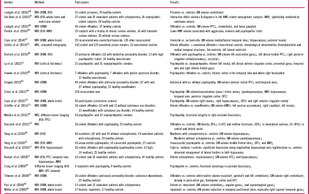
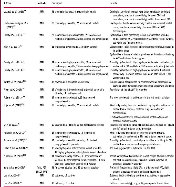
While there are to our knowledge (aside from the three exceptions reported above) no direct neuropathological or imaging investigations of the brains of terrorists or persons running amok, numerous structural and functional brain-imaging studies have meanwhile been performed in violent criminal offenders and in persons with antisocial personality disorder (APD) with/without psychopathy, a significant proportion of whom may be encountered among terrorists. The following sections provide an overview of the structural (see Table 1) and functional (see Table 2) imaging studies that investigated male adult violent samples published during the previous 10 years only (2008 to February of 2017). For earlier reviews, see, for example, Bufkin and Luttrell,Reference Bufkin and Luttrell 32 Raine et al.,Reference Raine and Yang 33 and Weber et al.Reference Weber, Habel, Amunts and Schneider 34
Structural imaging of aggression and violent behavior (studies investigating adult male samples with a history of violence, published between 2008 and 2017, in order by descending date)

AMY=amygdala; GM=gray matter; MRI=magnetic resonance imaging; OFC=orbitofrontal cortex; PFC=prefrontal cortex; ROI=region of interest; VBM=voxel-based morphometry.
Studies of functional imaging of aggressive and violent behavior (studies investigating adult male samples with a history of violence, published between 2008 and 2017, in order of descending date)

AMY=amygdala; fMRI=functional magnetic resonance imaging; MRS=magnetic resonance spectroscopy; OFC=orbitofrontal cortex; PET=positron emission tomography; PFC=prefrontal cortex; SPECT=single-photon emission computed tomography.
Structural Imaging Findings (CT and MRI)
A more global assessment of brain pathology in violent persons was performed in a retrospective investigation by Schiltz and colleagues,Reference Schiltz, Witzel, Bausch-Hölterhoff and Bogerts 35 who qualitatively rated brain tissue defects in magnetic resonance imaging (MRI) and computed tomography (CT) scans from a large sample of incarcerated violent offenders in comparison to nonviolent offenders and nonoffending healthy controls. Violent offenders compared to the two nonoffender groups displayed a considerable higher rate of brain abnormalities. The observed brain damage or atrophy was located mainly in the frontal and medial temporal brain regions.Reference Schiltz, Witzel, Bausch-Hölterhoff and Bogerts 35
Frontal lobe and violence
Frontal cortical pathology seems to play a major role in the pathophysiology of violent behavior. The frontal cortex is an anatomically and functionally heterogeneous brain structure involved, for example, in executive functioning, motor control, behavioral/emotional self-regulation, social behavior, and moral decision making.Reference Floden 36 – Reference Stuss 39 Frontal lobe abnormalities have been found consistently in morphometric neuroimaging studies.Reference Brower and Price 40 , Reference Müller, Gänßbauer and Sommer 41 Prefrontal cortex (PFC) abnormalities especially seem to be involved in aggressive and violent behavior.Reference Gregory 42 , Reference Leutgeb, Leitner and Wabnegger 43 There is an association between prefrontal volume reduction and aggression as well as antisocial behavior. This is also evident in subjects with both traumatic and neurodegenerative impairments of the prefrontal areas.Reference Brower and Price 40 PFC involvement in executive functioning could further be associated with deficits in decision making as well as problems in behavioral control.Reference Domenech and Koechlin 44 When comparing violent offenders with APD and psychopathy to violent offenders with APD but without psychopathy, a reduced amount of gray matter (GM) was found bilaterally in the anterior rostral medial PFC.Reference Gregory 42 Moreover, in antisocial offenders with high-psychopathic traits there was a significant volume reduction in the dorsomedial PFC.Reference Bertsch, Grothe and Prehn 45
One part of the prefrontal cortex, the orbitofrontal cortex (OFC), is relevant for processing of reward/punishment, emotion regulation, self-control, and behavioral inhibition,Reference Davidson 46 – Reference Rudebeck and Murray 48 and is interconnected with the limbic areas.Reference Schoenbaum, Roesch and Stalnaker 49 Specifically, OFC lesions are associated with an increase in aggression and impulsiveness, socially inadequate behavior, and enhanced disinhibition.Reference Brower and Price 40 , Reference Birbaumer, Veit and Lotze 50 It is a quite consistent finding that the orbitofrontal GM volume is reduced in male adult samples inclined to violence. OFC tissue reductions have been reported in unsuccessful psychopaths (prosecuted for their violent crimes) as compared to successful psychopaths (not prosecuted),Reference Yang, Raine, Colletti, Toga and Narr 51 but also in violent patients with schizophrenia,Reference Kumari, Barkataki, Goswami, Flora, Das and Taylor 52 as well as in violent offenders with APD as compared to healthy controls.Reference Tiihonen, Rossi and Laakso 53 Aside from volume reductions, a decreased orbitofrontal GM concentration was found in incarcerated men with psychopathy,Reference Ermer, Cope, Calhoun, Nyalakanti and Kiehl 54 and a 20% reduced GM density was reported in the OFC and frontal midline structures when comparing violent offenders with psychopathic and healthy controls.Reference Boccardi, Frisoni and Hare 55 However, there is also one study that reported an increased orbitofrontal GM volume in inmates with psychopathy.Reference Cope, Shane and Segall 56
Another focus of structural findings is the anterior cingulate cortex, which is situated beside the midline and is involved in the emotion-regulation circuitry. Abnormalities in this area are associated with an increased propensity for violent behaviorReference Davidson 46 and/or with psychopathy as compared to nonpsychopathic inmates.Reference Ly, Motzkin and Philippi 57 The posterior cingulate cortex is found to be impaired in persons inclined to violence, too. This brain area is linked to cognitive functions, including regulating attentional focus, and belongs to the so-called default mode network.Reference Buckner, Andrews-Hanna and Schacter 58 – Reference Leech and Sharp 60 In incarcerated men, psychopathy was associated with GM volume and concentration reductions in the posterior cingulate cortex.Reference Ermer, Cope, Calhoun, Nyalakanti and Kiehl 54 Furthermore, this region was smaller in another investigation of violent offenders versus healthy nonviolent ontrols.Reference Tiihonen, Rossi and Laakso 53 An atrophy was also reported in the frontopolar cortex.Reference Tiihonen, Rossi and Laakso 53 The right inferior frontal gyrus was found to be thinner in psychopathic versus nonpsychopathic inmates.Reference Ly, Motzkin and Philippi 57 The posterior part of this gyrus, the pars opercularis, belongs to the mirror neuron system and is involved in imitation as well as social reciprocity.Reference Yamasaki, Yamasue and Abe 61
Temporal lobe
The many functions of the temporal lobe involve emotional and social processes, including theory of mind, meaning the ability to recognize the thoughts and feelings of others, and to empathize.Reference Olson, Plotzker and Ezzyat 37 Many studies have shown that temporal lobe damage can cause aggressive and violent behavior.Reference Bannon, Salis and O’Leary 62 For example, in patients with temporal lobe epilepsy, a high incidence of behavior comparable to psychopathic habits was described—for example, hostility, diminished empathy, and even aggressive outbursts.Reference Kiehl 63 Furthermore, antisocial and socially inappropriate behavior were found to be associated with temporal brain alterations in frontotemporal dementia.Reference Miller, Darby, Benson, Cummings and Miller 64 , Reference Woermann, van Elst and Koepp 65
The temporal cortex was found to be thinner in offenders with psychopathy when compared with healthy noncriminals.Reference Howner, Eskildsen and Fischer 66 Furthermore, the temporal gray matter volume was reduced in forensic inpatients compared to healthy controls.Reference Müller, Gänßbauer and Sommer 41 In particular, the medial and lateral temporal regions were reported to have gray matter loss in incarcerated men who committed homicide compared to other crimes.Reference Cope, Ermer and Gaudet 67
Hippocampal formation, parahippocampal gyrus
Reduced GM volume and concentration in the hippocampal formation was found to occur in criminal psychopathy.Reference Ermer, Cope, Calhoun, Nyalakanti and Kiehl 54 A decreased hippocampal and parahippocampal volume was further described in a study comparing murderers with schizophrenia to nonschizophrenic murderers.Reference Yang, Raine, Han, Schug, Toga and Narr 68 This is in line with findings of a reduced volume of the parahippocampal gyrus in violent offenders compared to healthy men.Reference Tiihonen, Rossi and Laakso 53 Hippocampal tissue was found to be reduced bilaterally in incarcerated men who committed homicide compared to men sentenced for other crimes,Reference Cope, Ermer and Gaudet 67 but also in incarcerated men with psychopathy,Reference Ermer, Cope, Calhoun, Nyalakanti and Kiehl 54 , Reference Cope, Shane and Segall 56 as well as in violent patients with schizophrenia.Reference Puri, Counsell, Saeed, Bustos, Treasaden and Bydder 69 There are reports of increased impulsiveness in patients with schizophrenia inclined to repetitive violence, which was associated with reduced hippocampal volume.Reference Kumari, Barkataki, Goswami, Flora, Das and Taylor 52 When comparing high Psychopathy Checklist–Revised (PCL–RReference Hare 70 ) scorers with medium PCL–R scorers or healthy controls, a significant reduction along the hippocampal axis was described.Reference Boccardi, Ganzola and Rossi 71
The insula also belongs to the brain system involved in emotional experiences and empathy.Reference Eres, Decety, Louis and Molenberghs 72 The left insular cortex was reported to be thinner in psychopathic versus nonpsychopathic inmates.Reference Ly, Motzkin and Philippi 57 The left insular cortical tissue was also found to be reduced in violent offenders compared to nonoffenders,Reference Schiffer, Mueller and Scherbaum 73 whereas inmates with psychopathy had a smaller right insular GM volume in another study.Reference Cope, Shane and Segall 56 A third study reported insular GM volume reductions: incarcerated men who committed homicide in comparison to inmates sentenced for other crimes had decreased cortical tissue in the posterior insula.Reference Cope, Ermer and Gaudet 67
With regard to the pole of the temporal lobe, another paralimbic area, aggression and violent behavior are also associated with cortical thinning and volume reduction. Psychopathic compared to nonpsychopathic inmates had thinner bilateral temporal poles.Reference Ly, Motzkin and Philippi 57 In addition, psychopathy was associated with reduced temporal pole GM volume in two studies, one comparing antisocial personality-disordered violent offenders with and without psychopathyReference Gregory 42 and the other investigating incarcerated men with different PCL–R scores.Reference Ermer, Cope, Calhoun, Nyalakanti and Kiehl 54 The latter study also reported temporal pole GM concentration reductions in psychopaths.Reference Ermer, Cope, Calhoun, Nyalakanti and Kiehl 54 Cortical thinning has also been reported in the superior temporal gyrus.Reference Ly, Motzkin and Philippi 57
Amygdala
The amygdala is one of the key structures in recent neurobiological models of violence.Reference Siever 74 This medial temporal lobe structure is the central part of the limbic system and can be divided into three nuclear complexes: basolateral, centromedial, and cortical/superficial.Reference Price 75 , Reference Sah, Faber, Lopez De Armentia and Power 76 The amygdala is involved in fear conditioning, emotion expression and recognition, emotionally influenced memory, and moral reasoning.Reference Price 75 , Reference Blair 77 – Reference Phillips and LeDoux 79 Furthermore, it is a key structure for emotion regulation belonging to a circuitry that also consists of the orbital brain, the dorsolateral prefrontal cortex, and the anterior cingulate cortex.Reference Davidson 46 Amygdala impairments have been linked to a variety of psychiatric disorders characterized by aggressive behavior (e.g., psychopathy and antisocial personality disorder). Psychopathy has been found to be associated with reduced amygdala GM volume in a sample of 254 incarcerated men.Reference Ermer, Cope, Calhoun, Nyalakanti and Kiehl 54 Significantly reduced GM volume and cortical thickness of the amygdala were described in unsuccessful psychopaths compared to controls as well.Reference Yang, Raine, Colletti, Toga and Narr 51 The three subnuclei are obviously associated with different structural amygdalar impairments: in offenders with psychopathy, there was up to 30% reduced brain tissue in the basolateral nucleus, and the central and lateral nuclei tissue were enlarged by 10 to 30%.Reference Boccardi, Frisoni and Hare 55 Reduced amygdalar volume seems to be associated with aggression and violence in general.Reference Pardini, Raine, Erickson and Loeber 80 In violent but nonpsychotic subjects, violence was associated with volume reduction only in the left amygdala.Reference Del Bene, Foxe, Ross, Krakowski, Czobor and De Sanctis 81 However, there is also one study that reported increased amygdalar volume in violent offenders.Reference Schiffer, Mueller and Scherbaum 73
In psychopathic individuals, the white matter connection between the amygdala and frontal brain areas (e.g., ventromedial PFCReference Motzkin, Newman, Kiehl and Koenigs 82 and OFCReference Craig, Catani and Deeley 83 ) is also impaired. There is an association between psychopathy and reduced structural integrity in the right uncinate fasciculus (connection from ventromedial PFC to amygdala), which is important for aggression and emotion regulation.Reference Motzkin, Newman, Kiehl and Koenigs 82 Furthermore, fractional anisotropy has been found to be reduced in the uncinated fasciculus in psychopaths compared to controls, suggesting an abnormal connection between the amygdala and the OFC.Reference Craig, Catani and Deeley 83
Further brain regions
Some studies have reported that the volume of the cerebellum is increased in violent offenders compared to healthy controls.Reference Leutgeb, Leitner and Wabnegger 43 , Reference Leutgeb, Wabnegger and Leitner 84 Aside from its main function of extrapyramidal motor coordination, the cerebellum is also involved in cognition and emotion as well as empathy (for pain) and moral judgments.Reference Harada, Itakura and Xu 85 – Reference Schmahmann 87 It is anatomically connected with brain regions associated with emotion regulation, aggression, and moral decisions, like the amygdala, the hippocampus, and the PFC.Reference Demirtas-Tatlidede and Schmahmann 88 , Reference Turner, Paradiso and Marvel 89 However, one study found that cerebellar GM volume was reduced in violent patients with schizophrenia.Reference Puri, Counsell, Saeed, Bustos, Treasaden and Bydder 69 The cerebellum as well as the basal ganglia are relevant for motor aspects of impulsive acts.Reference Picazio and Koch 90 The findings for the basal ganglia, however, are more inconsistent. There are reports of increased as well as decreased gray matter volumes of the caudate, putamen, pallidum, and nucleus accumbens in violent,Reference Leutgeb, Leitner and Wabnegger 43 , Reference Schiffer, Mueller and Scherbaum 73 impulsive, or sensation-seekingReference Glenn, Raine, Yaralian and Yang 91 individuals. A few studies also found tissue reduction in the postcentral gyrus (lateral parietal lobe) in violent offenders or persons with psychopathic traits.Reference Bertsch, Grothe and Prehn 45 , Reference Tiihonen, Rossi and Laakso 53 When comparing violent and nonviolent patients with schizophrenia, reduced GM volume in the angular gyrus (anterolateral parietal lobe)Reference Puri, Counsell, Saeed, Bustos, Treasaden and Bydder 69 as well as in the supramarginal gyrus (parietal lobe) has been found.Reference Puri, Counsell, Saeed, Bustos, Treasaden and Bydder 69
Functional Imaging Studies in Violent Individuals
Frontal lobe
In addition to the structural deficits found in subregions of the frontal lobe, many functional imaging studies have described frontal lobe dysfunction in violent individuals. The lateral OFC of incarcerated psychopaths has been shown to be less activated by an empathy task.Reference Decety, Skelly and Kiehl 92 In addition, the inferior frontal gyrus of imprisoned psychopaths is less activated by a theory-of-mind task.Reference Mier, Haddad, Diers, Dressing, Meyer-Lindenberg and Kirsch 93 There is already some evidence that subjects show different functional responses in their emotional empathy while watching computer-generated actors compared to actors in live-action movies.Reference Vemuri and Surampudi 94 A metaanalysis consisting of 31 functional (and 12 structural) studiesReference Yang and Raine 95 came to the result that antisocial individuals have functional deficits in the right orbitofrontal, the left dorsolateral prefrontal cortex, and the right anterior cingulate cortex, and this effect persisted regardless of whether the subjects had to perform a cognitive or emotional task.
There is an impaired functional connectivity between the orbital brain and other brain areas. Criminal prisoners showed less functional connectivity between the orbital cortex and cerebellum,Reference Contreras-Rodriguez, Pujol and Batalla 96 and incarcerated psychopaths had less functional connectivity to the amygdala and the anterior insula.Reference Decety, Skelly and Kiehl 92
In the medial frontal cortex, criminal psychopaths showed decreased activation during performance of a moral-dilemma task.Reference Ly, Motzkin and Philippi 57 Spousal abusers showed less activation in the left medial frontal cortex during threat stimuli.Reference Lee, Chan and Raine 97 Criminal psychopaths showed increased functional connectivity within the dorsomedial frontal cortex but less gray matter volume in the PFC, in contrast to nonoffender controls.Reference Contreras-Rodriguez, Pujol and Batalla 96 Reduced activation in the ventromedial PFC was found in incarcerated psychopaths during empathizingReference Decety, Skelly and Kiehl 92 as well as during a moral judgment task.Reference Harenski, Harenski, Shane and Kiehl 98
Limbic areas: amygdala, hippocampus, cingulate cortex
Reduced functional connectivity between the PFC and the limbic (amygdala, hippocampus) as well as paralimbic areas was found in criminal psychopaths.Reference Contreras-Rodriguez, Pujol and Batalla 96 Imprisoned psychopaths had increased activation in the amygdala when they performed a theory-of-mind task, and functional connectivity between the amygdala and the superior temporal gyrus was impaired.Reference Mier, Haddad, Diers, Dressing, Meyer-Lindenberg and Kirsch 93 When psychopathic offenders had to perform an empathy task with people in pain, activation in the amygdala correlated negatively with factor 1 of the PCL–R scale.Reference Hare 70 Factor 1 consists of the interpersonal facet (e.g., superficial charm or manipulative behavior) and the affective facet (e.g., callousness). The more interpersonal and affective deficits the subjects had, the less activation of the amygdala was found.Reference Decety, Chen, Harenski and Kiehl 99 In nonpsychopathic offenders, a positive correlation between activation in the right amygdala and severity of moral judgment ratings was found. This implies that the higher activation in the right amygdala, the higher the severity of moral violation was rated, and this correlation was not found in psychopathic offenders.Reference Harenski, Harenski, Shane and Kiehl 98 In a cognitive control paradigm, spousal abusers showed increased activation of the right amygdala as well as of the hippocampus.Reference Lee, Chan and Raine 97 In contrast, the hippocampus was less activated in criminal psychopaths during a moral-dilemma task.Reference Pujol, Batalla and Contreras-Rodríguez 100
Functional deficits in the cingulate cortex have also been found in violent offenders. Activation in the right anterior cingulate cortex was reduced in a cognitive control paradigmReference Lee, Chan and Raine 97 and during empathizing.Reference Meffert, Gazzola, den Boer, Bartels and Keysers 101 The metaanalysis of Yang and RaineReference Yang and Raine 95 showed that antisocial individuals have functional deficits in the right anterior cingulate cortex regardless of whether the subjects had to perform a cognitive or emotional task. Interestingly, activation in the right anterior cingulate cortex during empathizing increased after offenders were instructed to empathize with the actors.Reference Meffert, Gazzola, den Boer, Bartels and Keysers 101 The anterior cingulate cortex as well as the anterior insula belong to the empathy-for-pain network,Reference Singer and Klimecki 102 which is obviously dysfunctional in psychopathic criminals. Activation of the posterior cingulate cortex decreased in moral decision makingReference Pujol, Batalla and Contreras-Rodríguez 100 but increased after presenting threat stimuli.Reference Lee, Chan and Raine 103 The functional connectivity between the left dorsal anterior cingulate cortex and the left insula was reduced,Reference Ly, Motzkin and Philippi 57 as well as the functional connectivity between the medial frontal cortex and the posterior cingulate cortex.Reference Pujol, Batalla and Contreras-Rodríguez 100
Other brain areas
Several studies have shown functional deficits during face processing in the fusiform gyrus in criminal offenders: either a bilateral decreased activation in the fusiform gyrus in incarcerated psychopathsReference Mier, Haddad, Diers, Dressing, Meyer-Lindenberg and Kirsch 93 , Reference Decety, Skelly, Yoder and Kiehl 104 or reduced activation only in the left fusiform gyrus in spousal abusers incarcerated because of domestic violence.Reference Lee, Chan and Raine 97
Activation in the insula of high-psychopathic inmates was increased during empathizing.Reference Decety, Skelly and Kiehl 92 Conversely, the more interpersonal and affective deficits the psychopathic inmates had (measured by factor 1 of the PCL–R), the less activation in the insula.Reference Decety, Chen, Harenski and Kiehl 99 Psychopathic offenders did not activate the insula spontaneously for vicarious pain representations, but, interestingly, activation in the insula increased after psychopaths were instructed to empathize with the actors.Reference Meffert, Gazzola, den Boer, Bartels and Keysers 101
The more psychopathic the inmates, the higher the activation in the left ventral striatum (which belongs to the reward systems in the brain) when receiving monetary rewards. Furthermore, the greater the interpersonal and affective deficits of high-psychopathic offenders (measured with factor 1 of the PCL–R), the higher the activation of the ventral striatum during empathizing with other people who are in pain.Reference Decety, Chen, Harenski and Kiehl 99
Conclusions
In the search for neurobiological correlates, aggressive and violent behaviors are usually subdivided into an impulsive/reactive subtype (aggressive rage) and an instrumental/proactive subtype (predatory attack). For the human spectrum of aggressive behavior, however, there is no strict subdivision in a categorical sense of these two types of violence; rather, they reflect a continuum with a broadly overlapping symptomatology between the two poles.Reference Rosell and Siever 23
As initially shown in this article, there is evidence from brain stimulation studies in experimental animals, as well as from stereotactic stimulations in humans, that different neuronal cell groups in the hypothalamus and amygdala can be regarded as phylogenetically very old “neuronal generators” of these different poles of aggression, which are controlled by inhibiting or activating fibers from the cortical, limbic, and paralimbic brain regions.
With regard to the possible neurobiological substrates of terrorism, the instrumental/proactive subtype, which can be encountered in many criminal offenders with psychopathy, seems to be more applicable to terrorists than the impulsive subtype. The latter might be more prevalent in persons running amok or in offenders who commit manslaughter in the heat of the moment.
Despite some inconsistencies due to methodological or sample differences, the structural and functional imaging studies in adult male samples with a history of violence reviewed here over the previous 10 years give the following general (statistical) pattern for brain dysfunctions for the two subtypes (poles) of violent individuals:
Impulsive/reactive violence: reduced volume in prefrontal, orbitofrontal and mesiotemporal structures (hippocampus, parahippocampus); diminished frontal activation and functional connectivity but increased limbic hippocampal and amygdala reactivity (depending on the activation paradigm).
Instrumental/proactive violence (mainly associated with psychopathy): decreased gray matter of orbitofrontal and prefrontal cortex, deceased volume of all temporolimbic structures (amygdala, hippocampus, parahippocampal gyrus) as well as of the insular cortex; reduced functional activation of the frontal and temporolimbic (amygdala, hippocampus, temporal pole, fusiform gyrus) and posterior cingulate cortex; diminished functional connectivity between the frontal cortical regions (orbitofrontal, ventromedial prefrontal, inferior frontal gyrus) with the limbic areas (amygdala, hippocampus), anterior insula, and posterior cingulate cortex.
To the best of our knowledge, there have been no imaging studies of the very small hypothalamic group of neurons that produce aggression by direct stimulation, because it is much more difficult to localize and delineate them accurately, even with the best available high-resolution imaging techniques. However, it is clear that the cortical regions essentially located at the basal frontal and medial temporal lobe projecting to the amygdala and thereby controlling aggression generating limbic and diencephalic networks show structural and functional defects in impulsive violent men. On the other hand, there is an impressive overlap between the regional pattern of structural and functional deficits in violent offenders of the psychopathic type with the brain regions that are crucial in the neuronal networks for empathy and compassion. These brain areas are involved in the vicarious suffering for unpleasant feelings like the pain of others (empathy) and for positive devotion and care for others, as well as a motivation to improve the other’s well-being (compassion).Reference Singer and Klimecki 102 There is no doubt that such individuals are overrepresented among terrorists. The malfunctioning brain areas in psychopaths involve the inferior frontal, subgenual, anterior insular, and cingulate cortex.
In addition, many terrorists might have feelings of superiority, dominance, and satisfaction when performing terroristic attacks, suggesting that a hedonistic component via activation of reward systems in the brain plays an additional role, especially during collective violent acts. However, as far as we know, as yet no functional imaging studies of brain reward structure systems in violent psychopathic perpetrators are available.
The present debate on the causes of terrorism is still dominated by social, psychodynamic, political, and economic arguments and perspectives, and, indeed, it seems plausible that under peculiar psychosocial conditions even normal people with no previous signs of psychopathology become willing to perform cruelties against others.Reference Browning 24 – Reference Möller-Leimkühler and Bogerts 29 However, our knowledge of the neurobiological basis of violence as well as the reported brain-imaging findings should enrich this debate by also taking aspects of brain pathology into account.