


Introduction
In the last decade, there has been a rapid expansion of research and clinical work on image cognitions. Over 30 years ago, Beck described “. . .maladaptive ideation. . .in pictorial form” (Beck, Reference Beck1976, p. 242), but the full significance of the potency and role of imagery has only recently been recognized. The experimental studies of Holmes and Mathews (Reference Holmes and Mathews2010) confirm that images are more potent, both negatively and positively, than thoughts of comparable content. In clinical groups, images are found to be intrusive, spontaneously occurring cognitive events with remarkable clarity, similarity and unchanging content (Hackmann and Holmes Reference Hackmann and Holmes2004; Holmes, Arntz and Smucker, Reference Holmes, Arntz and Smucker2007; Holmes and Mathews, Reference Holmes and Mathews2005, Reference Holmes and Mathews2010; Pratt, Cooper and Hackmann, Reference Pratt, Cooper and Hackmann2004). Their emotional potency does not appear to habituate. Studies of how these distressing images can be modified by a cognitive procedure called Rescripting are now appearing (Hackmann, Bennett-Levy and Holmes, 2011; Brewin et al., Reference Brewin, Wheatley, Patel, Fearon, Hackmann and Wells2009; Wild, Hackmann and Clark, Reference Wild, Hackmann and Clark2007).
Rescripting procedure requests patients to change their distressing index images into a preferred benign form. The patient is asked to describe their recurrent most distressing image in detail and is then encouraged to change the image and transform it to a preferred benign version. There are two distinguishable aspects of the rescripting procedure: sensory re-description and cognitive re-appraisal (Brewin et al., Reference Brewin, Wheatley, Patel, Fearon, Hackmann and Wells2009). The Index image (the most distressing/powerful image) loses some or all of its emotional potency by this relatively simple and straight forward cognitive method. Imagery rescripting techniques are now being incorporated into CBT methods of treating anxiety disorders (Hackmann et al., Reference Hackmann, Bennett-Levy and Holmes2011; PTSD: Hackmann, Ehlers, Speckens and Clark, Reference Hackmann, Ehlers, Speckens and Clark2004; Ehlers and Clark, Reference Ehlers and Clark2000; Depression: Brewin et al., Reference Brewin, Wheatley, Patel, Fearon, Hackmann and Wells2009; OCD: Rachman, personal communication; Social Phobia: Clark and Wells, Reference Clark, Wells, Heimberg, Liebowitz, Hope and Schneier1995; Agoraphobia: Day, Holmes and Hackmann, Reference Day, Holmes and Hackmann2004).
Images of people in pain (pain sufferers) have received scant interest, despite the recognition over 50 years ago of the influence of cognitive factors on pain experience (Melzack and Wall, Reference Melzack and Wall1965). Images experienced by pain sufferers have been reported by clinicians (Winterowd, Beck and Greuner, Reference Winterowd, Beck and Gruener2003; Jamani and Clyde, Reference Jamani and Clyde2008) and researchers (Potter, Reference Potter2007; Gillanders, Potter and Morris, in press; Philips, Reference Philips2011; Berna et al., Reference Berna, Vincent, Moore, Tracey, Goodwin and Holmes2011). However, the effect of distressing imagery upon the pain experience, and the systematic modification of images in pain management programs has not been addressed. An exception to this is the use of positive calming imagery to aid the induction of relaxation and to shift attention from pain (Philips and Rachman, Reference Philips and Rachman1996).
In a recent study of acute and chronic pain sufferers, 78% of the participants reported the presence of images associated with their pain experience (Philips, Reference Philips2011). Although a small numbers of images are of the pain itself (Potter, Reference Potter2007), the vast majority were found to be of past events (e.g. the accident that led to their pain onset) and/or of future projected events (e.g. being old alone and dependant). A Voluntary Image Exposure method (VIE) was used in which participants formed and held their Index images in their mind, while answering questions about their response to the images. This was shown to be easily accomplished by participants, and to be useful for investigating image effects (Philips, Reference Philips2011). The participants reported significant increases in anxiety, anger, sadness and pain (acute or chronic) during exposure to their Index images.
A number of appraisals were assessed in the pain sufferers while forming their Index images (Philips, Reference Philips2011): threat, responsibility for future events, and motivation to cope with their difficulties. These appraisals were significantly inter-correlated, and a factor analysis revealed a robust one factor solution, accounting for 63.7% of the variance. Recurrent disturbing images may increase and strengthen such beliefs.
These appraisals are consistent with the phenomenon of Mental Defeat found in chronic pain sufferers, described by Tang, Salkovskis and Hanna (Reference Tang, Salkovskis and Hanna2007). The phenomenon of Mental Defeat was originally described by Ehlers, Maercker and Boos (Reference Ehlers, Maercker and Boos2000) in studies in which PTSD sufferers described feelings of mental defeat resulting from their traumatic experience and the images it provoked. Tang et al. (Reference Tang, Salkovskis and Hanna2007) applied this phenomenon to pain sufferers, proposing that mental defeat occurs when repeated episodes of persistent and disabling pain trigger negative beliefs about the self in relation to pain. Research on mental defeat and pain has confirmed this hypothesis (Tang et al., Reference Tang, Salkovskis and Hanna2007). It seems likely that imagery provoked by pain can mediate the development of mental defeat. The relation between pain and mental defeat was therefore assessed.
The aims of the experiment were to assess the ability of pain sufferers to rescript their Index images, and to evaluate the effect of this procedure on their emotional reactions, appraisals, and pain levels. Specifically, we attempted to:
1. Establish the ability of pain sufferers (chronic and acute) to convert their Index images into preferred images;
2. Assess the effects of the rescripted images upon emotions, cognitive appraisals, and pain;
3. Consider the implications of image rescripting for cognitive-behavioural methods of pain management.
Method
Design
Following a structured interview to collected demographic and baseline data, participants were randomly allocated to two groups: a) repetition of the Rescripted Image (Group-R); or b) repetition of the Index Image (Group-I). Group-I was a control, in which the effects of mere repetition of the Index Images were assessed.
The Voluntary Image Exposure procedure enabled the assessment of the dependent variables at three time points.
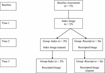
As can be seen in Figure 1, Group-R had the following exposures at the three time points: Time 1: Index; Time 2: Rescript; Time 3: Rescript. Group-I had the following exposures at the three time points: Time 1: Index; Time 2: Index; Time 3: Rescript.
Experimental design showing the Basal assessment and the Index image assessment at Time 1, followed by the division into two groups (G-Index and G-Rescript) with assessments at Time 2 and Time 3

Participants
Participants were recruited from a rehabilitation program of 8–10 weeks at an occupational rehabilitation centre in Vancouver, BC, Canada. The aim of the centre is to reduce physical disabilities and promote a return to work. A large majority of their clients report pain (from acute to chronic) as a major limiting symptom at in-take. Attendees at the Rehabilitation Centre had the following occupations: Trades, Construction and Transportation: 47%; Sales and Services: 31.5%; Health Care: 10.8%; and Other: 1.5%. They are referred predominantly by third party insurance companies. Clients with significant psychological problems are excluded and referred elsewhere to specialized centres (e.g. for alcohol, drug, PTSD, anxiety/depression).
Posters about the research were placed in the centre. Attendees at the Centre were approached by a member of staff to establish their interest in participating in a research study on: “how people think, when in pain”. There were 73 volunteers, of whom 57 (78%) met the study criteria for inclusion: presence of pain, and presence of imagery associated with their pain experience. The exclusionary criteria were inadequacy of English comprehension or brain damage. Two participants dropped out; they refused to form their Index images, and withdrew. All participants had been told at the onset that they were free to withdraw at any time without repercussions.
The exact number of individuals who were told of the study but did not wish to volunteer is not known. The predominant reasons given to rehabilitation centre staff for their failure to volunteer were either inadequate time given their rehabilitation program, or disinterest in participating in any research.
Ethical aspects
The participants were informed that the research study was voluntary and unrelated to their compensation claims or treatment at the Centre. Their contribution would be anonymous. The research aims were explained and each participant signed a consent form agreeing to be interviewed for 30–45 minutes, and to fill in questionnaires. The study was approved by the University of British Columbia Behavioural Psychological Ethics Committee. All participant data were coded and kept confidentially in a locked office.
Procedure
Demographic details, pain characteristics, and Index image characteristics were gathered during a structured interview (available from the first author). Baseline levels of the dependent variables using analogue scales were established at the onset. Self-report assessment scales were then given: Mental Defeat (PSPS), PTSD Symptom Check-list (PCL-C), Anxiety (HAD-A), and Depression (HAD-D).
All participants were asked to describe any images that accompanied their pain experience (see Philips, Reference Philips2011, for details of this method for obtaining image reports). They chose the most distressing/powerful of their images, which became their Index image for this study. Image characteristics (frequency, similarity on each occasion, clarity of the image, image perspective (field/observer), and chronicity of the image occurrence) were determined during the structured interview. Participants were asked a question about Index image meaning merely as an aid to the researcher in encouraging the sensory rescripting process.
Following this, all participants were asked to form their Index images (at Time 1) using the VIE procedure. The VIE procedure was used as in previous research (Philips, Reference Philips2011). After an image description had been detailed and recorded, participants were asked to form in their mind the image they had described, and to keep it in mind as they answered questions about it. This method was used to assess the image potencies (as in the previous study in 2011) at the three time points (T1-T3) separated by 6 minutes distraction. The distraction task involved the interviewer asking the participant to describe where he/she grew up, the area and their memories of it.
After the Time 1 evaluation of their Index image, participants were randomly allocated to one of the two groups: Group-R (Rescript Image) (n = 26) and Group-I (Index Image) (n = 29). The randomization was achieved using a random number table, whereby each subject was given the next number on the table. Those receiving odd numbers were placed in the Index Group, while those receiving even numbers were placed in the Rescript Group.
After the initial T1 exposure to their Index Image, participants in Group-R developed their preferred rescripted version of their Index image. To obtain their Rescripted version they were asked “How would you rather see the image?” or “How would you prefer it to have been?” and encouraged to describe in full what this image would entail. All details were recorded. They were then asked to form the new rescripted image and to evaluate it on the same scales used in the baseline assessment. After a 6-minute distraction, they repeated the rescripted image exposure with analogue scale re-assessments.
In comparison, after a 6-minute distraction, participants in Group-I were asked to form their Index image again (at Time 2) and complete the same analogue scales on the dependent measures. After a further 6-minute distraction, they were requested to develop a rescripted form of their Index image (method described above), and asked to form it and complete the same analogue scales (Time 3).
Data collection
Self report rating scales. The following variables were assessed for each participant: pain, emotions (anxiety, sadness, anger) and appraisals (fragility-physical, and fragility-emotional, health anxiety, and threat). Analogue scales from 0–100 were used on each of these variables, with 100 defined as the maximum level. In the pain-rating scale, 100 was defined as unendurable pain.
Self- report assessment scales. A group of reliable and validated questionnaires was administered to each participant at the onset:
Hospital Anxiety and Depression Scales (HAD; Zigmond and Snaith, Reference Zigmond and Snaith1983). HAD is a 14-item self-report measure of overall severity of anxiety and depression (7 items for each) in non-psychiatric medical settings. Each item was rated from 0–3 thus providing a total for each scale of 0–21. It has been shown to have good internal consistency. Scores of 8 or above for either anxiety or depression have around 80% sensitivity and specificity (Bjelland, Dahl, Haug and Neckelmann, Reference Bjelland, Dahl, Haug and Neckelmann2002).
Pain Self Perception Scale (PSPS; Tang et al., Reference Tang, Salkovskis and Hanna2007). PSPS is a 24-item instrument to assess mental defeat in relation to pain. Respondents are asked to recall a recent episode of intense pain and then rate the extent each item applied on a scale from 0–4 (0 = never/not at all to 4 = very strongly). The summed score ranges from 0–96. This scale has excellent internal consistency and test-retest reliability with chronic pain respondents.
PTSD Checklist (PCL-C: Ruggiero, Del, Scotti and Rabalais, Reference Ruggiero, Del, Scotti and Rabalais2003; Lang and Stein, Reference Lang and Stein2005). The PCL-C (PTSD Checklist) is a short self-report measure of DSM-IV symptoms of Post-Traumatic Stress Disorder. It was selected for its brief administration (2–5 minutes), established sensitivity, specificity and predictive values. It assesses symptom intensity but does not provide a diagnosis of PTSD. Blanchard, Jones-Alexander, Buckley and Forneris (Reference Blanchard, Jones-Alexander, Buckley and Forneris1996) reported that a cut-off criterion of over 44 is most efficient. However, given the overlapping symptomatology of pain and post-traumatic stress disorder on three specific items on this scale (e.g. sleep disturbance, irritability and concentration difficulties), a more conservative cut-off of 50 for pain sufferers suggested by Andrykowski, Cordova, Studts and Miller (Reference Andrykowski, Cordova, Studts and Miller1998) was used in this study as in the earlier study of Philips (Reference Philips2011).
Data analysis
Differences between particular categorical demographic data were assessed using the χ2 statistic. A multivariate analysis of variance (2 × 2 × 4 MANOVA) examined the differences between the two groups (Index vs. Rescript) over the four time intervals, for the subjects classified into acute vs. chronic pain sufferers. The latter was included to assess the extent that chronicity of pain was significantly affecting participant responses.
In addition, a 2 (group) by 4 (time) mixed model analysis of covariance (ANCOVA) was used to examine the differences in pain ratings over time between the two groups, controlling for changes in sadness and anxiety. Inter-correlation matrices assessed the relation between dependent measures at the four time points.
Finally, inter-correlations between cognitive appraisals and mental defeat (PSPS) were assessed for Index and Rescript images occasions, irrespective of group.
Results
Characteristics of sample
The participants were 24 males (43.6%) and 31 females (56.4%).The mean age was 44.5 yrs (SD 11.6).The average chronicity of reported pain was 54.14 weeks (SD 30.7) or 13.5 months. Divided into acute and chronic pain (acute: <24 wks, chronic: >24 weeks) the sample had 58% chronic (n = 31) and 41% acute (n = 22) participants. The average level of reported pain (0–100 with 100 = unendurable pain) was 54.1(SD 21.8).
The groups did not differ significantly in pain chronicity, average pain levels (ANOVA: both p > .005) or funding source and litigation status (χ2: both p > .05). Psychometric assessments provided an indication of the psychological state of the participants on a number of measuring instruments. The mean levels are shown in Table 1 for the Anxiety and Depression (HAD-A and -D), Mental defeat (PSPS), and PTSD symptoms (PCL-C). Anxiety is slightly above the cut-off (8) although this does not reach significance. Depression levels are below the cut-off (8). The two groups did not differ significantly in these measures of state anxiety and depression confirming the Rehabilitation Centre selection of clients without anxiety or depression problems. Support for this comes also with the PTSD symptom Check-list (PCL-C), which indicates results below the cut-off of 50 for pain sufferers on this measure. In fact the groups did not differ significantly and had levels comparable to non-pain sufferers.
Basal levels (Mean and SD) for the two groups on the psychometric scales

Notes: HAD-Depression and HAD-Anxiety = Hospital Anxiety and Depression Scale; PSPS = Pain Self -perception Scale; PCL-C = PTSD Checklist
Mental defeat scores (PSPS) averaged at 43.4 (25.10). When the participants were divided into acute and chronic pain sufferers, the PSPS scores were: 41.6 (25.9) and 44.7 (24.8) respectively. The scores are not significantly different.
Baseline levels of emotions, cognitive appraisals, and pain
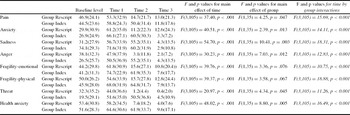
Baseline levels of the dependent variables were assessed to establish the prevailing levels of the emotions, pain and appraisal in this Rehabilitation Centre sample against which the Index and Rescripted image responses could be evaluated. In addition, the comparability of the two randomly collected groups could be assessed. Finally, the base-levels show the prevailing characteristics of this Rehabilitation sample. The basal levels of the key dependent variables are shown in Table 2.
Caption: mean levels for each dependent measure on analogue scales (0–100). MANOVA F and p values for main effects of Time, Group and Time × Group interactions

The mean levels (SD) on each dependent variable for each group at the baseline assessment are shown in column #1. It is evident that participants were experiencing considerable pain, negative emotions, and negative appraisals at the onset of the research interview. Their pain reports matched that reported for their average pain. There were no differences between the groups on any of the dependent variables (all p > .100) at the baseline assessment.
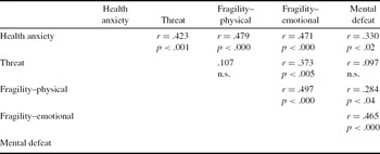
Table 3 shows the inter-correlation matrix of the appraisals: Health anxiety, Threat, Fragility (physical and emotional), and Mental defeat (PSPS) scores at the baseline assessment. The majority of these correlations between the appraisals are significant with sizeable r values. Of particular note is the significant association of Health anxiety with Threat, and with Fragility (emotional and physical). The two types of Fragility were highly correlated. Finally, the psychological construct of Mental defeat was particularly highly related to emotional Fragility but showed low, though significant, relationships with Health anxiety and Physical fragility.
Inter-correlation between cognitive appraisals at onset (baseline assessment): Health anxiety, Threat, Physical fragility, Emotional fragility and Mental defeat (PSPS)

Index image characteristics
The average number of images reported by participants when in pain was 3.2 (1.5). Selecting their Index image (most distressing/powerful), the average image clarity was 84.0 (SD 19.8). The frequency of repeated image intrusions was 43% (a few times/day), and 41.8% (a few times/week). Thus, 85% of participants reported having frequent, clear distressing images when in pain. They estimated the similarity of the Index image on each occurrence as 84/100 (where 100 = exactly the same). The perspective was 56.4% Field (through the participants’ own eyes) and 40% Observer (as seen by another).
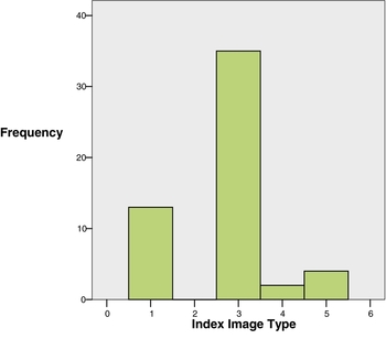
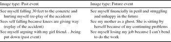
The Index images were classified by participants into five possible categories with respect to content type. The histogram shows their responses (Figure 2). The majority of the Index images were of an experienced event (n = 35; 63.6%), or of a future predicted event (n = 13; 23.6%). Only 2 participants (3.6%) selected the current event category. Although 5% (n = 3) reported images of the pain itself, only 2 selected these as their Index image. Examples of the Index image content of the two most common types of image are provided in Table 4.
Histogram showing the proportion of participants confirming Index image content categories: 1 = Future event, 2 = Imagined past event, 3 = Past event, 4 = Current event, 5 = Melded image of above categories

Examples of the two major types of Index images defined by content categories: past event and future event

Rescripted pain image
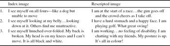
Examples of Index images with the rescripted alternative given by three of the participants “as (he/she) would have preferred the image to be” are found in Table 5. The Index images were reported to occur repeatedly and spontaneously. On the other hand, the Rescripted versions of the Index images are those that the participants would prefer to visualize. The chosen or preferred images were tailored by the participant.
Examples of Index images with the Rescripted alternative that the participant developed “as she/he would prefer it to be”

The time taken to develop the rescripted alternative to their distressing Index images was fast and appeared pleasurable to the participants. Importantly, not a single participant had any problem rescripting an alternative image. Occasionally, they would return to their description and add details to improve it further, once they recognized their opportunity to fine-tune the image. No discussions of image meanings were undertaken and only sensory changes were requested.
Using a Manova (2×2×4), the level of pain persistence (acute vs. chronic) of the two groups was compared across the four time points: Baseline (B); Index image (T1); Index or Rescript Image-dependant on group (T2); and Rescript image (T3). The VIE procedure allowed assessment of the eight dependent measures (of pain, emotions and appraisals) at each assessment point (B-T3). A comparable pattern is seen in all the variables. Specifically, multivariate tests revealed significant effects of time, F(24,12) = 15.81, p < .001, and a time by group interaction, F(24,12) = 7.56, p < .001. There were no significant effects of chronicity, F(8,28) = 1.71, p = .14, nor were there significant time by chronicity, F(24,12) = 0.68, p = 0.80, or time by group by chronicity, F(24,12) = 1.61, p = 0.20 interactions.
In Table 2, the follow-up univariate tests, along with mean responses on each of the variables at the four time points, and the group × time interactions are shown. All variables were found to be significantly different between the groups (all p = <.001). The Rescript group showed significant response elevation to the Index image on 5 of the 8 dependent variables. The exceptions were: Pain, Fragility-physical, and Health anxiety. Increases occurred but did not reach significance due to slightly higher onset levels in this group on these three measures. In the Index group, simple effect tests confirmed that all Index image responses were significantly elevated when compared to the baseline levels.
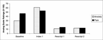
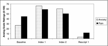
When Index and Rescripted image responses are compared, significant and immediate reductions occurred with the rescripted image on all measures. In Figure 3a and 3b, the sharp decrement in Pain and Anxiety levels during the Rescripted images is illustrated for each group. A comparable pattern is present for all the dependent measures.
Rescript group levels of anxiety and of pain on analogue scales (0–100) at each assessment time point: Baseline, Index 1, Rescript 1 and Rescript 2

Index group levels of anxiety and of pain on analogue scales (0–100) at each assessment time point: Baseline, Index 1, Index 2 and Rescript 1

During exposure to the Rescripted image, mean levels of pain (shown in Table 2) dropped to significantly low levels both statistically and clinically. The levels reached in both groups, while viewing their Rescripted images, are significantly lower than the level reported during basal assessment. Remarkably, 49% of participants reported 0 levels of pain while viewing their Rescripted image. (In contrast, during the Index image, only 11% of participants reported 0 levels of pain). These results highlight the power of rescripting procedures to change both the psychological reactions to distressing images as well as the level of pain experienced.
Image repetition-index and rescript
To control for the possibility that image repetition alone would itself result in decrements in potencies, the Rescript image in Group-R, and the Index images in Group-I were repeated. Comparing the responses on each of the variables, no significant difference was evident when the image repetitions (Index or Rescript) were compared (all p > .05). The stability of both Rescript and Index image responses was clear, providing evidence of no significant change in reaction with image repetition. Group-I acted as a control group for the effects of image repetition. It is evident that significant changes with rescripting cannot be explained as a function of image repetition (see Tables 6a and 6b).
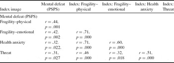
Inter-correlations between cognitive appraisals and Mental defeat during exposure to Index images

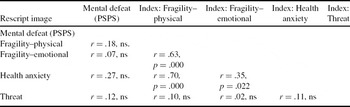
Inter-correlations between the cognitive appraisals and Mental defeat during exposure to Rescripted images

However, the inter-correlations between the cognitive appraisals did change with image repetition. For the Group-I group, the correlations between appraisals become stronger and more inclusive with image repetition. On the first exposure to the Index image, correlations were between r = .42 and .46, with a non-significant correlation between Mental defeat and Threat. On the second occasion when the Index image was viewed, Mental defeat and the four appraisals (Fragility emotional and physical, Threat, and Health anxiety) were all correlated significantly, with rs between .52 and .58.
This contrasts markedly with the lack of any significant correlations between any of the cognitive appraisals with the Rescripted image, on both occasions (Time 2 and Time 3).
Cognitive appraisals and image type
The appraisals of participants were also assessed when participants were viewing their Index and Rescripted images, irrespective of group allocation. With the Index image exposure (Time1), all the correlations between the cognitive measures (appraisals and Mental defeat scores) were significant. With the Rescripted image exposure, the correlations between the appraisals were predominantly low and insignificant. Mental defeat (PSPS) became unrelated to any of the cognitive appraisals during rescripting.
Discussion
Effects of rescripting of pain images
When the participants first formed their Index image, they experienced negative emotions and increased pain. However, after rescripting their images, they experienced substantial reductions in the negative emotions, pain and appraisals. Remarkably, 49% (of the 55 participants) reported no pain at all during the rescript. In the control group (G-I), repetition of the Index image was not accompanied by any of these reductions. All participants found the rescripting procedure easy to carry out, and pleasurable. The large majority had little hesitation in elaborating how they would prefer to see their Index images. A few needed some encouragement/suggestions, but no discussions of image meanings were undertaken. Only sensory details were suggested. A few participants resorted to magical realism ideas when they were unable to decide on a preferred sensory alternative.
The effects of the rescripting upon the measures of emotion, appraisal and pain experience were statistically significant and clinically substantial. The changes produced did not merely replicate the reduction in emotions reported in studies of other disorders using rescripting (Hackmann et al., Reference Hackmann, Ehlers, Speckens and Clark2004; Brewin et al., Reference Brewin, Wheatley, Patel, Fearon, Hackmann and Wells2009; Clark and Wells, Reference Clark, Wells, Heimberg, Liebowitz, Hope and Schneier1995; Ehlers et al., Reference Ehlers, Maercker and Boos2000) but also revealed a significant change in pain levels. The latter were shown to be independent of the significant reductions of anxiety and sadness. Thus a pain experience was demonstrated to be reduced by a relatively simple manipulation of a cognitive variable: image content. The pain decrements were fast, easily produced and dramatically large. These results are consistent with cognitive models of pain (i.e. Melzack and Wall, Reference Melzack and Wall1965; Turk and Rudy, Reference Turk and Rudy1992) that emphasize the power of the descending influences on pain experience and demonstrate central influences on pain experience.
It remains possible that the decline in pain levels is due to the distracting effects of the rescripted image. However, this seems unlikely to be the complete or predominant explanation. No declines in pain occurred with the Index image viewing on the first occasion or on the second repetition of the image. It could be argued that the potency of the Index image would act as a powerful distracter, but the pain levels rose significantly on both occasions when the Index images were formed. In addition, repetition of the rescripted image resulted in no return of pain even though, on the second occasion, the image would be familiar and less distracting.
The effects of distraction are characteristically fleeting. Distraction, unlike rescripting, is not revealing, does not lead to core cognitions, nor would it result in changes in appraisal.
The mechanism by which pain is reduced by rescripting of pain images is likely to be dependent upon the influence of cognitions upon pain levels. This is consistent with the writing of Melzack and Wall (Reference Melzack and Wall1965) who postulated this effect, and the subsequent research confirming their hypothesis (Turk and Rudy, Reference Turk and Rudy1992). The type of cognitions likely to be important in rescripting of pain images is both the positive image content (with its associated memories), and the sufferers’ appraisal or interpretation of the rescripted image.
This is reassuringly consistent with the major changes in clinical psychology that have produced an infusion of ideas about the influence of cognitive factors on psychological processes and problems (Clark, Reference Clark1986; Salkovskis, Reference Salkovskis1985). In addition, there is a growing literature on the potency of images upon emotions and appraisals (Hackmann et al., Reference Hackmann, Bennett-Levy and Holmes2011; Holmes and Mathews, Reference Holmes and Mathews2010). Finally the results of this study of unwanted imagery are consistent with findings on unwanted intrusions in obsessions (Rachman, Reference Rachman2007; Whittal, Woody, McLean and Robichaud, Reference Whittal, Woody, McLean and Robichaud2010). The mechanism operating in the rescripting of pain images needs further investigation.
Types of rescripting: sensory and cognitive appraisals
Rescripting procedures entail both sensory and cognitive changes in the Index images (Brewin et al., Reference Brewin, Wheatley, Patel, Fearon, Hackmann and Wells2009). The rescripting undertaken in this experiment was intentionally limited to sensory rescripting. Participants changed the sensory qualities of their Index images to make them “as they would prefer them to be”. The procedure avoided any attempt to influence or shift participants’ understanding of the meaning of their images in order to focus exclusively on the effects of sensory changes. It is comparable in this respect to the rescripting used in the study of the treatment of depression by Brewin et al. (Reference Brewin, Wheatley, Patel, Fearon, Hackmann and Wells2009). However, in the current experiment there was only one session for each participant.
It seems likely that a rescripting process may occur spontaneously in some pain sufferers. This possibility has not been investigated but positive coping images have been reported in pain sufferers (Berna et al., Reference Berna, Vincent, Moore, Tracey, Goodwin and Holmes2011; Philips, Reference Philips2011). Many years ago, the famous Soviet neuro-psychologist A. R. Luria (1902–1977) described a person (S) who reported a unique method to reduce his pain (quoted in Luria and Brunner, Reference Luria and Bruner1987). His description is an excellent example of naturally occurring sensory rescripting.
Let's say I am going to the dentist. I sit there and when the pain starts, I feel it. . . .It's a tiny orange red when the pain starts, I feel it. . . .It's a tiny orange red thread. I'm upset because I know that it turns into a dense mass. So I cut the thread, make it smaller and smaller until it's just a tiny pain. And the pain disappears.
This is a sensory rescripting similar to those evolved by participants in this study. The description includes many sensory details of just how the image appeared to S and what he changed to reduce/eliminate the pain. The frequency of this type of naturally occurring rescripting remains to be studied in pain sufferers.
Although the immediate effects of rescripting were robust and substantial, the long-term effects were not assessed in the present study. Brewin et al. (Reference Brewin, Wheatley, Patel, Fearon, Hackmann and Wells2009) have suggested that the most potent and enduring changes will follow rescripting that includes both sensory and cognitive re-appraisal of an Index image. With such changes, Brewin argues that the retrieval of the rescripted image will be primed and facilitated. When in pain, the new alternative image will be retrieved more readily and the spontaneous Index image weakened. Research on the longer term effects of sensory rescripting alone, and on the utility of adding changed cognitive appraisals, is necessary.
Stability of responses to imagery
Stability in the effects of both Index and Rescripted images was observed in this study. The interval between repetitions was short (6 minutes) but under these conditions there was no evidence of the potency fading, growing or changing. There is no suggestion of response decrements or increments with image repetition, despite image clarity remaining the same. In other words, there was no evidence of the reduction of potency merely by repeating the image. The significant decline in the potency of the rescripted image cannot be explained as a function merely of image repetition, or by a loss of image clarity.
The repetitive distressing effects of the Index image support the reports of pain sufferers that their intrusive images return with a high degree of similarity (Philips, Reference Philips2011). Image repetition does not lead to image disappearance even in chronic sufferers. Paralleling this, the positive potency of the rescripted image showed no sign of fading (after two repetitions).
Imagery and its effect on cognitive appraisals and mental defeat
An intriguing exception to the stability of both the Index and Rescripted responses was found in the inter-correlations of the appraisal dependent variables and Mental defeat (PSPS). The inter-correlation of participant appraisals changed with Index image repetitions. An evident tightening of the correlations between cognitive appraisals occurred, despite the fact that image potencies (emotions, pain and appraisals) remained the same. Although these preliminary data need replication and extension, they suggest that repetition of the distressing image increased its toxicity. In addition, Mental defeat became more strongly associated with the negative appraisals. This may provide initial data on the way in which Mental defeat develops in pain sufferers with Index imagery, and how this relationship is strengthened by other appraisals, common to those with disabling pain syndromes.
Implications of rescripting potency for CBT approaches to pain management
It is widely acknowledged that cognitions play an important role in pain experience (Melzack and Wall, Reference Melzack and Wall1965; Turk and Rudy, Reference Turk and Rudy1992). Cognitive-Behavioural treatment of pain has focused upon modifying catastrophic thoughts, predictions and beliefs (Morley, Eccleston and Williams, Reference Morley, Eccleston and Williams1999; Eccleston, Crombez, Aldrich and Stannard, Reference Eccleston, Crombez, Aldrich and Stannard1997; Sullivan, Bishop and Pivik, Reference Sullivan, Bishop and Pavik1995; Aldrich, Eccleston and Crombez, Reference Aldrich, Eccelston and Crombez2000). However insufficient attention has been paid to imagery in pain assessment and treatment. Discussions of the meanings of images may fast track the clarification of core beliefs. The appraisals of images are likely to be useful focuses of cognitive re-appraisal work during treatment. Most pain sufferers are unaware of the presence or power of their images, and are surprised by their emotional reactions (Philips, Reference Philips2011). The two drop-outs in this study were both due to an expressed desire to avoid further provoking the emotional reactions accompanying their index images.
During treatment, the use of the Voluntary Image Exposure procedure allows quantification of treatment changes. In addition, it could be a vehicle of change if rescripting is confirmed to be easy, repeatable, generalizable and powerful at affecting pain levels. The use of rescripting techniques to prevent pain-disrupting activities (physiotherapy, household tasks, work) will be a useful addition to treatment. Staff at the Rehabilitation Center requested that all participants receive rescripting instructions during their research session. The reason for this request was that they had observed that those having rescripting (during the pilot stage) returned to the Rehab gym more cheerfully and worked better at their rehabilitation than those having the Index image exposure only.
In addition, exploring the way in which the appraisals of Fragility and Mental defeat can be modified by image rescripting may prove extremely useful in reducing avoidance, safety behaviour, low self-esteem, and depression.
Limitations and implications for further research
A limitation of this study was the exclusive focus on sensory rescripting without assessing the meaning that the participants attached to their Index images. Now that significant and sizeable changes have been found with sensory rescripting alone, research on the re-appraisal of Index images and the effect of rescripting on image appraisal will be of interest. It is likely that combining cognitive re-appraisal with sensory rescripting will lead to even stronger results. It is predicted that the changes in potency of the Index image will be more persistent, leading to spontaneous replacement of Index images with benign alternatives. Positive changes in appraisal may act as a fixing agent in consolidating long term effects.
The present experiment did not include the collection of data on the short-term (7–10 days) effects of the rescripting. It would be interesting to know the reactions of the participants when they considered the comparative effect of their Index image, and their new preferred rescripted version. In addition, the durability of the effects of the rescripting needs to be clarified. A number of weekly sessions are likely to improve persistence and enhance the effects of the rescripted image. This research would also help distinguish rescripting from distraction. The latter would be predicted to have no lasting effects. Encouraging post-session practice of the rescripted benign alternative is likely to consolidate treatment progress, and reduce the frequency of distressing images.
As the experiment was undertaken in one session, participants made only one attempt at developing their preferred image. In a treatment context, and with cognitive reappraisal, it seems likely that participants would progressively refine their preferred image. A hint of this process occurred in this study. Having completed a rescripted image description and evaluated its effects on the analogue scales, one participant wanted to make an addition to his image. This illustrates that relatively small changes can have large effects on image potencies.
Index image: I see myself all alone . . .at home . . .unable to do anything. I am just watching the television.
Initial rescript: I see myself returning to my job. I'm not limping. I look strong and well. . .I am smiling and happy.
Addition: All my co-workers and pals are coming round me. My friend puts his arm around me to welcome me back.
Remarkable reductions in anxiety and sadness resulted from this addition. Meanings of the Index images were not being assessed, but from this potent addition one would predict that social isolation and exclusion consequent upon his injury were important aspects of his Index image. This had not been evident in the Index image description, in which co-workers were absent from the image.
A longitudinal evaluation of pain sufferers (acute to chronic stage) would provide useful information on the evolution of Index imagery as well as the incidence of spontaneously developing rescripted images. A number of participants reported higher frequency/more potent Index image at the onset (e.g. PTSD images). These had gradually reduced in the months that followed an accident. Others reported Index image onset associated with their doctor telling them they would “just have to live with it”, and referring them to a rehabilitation center.
As most of the participants reported more than one image (mean: 3.2 images), only their most powerful/distressing image was selected for this study. The generalizability of an established rescripting procedures (spontaneous and instructed) on other intrusive distressing images is worthy of investigation.
The voluntary image exposure procedure (VIE) used in this experiment can be developed into a useful assessment/treatment procedure. It allows evaluation of changing image potencies, and can be the basis for comparing the effects of different images and of image rescripting. Cognitive therapy focusing on image meanings and associated appraisals (re Fragility, Threat, and Health anxiety) would flow smoothly from this approach. In this research study, a number of participants remarked upon the ease with which they could alter the emotional impact of their pain and the pain itself by merely switching “how they viewed it”.
It is clear that in pain sufferers, spontaneous images are common, distressing and increase pain. However, simple rescripting was easily accomplished, fast and dramatically effective. Enhancing and prolonging the effects of rescripting will be a valuable research task that is likely to lead to useful therapeutic methods.
Acknowledgements
We are grateful for the support of the staff of the Rehabilitation Clinic, and all the attendees who volunteered to talk to me about their images. In addition, in the early days of planning this study, conversations with P. Salkovskis, C.R. Brewin, and A. Hackmann helped clarify the direction of the study. Drs Michael Papsdorf and A. Radomsky provided valuable help with the statistics and Dr S. Rachman provided useful comments on the manuscript.




Comments
No Comments have been published for this article.