Introduction
Bats (order Chiroptera) are one of the most diverse mammals and the only mammal that has developed the ability to fly. Albeit with differences in terms of roosting preferences and behaviours, bats can be found on almost every continent (Taylor et al., Reference Taylor, Clinton and Tuttle2019; Simmons and Cirranello, Reference Simmons and Cirranello2020). These animals play critical roles in the ecosystem by benefiting the forest and agricultural sectors (Kunz et al., Reference Kunz, Braun de Torrez, Bauer, Lobova and Fleming2011; Chen et al., Reference Chen, Shen, Lee, Wu, Zeng, Lu and Lin2017). However, they are also known to be a reservoir for a variety of infectious agents, such as Hendra virus (Murray et al., Reference Murray, Selleck, Hooper, Hyatt, Gould, Gleeson, Westbury, Hiley, Selvey, Rodwell and Ketterer1995), Nipah virus (Yob et al., Reference Yob, Field, Rashdi, Morrissy, van der Heide, Rota, Bin Adzhar, White, Daniels, Jamaluddin and Ksiazek2001), rabies virus and coronaviruses (Zhou et al., Reference Zhou, Yang, Wang, Hu, Zhang, Zhang, Si, Zhu, Li, Huang, Chen, Chen, Luo, Guo, Jiang, Liu, Chen, Shen, Wang, Zheng, Zhao, Chen, Deng, Liu, Yan, Zhan, Wang, Xiao and Shi2020; Bergner et al., Reference Bergner, Mollentze, Orton, Tello, Broos, Biek and Streicker2021b), Bartonella (Poofery et al., Reference Poofery, Narapakdeesakul, Riana, Arnuphapprasert, Nugraheni, Ngamprasertwong, Wangthongchaicharoen, Soisook, Bhodhibundit and Kaewthamasorn2021), Candidatus Mycoplasma haemohominis (Descloux et al., Reference Descloux, Mediannikov, Gourinat, Colot, Chauvet, Mermoud, Desoutter, Cazorla, Klement-Frutos, Antonini, Levasseur, Bossi, Davoust, Merlet, Goujart, Oedin, Brescia, Laumond, Fournier and Raoult2021), Rickettsia, Borrelia (Loftis et al., Reference Loftis, Gill, Schriefer, Levin, Eremeeva, Gilchrist and Dasch2005), Leptospira (Bevans et al., Reference Bevans, Fitzpatrick, Stone, Butler, Smith and Cheetham2020) and trypanosomes (Ramírez et al., Reference Ramírez, Hernández, Montilla, Zambrano, Flórez, Parra and Cucunubá2014). Furthermore, certain behaviours of bat species, such as roost switching, colony living and seasonal migration, have increased the possibility of infectious agent spill over to other mammals (Calisher et al., Reference Calisher, Childs, Field, Holmes and Schountz2006; Fan et al., Reference Fan, Zhao, Shi and Zhou2019).
Trypanosomes (order Trypanosomatida) are vector-borne parasitic protozoans that can be found in the blood and tissue of vertebrates (Hoare, Reference Hoare, Henle, Kikuth, Meyer, Nauck and Tomcsik1966; Lemos et al., Reference Lemos, Fermino, Simas-Rodrigues, Hoffmann, Silva, Camargo, Teixeira and Souto-Padrón2015; Ortiz-Baez et al., Reference Ortiz-Baez, Cousins, Eden, Chang, Harvey, Pettersson, Carver, Polkinghorne, Šlapeta, Rose and Holmes2020). Trypanosomes are uniflagellated protozoa that, similar to other organisms in the order Trypanosomatida, are distinguished by the kinetoplast, a modified mitochondrial genome. Despite the fact that several species can cause severe diseases in humans and animals, as well as economic losses (Ponte-Sucre, Reference Ponte-Sucre2016), these blood parasites have been largely neglected, especially those of wildlife origin. Recent studies have revealed the diversity and global distribution of trypanosomes in bats in America (Dos Santos et al., Reference Dos Santos, Lisboa, Xavier, Dario, Verde, Calouro, Roque and Jansen2018; Barros et al., Reference Barros, Lima, Schubach and Teixeira2019; Bergner et al., Reference Bergner, Becker, Tello, Carrera and Streicker2021a; Torres-Castro et al., Reference Torres-Castro, Cuevas-Koh, Hernández-Betancourt, Noh-Pech, Estrella, Herrera-Flores, Panti-May, Waleckx, Sosa-Escalante and Peláez-Sánchez2021), Africa (Lima et al., Reference Lima, Silva, Neves, Attias, Takata, Campaner, de Souza, Hamilton and Teixeira2012; Clément et al., Reference Clément, Dietrich, Markotter, Fasel, Monadjem, López-Baucells, Scaravelli, Théou, Pigeault, Ruedi and Christe2020), Europe (Baker, Reference Baker1973; Hamilton et al., Reference Hamilton, Cruickshank, Stevens, Teixeira and Mathews2012a), Asia (Mafie et al., Reference Mafie, Rupa, Takano, Suzuki, Maeda and Sato2018; Cai et al., Reference Cai, Wang, Zhang, Li, Gong, He and Zhang2019; Wang et al., Reference Wang, Han, Zhao, Liu, Luo, Wen, Qin, Zhou, Qi, Yu and Yu2019) and Australia (Barbosa et al., Reference Barbosa, Mackie, Stenner, Gillett, Irwin and Ryan2016; Austen et al., Reference Austen, Van Kampen, Egan, O'Dea, Jackson, Ryan, Irwin and Prada2020). To date, more than 30 trypanosome species have been identified from various bat species, with the majority of them being found in insectivorous bats (Molyneux, Reference Molyneux, Kreier and Baker1991; Cavazzana et al., Reference Cavazzana, Marcili, Lima, da Silva, Junqueira, Veludo, Viola, Campaner, Nunes, Paiva, Coura, Camargo and Teixeira2010; Lima et al., Reference Lima, Silva, Neves, Attias, Takata, Campaner, de Souza, Hamilton and Teixeira2012). Interestingly, phylogenetic analyses have led to the hypothesis that bats are ancestral hosts of Trypanosoma cruzi (Hamilton et al., Reference Hamilton, Cruickshank, Stevens, Teixeira and Mathews2012a, Reference Hamilton, Teixeira and Stevens2012b).
Trypanosoma requires a vector to complete its lifecycle (Esch and Petersen, Reference Esch and Petersen2013). Various haematophagous arthropods such as tsetse flies and reduviid bugs are common vectors for human trypanosomes (Bogitsh et al., Reference Bogitsh, Carter, Oeltmann, Bogitsh, Carter and Oeltmann2019). Some of the potential vectors for trypanosomes that have been reported before include Cavernicola pilosa (Marinkelle, Reference Marinkelle1982), Cimex pipistrelle, Cimex lectularius, sandflies (Phumee et al., Reference Phumee, Tawatsin, Thavara, Pengsakul, Thammapalo, Depaquit, Gay and Siriyasatien2017; Srisuton et al., Reference Srisuton, Phumee, Sunantaraporn, Boonserm, Sor-Suwan, Brownell, Pengsakul and Siriyasatien2019) and bat flies (Szentiványi et al., Reference Szentiványi, Markotter, Dietrich, Clément, Ançay, Brun, Genzoni, Kearney, Seamark, Estók, Christe and Glaizot2020). Determination of the potential vectors of other bat trypanosomes is crucial to understand their transmission and to control the disease.
Despite growing knowledge and the rapid development of diagnostic methods for identifying new pathogens, several of the bat hosts that can be infected by trypanosomes, as well as their geographic distribution, remain largely unknown. Surveillance of these parasites in Asia has mainly been conducted in China, Taiwan and Japan (Mafie et al., Reference Mafie, Rupa, Takano, Suzuki, Maeda and Sato2018; Cai et al., Reference Cai, Wang, Zhang, Li, Gong, He and Zhang2019; Wang et al., Reference Wang, Han, Zhao, Liu, Luo, Wen, Qin, Zhou, Qi, Yu and Yu2019; Clément et al., Reference Clément, Dietrich, Markotter, Fasel, Monadjem, López-Baucells, Scaravelli, Théou, Pigeault, Ruedi and Christe2020). These studies have highlighted the variety of trypanosomes found in bats from various regions, as well as the importance of bats as a natural reservoir. However, there is inadequate information concerning the occurrence and distribution of trypanosomes in Thai bats, which account for 37% of bat species found in southeast Asia (Simmons and Cirranello, Reference Simmons and Cirranello2020). In the current study, the authors explore the diversity of trypanosomes, including their bat host ranges, geographical distribution and phylogenetic relationships, to acquire a better understanding of trypanosomes in 23 bat species in Thailand.
Materials and methods
Sample collection
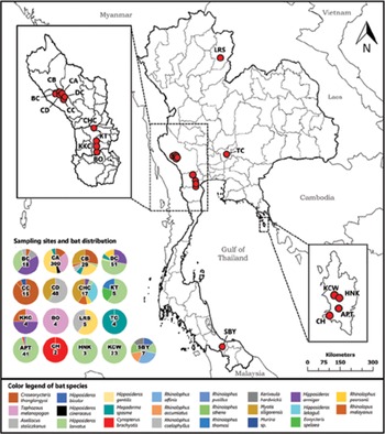
Multiple surveys were carried out from February 2018 to December 2020. Bats were captured across 17 sampling sites located in Kanchanaburi, Songkhla, Saraburi, Trat, Phetchaburi, Ratchaburi and Nan provinces (Fig. 1 and Table S1). The sampling sites in the current study were chosen according to arrangement and collaboration with the local authorities from the Department of National Parks, Wildlife and Plant Conservation. Bats were caught using harp traps and tunnel nets set up in front of cave entrances. Inside the caves, bats were caught using a hand net. Where possible, harp traps and mist nets were set over forest trails and streams to capture commuting bats. Captured bats were transferred individually from the trap to the clothing bag prior to identification and sample collection. Identification of each individual was performed based on the morphological keys in a field guide to the mammals of southeast Asia (Francis, Reference Francis2008) and with echolocation calls if necessary. Standard measurements such as sex, reproductive status, forearm length and body weight were documented to confirm species identification. Blood was collected using 21–27 G needles pricking the uropatagial vein (interfemoral membrane) and transferred into acid citrate dextrose tubes (BD Franklin Lakes, USA) via capillary tubes. An average of 5–20 μL of blood was drawn from bats weighing more than 2.5 g. Samples were transported to a laboratory and maintained at −20°C until further processing. Bats were released alive at their captured sites immediately after blood collection.
Sampling sites in the current study. Each sampling site's letter abbreviation is as follows: Bo cave (BO), Bat cave (BC), Chompon cave (CHC), Daowadung cave (DC), cave A (CA), cave B (CB), cave C (CC), cave D (CD), Tiger cave (TC), Khun Kunchon cave (KKC), Kunchorn temple (KT), Saba Yoi (SBY), Ao Phrao temple (APT), Khlong Chao waterfall (KCW), Huang Nam Khiao waterfall (HNK), Cham's house (CH) and Lainan Research Station (LRS).

Microscopic examination
The remaining blood from the capillary tube was used to prepare a thin blood film onsite, which was air-dried, stored and transported to the laboratory. Slides were fixed with 100% methanol for 3 min and stained with a 10% (v/v) Giemsa solution (Merck, Germany) in phosphate buffer (pH 7.2) for 30–45 min. Screening of blood films was carried out at high magnification (1000×) with oil immersion using a light microscope (Olympus CX31, Tokyo, Japan). The examination was carried out twice to avoid false-negative results. Images were taken using an Olympus DP21-SAL digital camera (Tokyo, Japan). Total body length and free flagellum length of each trypomastigote observed in the blood films were measured using ImageJ v1.53a (Schneider et al., Reference Schneider, Rasband and Eliceiri2012). Mean, standard error and range were calculated according to the results using SPSS v22.0.
DNA extraction
Genomic DNA was extracted from the blood using a NucleoSpin® blood kit (Macherey-Nagel, Düren, Germany) following the manufacture's protocol. The protocol was modified in the final elution buffer which was reduced from 50 to 25 μL (Arnuphapprasert et al., Reference Arnuphapprasert, Riana, Ngamprasertwong, Wangthongchaicharoen, Soisook, Thanee, Bhodhibundit and Kaewthamasorn2020). Genomic DNA was stored at −20°C until polymerase chain reaction (PCR) amplification.
18S rRNA gene amplification and sequencing
Detection of trypanosomes was carried out using nested-PCR amplification. PCR amplification was carried out by targeting approximately 560 bp of the 18S rRNA gene following the primers and protocols previously described by Noyes et al. (Reference Noyes, Stevens, Teixeira, Phelan and Holz1999, Reference Noyes, Stevens, Teixeira, Phelan and Holz2000) and Cai et al. (Reference Cai, Wang, Zhang, Li, Gong, He and Zhang2019). The first-round PCR was performed using the primers TRY927F and TRY927R (Noyes et al., Reference Noyes, Stevens, Teixeira, Phelan and Holz1999, Reference Noyes, Stevens, Teixeira, Phelan and Holz2000) in a total volume of 12.5 μL containing 1× PCR buffer KOD FX Neo, 0.4 mm dNTPs, 0.3 μ m each primer, 0.25 units KOD FX Neo DNA polymerase (Toyobo, Japan), 1.75 μL sterile distilled water and 1 μL of genomic DNA as a template. The primers SSU561F and SSU561R were used for the secondary PCR (Cai et al., Reference Cai, Wang, Zhang, Li, Gong, He and Zhang2019). The undiluted PCR product from the first round was used as a template in the second amplification with a total volume of 12.5 μL reaction mixture, which consisted of 7.5625 μL sterile distilled water, 1× Ex Taq buffer, 0.2 mm dNTP mixture, 0.25 μ m each primer, 0.3125 units of Takara Ex Taq (Takara Bio, USA) and 2 μL first-round PCR product.
During all amplifications, the genomic DNA of T. cruzi and sterile distilled water were used as positive and negative controls, respectively. PCR amplifications were performed in Axygen® MaxyGene II thermal cycler (Life Sciences, USA). Secondary PCR products were analysed in 1.5% agarose gel stained with RedSafe™ (Intron Biotechnology, South Korea). Gel electrophoresis was set at 100 V for 40 min and visualized in a UV transilluminator. First-round PCR products of positive samples from different bat hosts were chosen as representatives and reamplified with primers SSU561F/R. To ensure that there was enough DNA for sequencing, the PCR mixture was re-prepared in a larger volume (50 μL). The PCR products with non-specific bands were purified using NucleoSpin® gel and PCR cleanup (Macherey-Nagel, Germany) kits. Gel purification was performed following the manufacturer's instructions and, in the final step, the PCR products were eluted with 25 μL elution buffer, while PCR products with a single band were treated with ExoSAP-IT™ (USB Corporation, USA) following the manufacturer's protocol. The concentration and quality of the purified PCR products were measured using spectrophotometry analysis (NanoDrop, Thermo ScientificTM, USA). The purified PCR products were sent to Pacific Science (http://www.pacificscience.co.th/) for sequencing using the same set of primers as the second amplification (SSU561F/R).
Glycosomal glyceraldehyde-3-phosphate dehydrogenase (gGAPDH) gene amplification and sequencing
The gGAPDH gene was amplified in semi-nested PCR with primers GAPDHF/GAPDHR (McInnes et al., Reference McInnes, Gillett, Ryan, Austen, Campbell, Hanger and Reid2009) and GAPDHF/g4a (Hamilton et al., Reference Hamilton, Stevens, Gaunt, Gidley and Gibson2004) for the first and second rounds, respectively. First-round PCR was performed using KOD FX Neo (Toyobo, Japan) with PCR as described above using 1 μL genomic DNA as a template. The PCR conditions consisted of an initial denaturing condition for 2 min at 94°C, followed by 40 cycles of 10 s at 98°C, 30 s at 55°C and 30 s at 68°C. A final extension was done for 5 min at 68°C. Non-diluted PCR products from the first round were used as a template in the second-round PCR using Takara Ex Taq (Takara Bio, USA) with an initial denaturing condition of 4 min at 94°C. This was followed by 40 cycles of 30 s at 94°C, 30 s at 57°C, 1 min at 68°C and a final extension of 5 min at 69°C. All amplifications were performed in a MiniAmp™ thermal cycler (Thermo Fisher Scientific, USA). Samples that failed to amplify with the semi-nested PCR primer sets were retested with primers G3/G5 in the first round and G1a/G4b in the second round (Hamilton et al., Reference Hamilton, Stevens, Gaunt, Gidley and Gibson2004). All positive samples were re-amplified with either GAPDHF/g4a or G1a/G4b in preparation for sequencing. DNA sequencing was achieved as described previously.
Sequence analyses
The sequence results of partial 18S rRNA and gGAPDH genes were analysed and edited manually where necessary based on chromatograms obtained from both directions, and aligned using BioEdit v7.0.5.3 (Hall, Reference Hall1999). The edited sequences were subjected to BLAST analysis using BLASTn (https://blast.ncbi.nlm.nih.gov/Blast.cgi) against the nucleotide collection database to identify the most similar species. Our analyses included sequences from the current study (Table S2) and a representative of known bat Trypanosoma species from the GenBank database (Table S3). Trypanosoma lewisi (AJ009156 and AJ223566) was used as the out-group species. The 18S rRNA and gGAPDH genes were concatenated into one data matrix in MEGA X (Kumar et al., Reference Kumar, Stecher, Li, Knyaz and Tamura2018). Final datasets of 1478 bp (568 bp of 18S rRNA; 820 bp of gGAPDH) were used to construct phylogenetic trees using both maximum likelihood (ML) and Bayesian inference (BI). The dataset was partitioned into six data blocks, and the best partition schemes and substitution models were determined using PartitionFinder v2 (Lanfear et al., Reference Lanfear, Calcott, Ho and Guindon2012, Reference Lanfear, Frandsen, Wright, Senfeld and Calcott2017).
The GTR + G + I was chosen as the best model based on the lowest Bayesian information criterion score in each partition. Phylogenetic tree by ML was analysed in the IQ TREE v1.6-12 (Nguyen et al., Reference Nguyen, Schmidt, von Haeseler and Minh2015; Chernomor et al., Reference Chernomor, von Haeseler and Minh2016) using 1000 bootstrap (BS) replicates. For the BI analysis, the data implemented were in MrBayes v3.2.7 (Huelsenbeck and Ronquist, Reference Huelsenbeck and Ronquist2001; Ronquist and Huelsenbeck, Reference Ronquist and Huelsenbeck2003) using the Markov chain Monte Carlo method. Two independent runs of 10 million generations were used with four chains (heated = 3, cold = 1, temperature = 0.01). The first 25% of trees were discarded as burn-in. Convergence of runs and mixing and effective sample sizes (>200) were evaluated using Tracer v1.6 (Rambaut et al., Reference Rambaut, Drummond, Xie, Baele and Suchard2018). Phylogenetic trees were visualized in FigTree v1.4.3 (available at http://tree.bio.ed.ac.uk/software/figtree/). Bootstrap support (BS) and posterior probability (PP) were indicated at the major nodes when the values were more than 50% and 0.5, respectively.
The genetic diversity of the sequences from the current study and references was assessed in DNaSP v6.12.03 (Rozas et al., Reference Rozas, Ferrer-Mata, Sánchez-DelBarrio, Guirao-Rico, Librado, Ramos-Onsins and Sánchez-Gracia2017). Haplotype networks using the 18S rRNA gene were visualized in Population Analysis with Reticulate Trees software using the median-joining method, in which colour represented countries (Leigh and Bryant, Reference Leigh and Bryant2015). Phylogenetic trees and haplotype networks were finalized using Adobe Illustrator v25.2.1 (Adobe Inc., San José, California, USA).
Statistical analyses
Statistical analyses were performed in SPSS v22.0. The chi-squared test was used to determine the association between family, sex, status, sampling sites and season with the infection rates. Seasons were divided into two groups: rainy season (May–October) and dry season (November–April) (Nawata et al., Reference Nawata, Nagata, Sasaki, Iwama and Sakuratani2005; Takahashi and Yasunari, Reference Takahashi and Yasunari2006). The results were considered statistically significant if the P value was <0.05.
Results
Bat samples
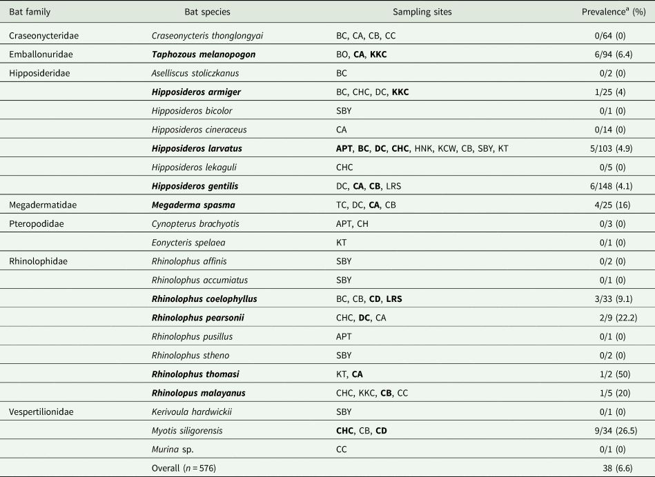
A total of 576 bats from 23 species and seven families were collected (Table 1 and Table S1). The bats in the family Rhinolophidae had the most diversity, with eight different species captured in the current study. The majority of the samples, however, came from the Hipposideridae family. Furthermore, Kanchanaburi had the highest bat species diversity among the seven provinces studied. We detected Trypanosoma in 2 out of 524 samples by microscopy and 37 out of 576 samples by PCRs, corresponding to overall infection rates of 0.38 and 6.42%, respectively. Trypanosoma parasites were discovered in ten different bat families, including the Emballonuridae, Hipposideridae, Megadermatidae, Rhinolophidae and Vespertilionidae. The bat family Vespertilionidae had the highest infection rate of 25% (9/36). In contrast, it was found that there was no infected bat in the Pteropodidae and Craseonycteridae families.
List of bats captured in the current study and the prevalence of trypanosomes

Each sampling site's letter abbreviation is as follows: Bo cave (BO), Bat cave (BC), Chompon cave (CHC), Daowadung cave (DC), cave A (CA), cave B (CB), cave C (CC), cave D (CD), Tiger cave (TC), Khun Kunchon cave (KKC), Kunchorn temple (KT), Saba Yoi (SBY), Ao Phrao temple (APT), Khlong Chao waterfall (KCW), Huang Nam Khiao waterfall (HNK), Cham's house (CH) and Lainan Research Station (LRS). Bat species and sampling sites in bold indicate trypanosome positive.
a Positive results were obtained using either microscopy or PCR.
The authors investigated the association between the infection rates and a variety of factors such as season, bat family, sampling sites, sex and bat status. The infection rates appeared to be associated with the bat family group (χ 2 = 34.732, df = 6, P < 0.0001). Further analyses revealed that Vespertilionidae was the most susceptible to infection. Although males had higher infection rates compared to females, no statistically significant difference in Trypanosoma infection rates was found (χ 2 = 2.023, df = 1, P = 0.155). Moreover, the chi-squared analysis revealed that there was no relationship between the infection rates and season (χ 2 = 0.072, df = 1, P = 0.788) or bat status (χ 2 = 3.181, df = 1, P = 0.074). Furthermore, it appeared that there was a statistically significant association between the sampling site and prevalence (χ 2 = 35.57, df = 16, P = 0.003); 15 out of 38 positive bats were captured in cave A.
Morphology of the trypanosomes
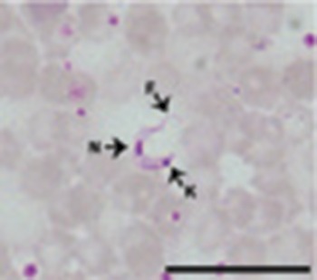
According to microscopic examination, two blood films from Hipposideros gentilis (sample IDs: THBat19-195 and THBat20-311) were positive for Trypanosoma. Trypomastigotes in blood films had similar morphology, with a C or U shape. The trypomastigote in H. gentilis bat is relatively small, with a large kinetoplast, a narrow undulating membrane and a free flagellum, as shown in Fig. 2. The body length was in the range of 13.5–16.3 μm (15.04 ± 0.95 μm), whereas the free flagellum length was in the range of 3.7–5.7 μm (4.42 ± 0.59 μm). The kinetoplast is rounded and positioned in the posterior end, far from the nucleus. These characteristics resembled the characteristics of Trypanosoma belonging to the subgenus Schizotrypanum.
Micrograph depicting trypomastigotes of Trypanosoma sp. from Hipposideros gentilis. N, nucleus, K, kinetoplast and F, flagellum; scale bar = 20 μm.

Sequence analyses, genetic diversity and phylogenetic analyses
The partial amplified Trypanosoma PCR products from the 18S rRNA and gGAPDH regions were compared to the GenBank database with BLASTn results identifying six Trypanosoma species, with two of them corresponding to known species, namely Trypanosoma dionisii and Trypanosoma noyesi (Table 2). The majority of sequences from this study could not be assigned to known species, but matched to an undescribed Trypanosoma species. Therefore, these 13 sequences were clustered into four lineages according to BLASTn results, genetic distance, haplotype network and phylogenetic analyses.
BLASTn search results of the trypanosomes in the current study

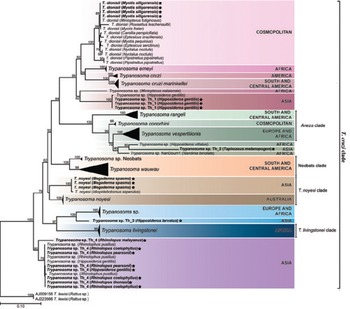
Phylogenetic analyses based on concatenated Trypanosoma sequences of the 18S rRNA and gGAPDH genes grouped the Trypanosoma isolates from Thailand bats into the T. cruzi clade of bat trypanosomes. Phylogenetic trees of ML and BI are shown in Fig. 3 and Fig. S1, respectively. The Thailand Trypanosoma isolates fall within the main T. cruzi clade as well as forming a new subclade referred to as the Asian clade that was basal to the Trypanosoma livingstonei clade. Interestingly, a few novel bat Trypanosoma genotypes from this study grouped with previously undescribed Trypanosoma isolates from Africa, Asia and Europe.
Phylogenetic tree of Trypanosoma inferred by ML based on 18S rRNA and gGAPDH genes using a final dataset of 1478 bp (658 bp of 18S rRNA and 820 bp of gGAPDH). Node labels correspond to BS value (>50). Sequences found in the current study are in bold font. Each trypanosome isolate's name is derived from the host binomial nomenclature. The host and country of origin (where available) for each trypanosome taxon are indicated.

The first main clade belongs to Schizotrypanum subgenus containing Trypanosoma erneyi, T. dionisii, T. cruzi marinkellei, T. cruzi and Trypanosoma sp. Four Thailand T. dionisii isolates from Myotis siligorensis bats grouped within the Schizotrypanum clade along with other T. dionisii isolates from Japan, China, Brazil, Taiwan, the UK and Switzerland. The four T. dionisii isolates were 100% similar to one another and were most closely related to a T. dionisii isolate (LC326397) from Japan and an isolate from Taiwan (MN956690) with a 0.00% genetic at the 18S rRNA locus (Table S4). In addition, at the gGAPDH locus it showed 0.12% genetic distance to the T. dionisii isolate (LC326399) from Japan (Table S5). We did not detect any T. erneyi, T. cruzi or T. cruzi marinkellei in the current samples. In the basal position of the Schizotrypanum clade, Trypanosoma sp. Th_1 from H. gentilis clustered with a sequence from H. gentilis, China, and was a sister clade to Trypanosoma sp. 2 from Miniopterus natalensis, South Africa. Three sequences of Trypanosoma sp. Th_1 from H. gentilis at 18S rRNA gene showed high similarity and 0.0% genetic distance with an isolate of Trypanosoma sp. from H. gentilis from China (MK625435). According to the gGAPDH gene, the closest bat trypanosome was T. dionisii from Eptesicus serotinus from the UK (FN599056) with a 7.2% genetic distance.
The second clade belongs to Aneza containing Trypanosoma vespertilionis, Trypanosoma conorhini and Trypanosoma rangeli. Trypanosoma sp. Th_2 (MZ606798) formed a distinct branch and was closely related to Trypanosoma sp. ex Hipposideros vittatus from Zambia with good support (BS = 80, PP = 0.96). Moreover, both analyses placed this clade as a sister clade to Trypanosoma sp. HochNdi1 and appeared to be closely related to other Trypanosoma within the Aneza clade. According to 18S rRNA gene of Trypanosoma sp. Th_2 occurred in Taphozous melanopogon, displayed a low percentage identity (90.74%) and genetic distance at 9.6% with undescribed Trypanosoma sp. from H. vittatus, Zambia (LC415424). Unfortunately, we were not able to amplify the gGAPDH gene from this sample.
Three T. noyesi isolates from Megaderma spasma clustered within the T. noyesi clade. These isolates shared clade with T. noyesi from Idiophlebotomus asperulus, Thailand. In addition, T. noyesi from Thailand appeared to be closely related to T. noyesi from Australia. At the 18S rRNA locus, T. noyesi had 0.20% genetic distance from the T. noyesi isolate (MK603815) from Thailand (Table S4). Moreover, based on the gGAPDH locus the closest Trypanosoma species was T. noyesi (JN315397 and AJ620276) from Australia with a genetic distance of 8.20 and 8.34%, respectively (Table S5).
The fourth main clade consists of T. livingstonei and Trypanosoma sp. from European bats and bat flies. The ML and BI analyses revealed Trypanosoma sp. Th_3 fell within the T. livingstonei clade. Trypanosoma sp. Th_3 from Hipposideros larvatus appeared to have a 1.1% genetic distance to Trypanosoma sp. from Italy (MT956923, MN956684), Spain (MT956924, MT956926), Hungary (MT956930) and South Africa (MT956927) at the 18S rRNA locus. However, based on the gGAPDH gene, this isolate showed a low-genetic distance of 8.5% to Trypanosoma sp. from Italy (MN969659).
Basal to all bat trypanosomes represented Trypanosoma sp. Th_4 from Hipposideros and Rhinolopus hosts from the current study, along with isolates from China. This well-supported clade corresponds to undescribed Trypanosoma sp. in Asian Hipposideros and Rhinolopus bats. Corresponding to the 18S rRNA locus, these sequences had 0.0% genetic distance to undescribed Trypanosoma sp. from China (MK625436, MK625442, MK625445, MK625447) and percentage similarity was 98.10–99.62%. At the gGAPDH locus, Trypanosoma sp. from Bettongia penicillate (KU354264) and Macropus giganteus (AJ620276) were the closest relative to these sequences with a genetic distance of 9.3 and 9.4%, respectively.
Haplotype network
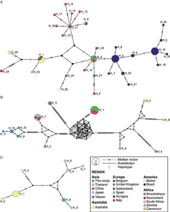
We investigated the population structure of Trypanosoma species from this study along with global isolates. A haplotype network analysis based on partial 18S rRNA gene of T. dionisii, Trypanosoma sp. and T. noyesi is shown in Fig. 4. Haplotypes from this study are depicted in green colour. Additionally, partial 18S rRNA gene sequences were analysed further to observe the genetic diversity in comparison with previously published sequences (Table 3). Four T. dionisii isolates from Thai bats appeared to be identical in the genetic diversity analyses. Furthermore, all isolates from Africa, Asia, Australia, Europe and Brazil had low polymorphisms and nucleotide diversity. The highest polymorphic sites, haplotype and nucleotide diversity were found in African bats (S = 15; H d = 1.000; π = 0.029). Overall, the nucleotide diversity (π) of T. dionisii was in the range of 0.000–0.029. Median-joining network revealed 30 haplotypes from 69 sequences (473 bp) of T. dionisii. In our T. dionisii haplotype network, we found two dominant haplotypes, H_3 and H_18, each with 11 sequences. Trypanosoma dionisii from Brazilian bats was found in both haplotypes. In addition, two other common haplotypes are H_1 and H_4, each of which contains eight sequences. The sequences from this study were clustered into H_4, together with isolates from the UK, Japan, China and Taiwan. Meanwhile, H_1 was discovered in T. dionisii from Belgium, the UK, Switzerland and Australia. According to the results of the analysis, H_4 is more similar to isolates from Switzerland, Brazil and China.
Median-joining haplotype network of partial 18S rRNA gene sequences of Trypanosoma dionisii (A), Trypanosoma sp. (B) and Trypanosoma noyesi (C). Each circle illustrates a haplotype. Bar on the branches show the number of mutations. Colours represent geographical origin. The node size indicates the frequency or number of samples in each haplotype. Black circles represent missing haplotype or median vector.

Genetic diversity of Trypanosoma spp. based on 18S rRNA gene

N, number of sequences; S, number of polymorphic sites; H, number of haplotypes; H d, haplotype diversity; π, nucleotide diversity (Pi); s.d., standard deviation.
Similar to T. dionisii, T. noyesi isolates from the current study were indistinguishable. In contrast, the highest nucleotide diversity was observed in four Thai T. noyesi sequences (π = 0.013; H d = 1.000; S = 25; H = 4). Our analysis used 17 T. noyesi sequences (515 bp), and the median-joining network revealed eight distinct haplotypes. Trypanosoma noyesi from Australian marsupials and Asian bats were separated and could be distinguished from the network. From the analysis, the sequences from Thai bats were clustered into one haplotype (H_8) and separated from other T. noyesi in Thailand. Moreover, the closest haplotype observed in the network was H_5. Additionally, the most dominant haplotype was H_1 from Australian marsupials.
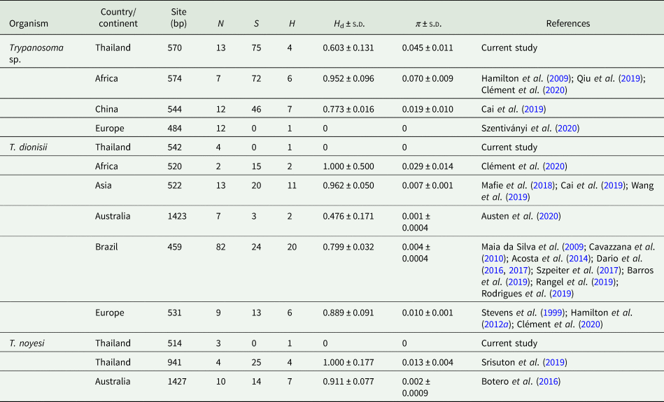
The haplotype diversity of Trypanosoma spp. obtained in the current study was 0.603, which was the lowest compared to other studies. The highest polymorphic sites were observed in the sequences from the current study (S = 75). The highest nucleotide diversity was found in African isolates (π = 0.070), followed by the isolates from Thailand (π = 0.045) and China (π = 0.019). In contrast, Trypanosoma sp. sequences from European bats and bat flies were identical which later clustered into one haplotype.
A total of 12 unique haplotypes representing 36 sequences (542 bp) of Trypanosoma sp. were found in our analysis (Table S6). Most sequences in the current study clustered in H_1, together with sequences from Yunnan, China. This haplotype appeared to be the most dominant haplotype representing Trypanosoma sp. from Hipposideros and Rhinolopus bats. Next is H_2, this haplotype appeared to be isolated from other previously reported Trypanosoma sp. In addition, H_7 was found to be the closest Trypanosoma sp. to H_2. Another distinct haplotype is H_3, which comprised of three sequences of Trypanosoma sp. from H. gentilis. This haplotype is shared with a sequence from H. gentilis, China. Similar to H_2, the H_4 was found to be separated with several mutations and median vectors from other isolates. Genetically, the closest Trypanosoma sp. would be H_5, H_6 and H_7 of Trypanosoma sp. from Cameroon and Zambia.
Discussion
The increase in research in recent decades has highlighted the importance of bats as a natural reservoir for several zoonotic pathogens, including Trypanosoma. Bats play an important epidemiological role in the transmission of zoonotic diseases in the context of One Health (Calisher et al., Reference Calisher, Childs, Field, Holmes and Schountz2006; Letko et al., Reference Letko, Seifert, Olival, Plowright and Munster2020; Austen and Barbosa, Reference Austen and Barbosa2021). This discovery has sparked the interest and concern of experts. Recently, T. cruzi has been found in the saliva of haematophagous bats (Diaemus youngi, Desmodus rotundus and Diphylla ecaudata), increasing concerns about the possible route of transmission to humans via bites or the contamination of bat saliva (Villena et al., Reference Villena, Gomez-Puerta, Jhonston, Del Alcazar, Maguina, Albujar, Laguna-Torres, Recuenco, Ballard and Ampuero2018; Bergner et al., Reference Bergner, Becker, Tello, Carrera and Streicker2021a). However, further research is required to prove this alternative transmission route. Furthermore, the fact that bats may roost in close proximity to humans or terrestrial animals increases the possibility of transmission to other hosts (Kessler et al., Reference Kessler, Becker, Peel, Justice, Lunn, Crowley, Jones, Eby, Sánchez and Plowright2018). Insect vectors and ticks that feed on bats may occasionally feed on humans or animals and spread parasites (Sándor et al., Reference Sándor, Mihalca, Domşa, Péter and Hornok2021).
Trypanosoma has been reported to exist in several parts of the world, and several species, including T. livingstonei, T. rangeli and T. dionisii, have been well described (Lima et al., Reference Lima, Espinosa-Álvarez, Hamilton, Neves, Takata, Campaner, Attias, de Souza, Camargo and Teixeira2013, Reference Lima, Espinosa-Álvarez, Pinto, Cavazzana, Pavan, Carranza, Lim, Campaner, Takata, Camargo, Hamilton and Teixeira2015; Barros et al., Reference Barros, Lima, Schubach and Teixeira2019; Clément et al., Reference Clément, Dietrich, Markotter, Fasel, Monadjem, López-Baucells, Scaravelli, Théou, Pigeault, Ruedi and Christe2020). In contrast, the occurrence of these parasites in Asia is still poorly understood. The Australian terrestrial clade of trypanosomes appeared relatively recently and is a result of transmission from a bat host. To the best of the authors' knowledge, this is the first study of trypanosomes in a variety of Thai bats with their evolutionary relationships. The new evidence presented in this study appears to support the bat seeding theory proposed by Hamilton et al. (Reference Hamilton, Teixeira and Stevens2012b). The current study provides the first investigation on trypanosomes in a wide range of Thai bat species with an overall prevalence of 6.6% (38/576). Compared to previous studies in Asia, the current prevalence was comparable to a report from Yunnan, China (6.17%), though (Cai et al., Reference Cai, Wang, Zhang, Li, Gong, He and Zhang2019) lower than another study in Shandong, China (10.3%) (Wang et al., Reference Wang, Han, Zhao, Liu, Luo, Wen, Qin, Zhou, Qi, Yu and Yu2019). Additionally, the prevalence was still higher than the prevalence in Japan (2.13%) (Mafie et al., Reference Mafie, Rupa, Takano, Suzuki, Maeda and Sato2018). The authors were unable to discover Trypanosoma in the samples taken from Songkhla, Saraburi and Phetchaburi, which could be due to the limited sample sizes.
Trypanosomes have been found in a variety of bat species in Asia, including Miniopterus fuliginosus, Rhinolophus pusillus, Rhinolopus leschenaultia, Myotis pequinius and other species (Mafie et al., Reference Mafie, Rupa, Takano, Suzuki, Maeda and Sato2018; Cai et al., Reference Cai, Wang, Zhang, Li, Gong, He and Zhang2019; Wang et al., Reference Wang, Han, Zhao, Liu, Luo, Wen, Qin, Zhou, Qi, Yu and Yu2019). In addition, the current study identified ten different species that serve as trypanosome hosts. All positive bats are cave-dwelling and insectivorous bats. Although the transmission route of Trypanosoma in bats remains unclear, several reports have suggested that transmission can occur from vector consumption while grooming, or from the diet (Gardner and Molyneux, Reference Gardner and Molyneux1988b; Cavazzana et al., Reference Cavazzana, Marcili, Lima, da Silva, Junqueira, Veludo, Viola, Campaner, Nunes, Paiva, Coura, Camargo and Teixeira2010; Lima et al., Reference Lima, Silva, Neves, Attias, Takata, Campaner, de Souza, Hamilton and Teixeira2012; Lima et al., Reference Lima, Espinosa-Álvarez, Hamilton, Neves, Takata, Campaner, Attias, de Souza, Camargo and Teixeira2013; Bergner et al., Reference Bergner, Becker, Tello, Carrera and Streicker2021a). Therefore, behaviour, diet and vector abundance in caves might influence the prevalence of this parasite.
Statistical analyses were used to determine the association between the infection rates with several variables such as bat family, sex, bat status and season. Bat families and sampling sites were the only factors that influenced infection rates in the current study, with the Vespertilionidae family being the most vulnerable. In terms of sampling sites, 15 of the 38 positive bats were captured in cave A, indicating that trypanosome diversity and infection remain in this cave. Investigation of the potential vectors of Trypanosoma in this cave may enable us to understand how the parasite is transmitted between bats. The current result corroborates that trypanosome infection is common in insectivorous bats, as previously reported by Lima et al. (Reference Lima, Silva, Neves, Attias, Takata, Campaner, de Souza, Hamilton and Teixeira2012).
The blood stages of Trypanosoma found in the current samples were morphologically identical, especially with the members of subgenus Schizotrypanum, such as Trypanosoma diosinii and T. cruzi (Hoare, Reference Hoare1972; Maia da Silva et al., Reference Maia da Silva, Marcili, Lima, Cavazzana, Ortiz, Campaner, Takeda, Paiva, Nunes, Camargo and Teixeira2009). The authors observed small trypomastigotes with round kinetoplasts in the blood films. The nucleus was elongated with a large kinetoplast, and the posterior exhibited a pointed end. These characteristics resembled the description of Schizotrypanum (Maia da Silva et al., Reference Maia da Silva, Marcili, Lima, Cavazzana, Ortiz, Campaner, Takeda, Paiva, Nunes, Camargo and Teixeira2009).
Previous studies on Trypanosoma have focused on mini-exon, 24S RNA, MSH2 and TcSD5D genes to investigate their genetic diversity (Dumonteil et al., Reference Dumonteil, Elmayan, Majeau, Tu, Duhon, Marx, Wolfson, Balsamo and Herrera2020; Maiguashca Sánchez et al., Reference Maiguashca Sánchez, Sueto, Schwabl, Grijalva, Llewellyn and Costales2020; Majeau et al., Reference Majeau, Murphy, Herrera and Dumonteil2021). In the current study, the authors targeted the partial 18S rRNA and gGAPDH genes, which are the most common targets for studying Trypanosoma phylogenetic relationships. Using these genes, the authors were able to identify and distinguish different species as well as study their phylogenetic relationship. Moreover, concatenated sequences of these genes were able to generate a more robust and well-resolved phylogenetic tree to demonstrate phylogenetic relationship and classification of Trypanosoma species (Haag et al., Reference Haag, O'HUigin and Overath1998, Stevens et al., Reference Stevens, Noyes and Gibson1998, Reference Stevens, Noyes, Dover, Dover and Gibson1999; Noyes et al., Reference Noyes, Stevens, Teixeira, Phelan and Holz1999; Cavazzana et al., Reference Cavazzana, Marcili, Lima, da Silva, Junqueira, Veludo, Viola, Campaner, Nunes, Paiva, Coura, Camargo and Teixeira2010). The current analyses on the 18S rRNA gene of Trypanosoma revealed a great number of polymorphic sites, but low nucleotide diversity. In addition, T. dionisii and T. noyesi sequences in our study were identical within each species, whereas sequences from other continents appeared to have low nucleotide diversity and polymorphisms. These results suggest that the 18S rRNA gene appeared to be highly conserved.
In the current study, only two of the six trypanosome species found in Thailand bats are known species, with the four remaining taxa being uncharacterized. According to BI and ML analyses, the remaining sequences were assigned to multiple bat trypanosome lineages. The current findings revealed a monophyletic clade consisting of members of the subgenus Schizotrypanum, which agrees with previous studies (Cavazzana et al., Reference Cavazzana, Marcili, Lima, da Silva, Junqueira, Veludo, Viola, Campaner, Nunes, Paiva, Coura, Camargo and Teixeira2010; Clément et al., Reference Clément, Dietrich, Markotter, Fasel, Monadjem, López-Baucells, Scaravelli, Théou, Pigeault, Ruedi and Christe2020; Austen and Barbosa, Reference Austen and Barbosa2021). Trypanosoma cruzi is the only pathogenic Schizotrypanum species, while other species are restricted to bats such as T. dionisii have been shown to not infect BALB/c mice (Lima et al., Reference Lima, Silva, Neves, Attias, Takata, Campaner, de Souza, Hamilton and Teixeira2012). On the contrary, Dario et al. (Reference Dario, Rodrigues, Barros, Xavier, D'Andrea, Roque and Jansen2016) detected the presence of T. dionisii DNA in the cardiac tissue of a patient in Espírito Santo state, Brazil. This discovery increased concerns about pathogenicity of T. dionisii and potential transmission to humans and other animals (Austen and Barbosa, Reference Austen and Barbosa2021). Sequences from the current study clustered together with T. dionisii from Japan (Mafie et al., Reference Mafie, Rupa, Takano, Suzuki, Maeda and Sato2018), China (Cai et al., Reference Cai, Wang, Zhang, Li, Gong, He and Zhang2019; Wang et al., Reference Wang, Han, Zhao, Liu, Luo, Wen, Qin, Zhou, Qi, Yu and Yu2019), Brazil (Cavazzana et al., Reference Cavazzana, Marcili, Lima, da Silva, Junqueira, Veludo, Viola, Campaner, Nunes, Paiva, Coura, Camargo and Teixeira2010), UK (Hamilton et al., Reference Hamilton, Cruickshank, Stevens, Teixeira and Mathews2012a) and Switzerland and Taiwan (Clément et al., Reference Clément, Dietrich, Markotter, Fasel, Monadjem, López-Baucells, Scaravelli, Théou, Pigeault, Ruedi and Christe2020), but were separated from two isolates from Europe (Stevens et al., Reference Stevens, Noyes, Dover, Dover and Gibson1999). In general, T. dionisii has been detected in several bat hosts, from the Old World (Mafie et al., Reference Mafie, Rupa, Takano, Suzuki, Maeda and Sato2018; Wang et al., Reference Wang, Han, Zhao, Liu, Luo, Wen, Qin, Zhou, Qi, Yu and Yu2019; Austen et al., Reference Austen, Van Kampen, Egan, O'Dea, Jackson, Ryan, Irwin and Prada2020; Clément et al., Reference Clément, Dietrich, Markotter, Fasel, Monadjem, López-Baucells, Scaravelli, Théou, Pigeault, Ruedi and Christe2020) and the New World (Cavazzana et al., Reference Cavazzana, Marcili, Lima, da Silva, Junqueira, Veludo, Viola, Campaner, Nunes, Paiva, Coura, Camargo and Teixeira2010; Espinosa-Álvarez et al., Reference Espinosa-Álvarez, Ortiz, Lima, Costa-Martins, Serrano, Herder, Buck, Camargo, Hamilton, Stevens and Teixeira2018; Barros et al., Reference Barros, Lima, Schubach and Teixeira2019; Rangel et al., Reference Rangel, Lisboa, Novaes, Silva, Souza, Jansen, Moratelli and Roque2019), and is commonly transmitted by cimicid bugs (Gardner and Molyneux, Reference Gardner and Molyneux1988a). This indicates a broad range of hosts and geographical distributions of T. dionisii, similar to previous studies (Cavazzana et al., Reference Cavazzana, Marcili, Lima, da Silva, Junqueira, Veludo, Viola, Campaner, Nunes, Paiva, Coura, Camargo and Teixeira2010; Hamilton et al., Reference Hamilton, Cruickshank, Stevens, Teixeira and Mathews2012a; Hodo et al., Reference Hodo, Goodwin, Mayes, Mariscal, Waldrup and Hamer2016; Szpeiter et al., Reference Szpeiter, Ferreira, Assis, Stelmachtchuk, Peixoto, Ajzenberg, Minervino, Gennari and Marcili2017; Mafie et al., Reference Mafie, Rupa, Takano, Suzuki, Maeda and Sato2018; Austen et al., Reference Austen, Van Kampen, Egan, O'Dea, Jackson, Ryan, Irwin and Prada2020; Clément et al., Reference Clément, Dietrich, Markotter, Fasel, Monadjem, López-Baucells, Scaravelli, Théou, Pigeault, Ruedi and Christe2020). Clément et al. (Reference Clément, Dietrich, Markotter, Fasel, Monadjem, López-Baucells, Scaravelli, Théou, Pigeault, Ruedi and Christe2020) hypothesized that T. dionisii might have originated from Africa, then spread to South America and Europe through several dispersion events. The absence of host specificity in T. dionisii is one of the factors that contribute to the dispersion of this blood parasite in different bat populations and its wide distribution (Hamilton et al., Reference Hamilton, Gibson and Stevens2007).
Trypanosoma noyesi was discovered in Australian marsupials (Bettongia penicillata) for the first time using phylogenetic inferences from three gene regions (18S rDNA, gGAPDH and CytB) (Botero et al., Reference Botero, Cooper, Thompson, Clode, Rose and Thompson2016). In addition, T. noyesi DNA was detected in tabanid and biting midges, suggesting that these blood sucking insects could be potential vectors (Botero et al., Reference Botero, Cooper, Thompson, Clode, Rose and Thompson2016). Previous T. noyesi analyses placed this species within the T. cruzi clade, suggesting that T. noyesi was more distantly related to Australian trypanosomes (Trypanosoma copemani, Trypanosoma irwini, Trypanosoma gilletti and Trypanosoma vegrandis) and closely related to T. cruzi members such as T. rangeli, T. vespertilionis and Trypanosoma wauwau (Botero et al., Reference Botero, Cooper, Thompson, Clode, Rose and Thompson2016). In the current study, T. noyesi was detected in bats for the first time. In Thailand, T. noyesi had been previously detected from I. asperulus (Srisuton et al., Reference Srisuton, Phumee, Sunantaraporn, Boonserm, Sor-Suwan, Brownell, Pengsakul and Siriyasatien2019). In the current study, T. noyesi isolates from M. spasma were grouped with T. noyesi from I. asperulus and formed a sister clade to T. noyesi from Australian marsupials and flies. This placement is similar to the previous study by Clément et al. (Reference Clément, Dietrich, Markotter, Fasel, Monadjem, López-Baucells, Scaravelli, Théou, Pigeault, Ruedi and Christe2020). Other studies, however, have consistently identified T. noyesi as a sister clade to T. wauwau (Espinosa-lvarez et al., Reference Espinosa-Álvarez, Ortiz, Lima, Costa-Martins, Serrano, Herder, Buck, Camargo, Hamilton, Stevens and Teixeira2018; Austen and Barbosa, Reference Austen and Barbosa2021). The different placement of T. noyesi could be attributed to the different target genes used in each study. Furthermore, the length of a sequence as well as the conserved and variable regions within a gene have a significant impact on the topology of a phylogenetic tree.
In the current study, four uncharacterized trypanosome taxa from Thailand were analysed using genetic distance, network phylogeny and phylogenetic relationships. The genetic distance between the 18S rRNA and gGAPDH loci was in the range of 0.0–9.6% and 7.2–9.4%, respectively. According to McInnes et al. (Reference McInnes, Hanger, Simmons, Reid and Ryan2011), the genetic distance (±s.e.) between T. gilletti and T. copemani (wombat) at the 18S rDNA locus was 2.7 ± 0.5%. Genetic distance of T. gilletti from T. copemani (wombat) at the gGAPDH locus was 8.7 ± 1.1%. A recent study on Trypanosoma madeirae, a novel bat trypanosome from D. rotundus, classified this isolate into a new species based on its morphological and biological features, xenodiagnoses, phylogenetic position and genetic distance based on SSU rRNA and gGAPDH (Barros et al., Reference Barros, Lima, Schubach and Teixeira2019). Trypanosoma madeirae had 0.4% sequence divergence to Trypanosoma sp. Neot 1 at the V7V8 SSU rRNA and 10% to T. wauwau at the gGAPDH gene, according to Barros et al. (Reference Barros, Lima, Schubach and Teixeira2019). Prior studies on Theileria and Babesia suggested that genetic distances at the 18S rRNA locus greater than 0.7% for Theileria and 3.4% for Babesia should be considered different species (Schnittger et al., Reference Schnittger, Yin, Gubbels, Beyer, Niemann, Jongejan and Ahmed2003). The threshold for genetic distance or percentage similarity must be resolved to assign newly discovered trypanosomes into distinct species. Furthermore, when combined with morphological and biological characteristics, this molecular method could help justify its effectiveness in classifying uncharacterized trypanosomes.
Trypanosoma sp. Th_1 was found in the same clade as a previously undescribed Trypanosoma sp. from Yunnan, China (Cai et al., Reference Cai, Wang, Zhang, Li, Gong, He and Zhang2019). This trypanosome was exclusively found in the Hipposideridae family, which could indicate host specificity. So far, the number of records of bat trypanosomes has increased significantly, and more new lineages have been discovered. Clément et al. (Reference Clément, Dietrich, Markotter, Fasel, Monadjem, López-Baucells, Scaravelli, Théou, Pigeault, Ruedi and Christe2020) found a new Trypanosoma sp. lineage (genotype T. sp. 2) from M. natalensis in South Africa, which was found to be at the basal position of T. cruzi. The current phylogeny yielded a similar result, with Trypanosoma sp. from M. natalensis clustered on the edge of the T. cruzi clade. Surprisingly, this sequence appeared to be genetically similar to Trypanosoma sp. Th_1.
In a previous study, Trypanosoma sp. from H. vittatus was found to be clustered with T. conorhini (Qiu et al., Reference Qiu, Kajihara, Harima, Hang'ombe, Nakao, Hayashida, Mori-Kajihara, Changula, Eto, Ndebe, Yoshida, Takadate, Mwizabi, Kawabata, Simuunza, Mweene, Sawa, Takada and Sugimoto2019). The current findings, on the contrary, revealed that Trypanosoma sp. from H. vittatus is a sister clade to Trypanosoma sp. Th_2. The placement also revealed a close relationship between Trypanosoma sp. Th_2 and Trypanosoma sp. Nandoum1 from Nandinia binotata from Cameroon (African palm civet) (Hamilton et al., Reference Hamilton, Adams, Njiokou, Gibson, Cuny and Herder2009; Qiu et al., Reference Qiu, Kajihara, Harima, Hang'ombe, Nakao, Hayashida, Mori-Kajihara, Changula, Eto, Ndebe, Yoshida, Takadate, Mwizabi, Kawabata, Simuunza, Mweene, Sawa, Takada and Sugimoto2019). It remains unclear how palm civet trypanosome falls within the T. cruzi clade, which is dominated by bat trypanosomes. According to Hamilton et al. (Reference Hamilton, Adams, Njiokou, Gibson, Cuny and Herder2009), trypanosome lineages in terrestrial mammals such as monkeys and palm civets may have evolved from bat lineages via host jumping. The discovery of new lineages within the T. cruzi clade suggests that this clade may contain more species than previously thought. The current study adds to the growing body of knowledge regarding bat trypanosomes, particularly in terms of their diversity and phylogenetic relationships.
Another uncharacterized Trypanosoma sp., Th_3, was found in the same clade as T. livingstonei, along with Trypanosoma sp. from African and European bats and bat flies (Clément et al., Reference Clément, Dietrich, Markotter, Fasel, Monadjem, López-Baucells, Scaravelli, Théou, Pigeault, Ruedi and Christe2020; Szentiványi et al., Reference Szentiványi, Markotter, Dietrich, Clément, Ançay, Brun, Genzoni, Kearney, Seamark, Estók, Christe and Glaizot2020). Trypanosoma livingstonei is a Mozambican species that is endemic to the Rhinolopus and Hipposideros bat genera. So far, T. livingstonei appears to be restricted to sub-Saharan African bats, even though these bat species are geographically widespread in Asia, Europe and Oceania (Lima et al., Reference Lima, Espinosa-Álvarez, Hamilton, Neves, Takata, Campaner, Attias, de Souza, Camargo and Teixeira2013). Clément et al. (Reference Clément, Dietrich, Markotter, Fasel, Monadjem, López-Baucells, Scaravelli, Théou, Pigeault, Ruedi and Christe2020) and Szentiványi et al. (Reference Szentiványi, Markotter, Dietrich, Clément, Ançay, Brun, Genzoni, Kearney, Seamark, Estók, Christe and Glaizot2020) were the first to identify Trypanosoma sp., which was later linked to T. livingstonei. This parasite has been found in both African and European bats, as well as in bat flies. The Trypanosoma sp. Th_3 belongs to the T. livingstonei group, which is consistent with previous reports by Clément et al. (Reference Clément, Dietrich, Markotter, Fasel, Monadjem, López-Baucells, Scaravelli, Théou, Pigeault, Ruedi and Christe2020) and Szentiványi et al. (Reference Szentiványi, Markotter, Dietrich, Clément, Ançay, Brun, Genzoni, Kearney, Seamark, Estók, Christe and Glaizot2020). The placement of Trypanosoma sp. Th_3 suggests that this parasite is genetically related to T. livingstonei and Trypanosoma sp.
The last clade is a new clade that exclusively included Trypanosoma sp. Th_4 isolates from Hipposideros in the current study and Rhinolopus bats from China (Cai et al., Reference Cai, Wang, Zhang, Li, Gong, He and Zhang2019). In the current study, this group comprised of dominant trypanosomes and formed a distinct bat-associated clade. Cai et al. (Reference Cai, Wang, Zhang, Li, Gong, He and Zhang2019) previously identified sequences from Yunnan, China, as a sister clade of T. wauwau. The current findings resulted in the clustering of these sequences into a single clade at the bottom of a tree that did not nest to any known species. Previous studies have placed T. livingstonei in the edge clade of the T. cruzi evolutionary tree (Lima et al., Reference Lima, Espinosa-Álvarez, Hamilton, Neves, Takata, Campaner, Attias, de Souza, Camargo and Teixeira2013; Clément et al., Reference Clément, Dietrich, Markotter, Fasel, Monadjem, López-Baucells, Scaravelli, Théou, Pigeault, Ruedi and Christe2020). As a result of our findings, we added new bat trypanosome isolates that are even more basal than T. livingstonei. The current analyses were limited to a final dataset of 1478 bp (658 bp of 18S rRNA and 820 bp of gGAPDH), which might not be sufficient to show the phylogenetic relationships of Trypanosoma. Moreover, short sequences might have reduced the information needed to show the relationship of Trypanosoma sp. To corroborate this placement, a larger investigation focusing on more loci and covering more taxa is required. Phylogenetic analyses were unable to provide enough evidence to support the migration of bats. However, the association of several trypanosome sequences in the current study, which were previously found only in African bats, suggests that trypanosomes may disperse along with the geographical mobility of bats (Hamilton et al., Reference Hamilton, Adams, Njiokou, Gibson, Cuny and Herder2009, Reference Hamilton, Cruickshank, Stevens, Teixeira and Mathews2012a).
Together with previous studies, a number of new bat species have been added to the list of trypanosome hosts. As a result, the new findings suggest that Trypanosoma may have more species and can be identified in more bat species than previously thought. Further studies covering wider regions in Asia are crucial to discover underrated Trypanosoma species in bats and to gain a better understanding on their phylogeographical and evolutionary history.
Supplementary material
The supplementary material for this article can be found at https://doi.org/10.1017/S0031182022000117.
Data
DNA sequences obtained from the trypanosomes described in the current study have been submitted to the GenBank database and can be found under accession numbers MZ606781-800 (18S rRNA) and OL691559-69 (gGAPDH) genes.
Acknowledgements
The authors acknowledge the Agriculture and Cooperatives Division, Military Development Office, Armed Forces Development Command for permission to access the areas and facilities. The Ph.D. students Aung Aung, Hoang Lan Anh Nguyen, Trang Nguyen and Yudhi Ratna aided us in the fieldwork. The following DNA sample was obtained through BEI Resources, NIAID, NIH: Trypanosoma cruzi, NR-49382.
Author contributions
MK conceived and designed the study. ER, AA, DN, TN, MW, PS, PB and MK carried out surveys and sample acquisitions. ER conducted data gathering and performed statistical analyses. ER and MK wrote the article and incorporated other author's comments. All authors read and approved the final manuscript.
Financial support
ER was supported by the Graduate Scholarship Program for ASEAN countries and the 90th Anniversary of Chulalongkorn University under the Rachadapisek Somphot Fund (GCUGR1125641048M). AA was supported by The Royal Golden Jubilee (RGJ) Ph.D. Program (grant number PHD/0028/2561). DN was supported by the Second Century Fund (C2F), Chulalongkorn University. TN was supported by the Plant Genetic Conservation Project under the Royal Initiative of Her Royal Highness Princess Maha Chakri Sirindhorn (RSPG) replied by Chulalongkorn University. PS was supported by Thailand Research Fund (grant number DBG6180028). This research was funded by Thailand Science Research and Innovation Fund Chulalongkorn University to MK.
Conflict of interest
The authors declare there are no conflicts of interest.
Ethical standards
The Department of National Parks, Wildlife and Plant Conservation granted permission to conduct the surveys (permit nos. DNP 0909.6/22928 to TN, DNP 0907.4/8179 to PS and DNP 0907.4/29038 to MK). The protocols were reviewed and approved by the Faculty of Veterinary Science's Institutional Biosafety Committee and the Institutional Animal Care and Use Committee (IBC no. 2031032; IACUC no. 1931090).