Introduction
Down syndrome occurs as a consequence of trisomy at chromosome 21. It is one of the most common genetic disorders, affecting approximately 1 in every 700 live births.Reference Parker, Mai, Canfield, Rickard, Wang and Meyer 1 Its incidence is related to advanced maternal age.Reference Vundinti and Ghosh 2 Individuals with Down syndrome demonstrate mild to moderate intellectual disability and exhibit significant phenotypic variance in associated health findings. Common sequelae include congenital heart disease, endocrine disorders, leukaemia, immunodeficiency, Alzheimer's disease and gastrointestinal abnormalities.Reference Asim, Kumar, Muthuswamy, Jain and Agarwal 3
With improved awareness and detection methods for Down syndrome and its related conditions, the life expectancy of affected individuals continues to increase. As a result, the prevalence of otolaryngological disease among this population is also on the rise. In fact, Down syndrome is the most commonly encountered genetic disorder by otolaryngologists. Persons with Down syndrome experience increased rates of craniofacial dysmorphism, pharyngolaryngeal abnormalities, and otic anomalies including impairments in hearing.Reference Venail, Gardiner and Mondain 4
Hearing loss has been reported in 40–90 per cent of individuals with Down syndrome,Reference Fulton and Lloyd 5 – Reference McPherson, Lai, Leung and Ng 9 which is markedly higher than the prevalence of 2.5 per cent among the general population.Reference Marttilla 10 The dysfunction in hearing encountered in this population often further exacerbates the acquisition and development of speech and language.Reference Yoshinaga-Itano, Sedey, Coulter and Mehl 11 Hearing impairments in Down syndrome can be a consequence of conductive, sensorineural and mixed aetiologies.
Conductive hearing loss is the most commonly encountered type of hearing impairment in this population, and typically occurs secondary to abnormalities in the external and middle ear.Reference Park, Wilson, Stevens, Harward and Hohler 12 In particular, up to 40–50 per cent of newborns with Down syndrome exhibit stenotic ear canals that frequently lead to cerumen impaction and thus complicate the accuracy in diagnosis of middle-ear disease in this population.Reference Strome 13 Hypoplasia of the mid-face is also common among individuals with Down syndrome and can predispose them to Eustachian tube dysfunction.Reference Shibahara and Sando 14 These structural abnormalities lead to an increased prevalence of chronic otitis media and chronic middle-ear effusions among the Down syndrome population, which can further exacerbate conductive hearing loss.Reference Shott 15 Sensorineural hearing loss in the Down syndrome population is believed to result from structural anomalies in inner-ear structures.Reference Blaser, Propst, Martin, Feigenbaum, James and Shannon 16
Much attention has been paid to the histopathological abnormalities of the inner and middle ear in individuals with Down syndrome. A prior histological study of the Eustachian tubes in this population revealed a decrease in the density of cartilage cells, which is thought to be correlated with poor aeration of the middle-ear cavity.Reference Yamaguchi, Sando, Hashida, Takahashi and Matsune 17 Other studies of the temporal bones in Down syndrome patients have demonstrated dysplastic inner-ear structures, including small lateral semicircular canals, a reduction in the number of spiral ganglion cells, short cochlea and Mondini malformation.Reference Igarashi, Takahashi, Alford and Johnson 18 – Reference Harada and Sando 21 Although the histological evaluation of inner-ear structures in this population has been relatively extensive, there remains a paucity of studies evaluating radiological imaging findings.
To the best of our knowledge, only three prior studies have investigated the radiological imaging findings of the inner ear in individuals with Down syndrome.Reference Blaser, Propst, Martin, Feigenbaum, James and Shannon 16 , Reference Intrapiromkul, Aygun, Tunkel, Carone and Yousem 22 , Reference Saliba, Sbeity, El-Zir, Yammine, Noun and Haddad 23 Two of these studies found that compared to matched controls and normative data, Down syndrome subjects had significantly larger vestibular aqueducts.Reference Blaser, Propst, Martin, Feigenbaum, James and Shannon 16 , Reference Saliba, Sbeity, El-Zir, Yammine, Noun and Haddad 23 This has significant implications, as dilation of the vestibular aqueducts has been correlated with sensorineural hearing loss that is progressive in nature.Reference Arcand, Desrosiers, Dube and Abela 24 More importantly, the decline in hearing in persons with enlarged vestibular aqueducts is to some extent preventable, as patients can be instructed to avoid situations that lead to barotrauma.Reference Griffith and Wangemann 25 Thus, the awareness and identification of this structural anomaly is imperative.
Accordingly, our study aimed to evaluate the prevalence of inner-ear abnormalities and measure the widths of the vestibular aqueducts on temporal bone computed tomography (CT) images of individuals with Down syndrome.
Materials and methods
Subjects
A total of 274 patients who had previously been seen in our otolaryngology clinic and had a clinical diagnosis of Down syndrome were identified in our electronic medical records. All prior imaging studies were reviewed for each patient to determine if the patient had ever undergone thin-cut, axial, high-resolution CT scanning (slice thickness of less than 1.0 mm) in which the temporal bones were visible. A total of 38 patients (75 ears, with 1 ear determined to be not assessable) were found to have CT scans meeting these criteria.
This study was reviewed and approved by the Institutional Review Board at the Penn State Milton S Hershey Medical Center. The Institutional Review Board determined this study to be exempt from informed consent and in compliance with Health Insurance Portability and Accountability Act (‘HIPAA’) regulations.
Radiological evaluation
A board-certified neuroradiologist reviewed the temporal bone CT images for each patient who met the inclusion criteria. The widths of the vestibular aqueducts for each patient were measured on axial views at the point of greatest middle anterior–posterior diameter using a measurement tool in Picture Archiving and Communication System software (GE Healthcare Imagecast™ PACS), and measurements were recorded. In accordance with previously established criteria, vestibular aqueduct enlargement was defined as a width measuring greater than 1.5 mm.Reference Valvassori and Clemis 26
Axial CT images were also evaluated for the presence of other inner-ear abnormalities, and findings were recorded for each patient. Additional abnormalities included cystic malformations of the lateral semicircular canals, and variations in internal auditory canal and vestibule widths. Normative data from Stimmer et al. were used as a reference for determinations in internal auditory canal width, with measurements greater than 7.8 mm on the right and 7.7 mm on the left in the axial plane signifying dilation.Reference Stimmer, Nidermeyer, Kehl and Rummeny 27 Vestibule widths were assessed based on normative data established by Purcell et al.Reference Purcell, Johnson, Fischbein and Lalwani 28
Mastoid air cell and middle-ear anomalies, if present, were also noted. Mastoid air cell abnormalities included under-pneumatisation, opacification, sclerosis and the presence of fluid. Middle-ear anomalies included decreased middle-ear cavity size and middle-ear cavity opacification.
Statistical analysis
Descriptive statistics, including means, standard deviations (SDs) and confidence intervals (CIs), were calculated for vestibular aqueduct widths. Proportions of study subjects exhibiting inner-ear abnormalities, including enlarged vestibular aqueducts, a dilated internal auditory canal, cystic malformations of lateral semicircular canals and dilated vestibules, were also calculated. In addition, proportions were determined for: mastoid air cell anomalies, including under-pneumatisation, opacification, sclerosis and fluid presence; and middle-ear anomalies, including decreased middle-ear cavity size and middle-ear cavity opacification. Microsoft Excel® was used to compute these values.
Results
A total of 38 patients (75 ears, with 1 ear found to be not assessable) with a diagnosis of Down syndrome who had undergone thin-cut, axial CT scanning (with a slice thickness of 1.0 mm), with visible temporal bones, were identified. Mean patient age was 24 ± 3 years.
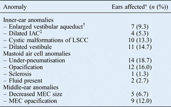
Enlarged vestibular aqueducts (width of more than 1.5 mm) were observed in 9.3 per cent of ears (7 of 75 ears). The mean vestibular aqueduct width was 1.20 mm (95 per cent CI = 1.15–1.25 mm) with an SD of 0.23 mm.
Other abnormalities of the inner ear, including a dilated internal auditory canal, cystic malformations of the lateral semicircular canals, dilated vestibules and enlarged vestibular aqueducts, were identified in 52.6 per cent of patients (20 of 38 patients). A dilated internal auditory canal was identified in 5.3 per cent of ears (4 of 75 ears). Cystic malformations of the lateral semicircular canals were observed in 13.3 per cent of ears (10 of 75 ears). Dilated vestibules were seen in 14.7 per cent of ears (11 of 75 ears).
Mastoid air cell abnormalities, including under-pneumatisation, opacification, sclerosis and fluid, were observed in 44.7 per cent of patients (17 of 38 patients). Under-pneumatisation of the mastoid air cells was identified in 18.7 per cent of mastoid processes (14 out of 75). Opacification of the mastoid air cells was observed in 16.0 per cent of mastoid processes (12 out of 75). Sclerosis of the mastoid air cells was observed in 1.3 per cent of mastoid processes (1 out of 75). Fluid in the mastoid air cells was identified in 2.7 per cent of mastoid processes (2 out of 75).
Radiological anomalies of the middle ear were observed in 26.3 per cent of patients (10 of 38 patients). Decreases in middle-ear cavity size were observed in 6.7 per cent of ears (5 of 75 ears). Middle-ear cavity opacification was seen in 12.0 per cent of ears (9 of 75 ears). Table I highlights the percentage of ears exhibiting each inner-ear, mastoid air cell and middle-ear anomaly.
Proportion of ears exhibiting each anomaly on CT in down syndrome participants

*Total n = 75. †Width of more than 1.5 mm; ‡width of more than 7.8 mm on right and 7.7 mm on left. CT = computed tomography; IAC = internal auditory canal; LSCC = lateral semicircular canal; MEC = middle-ear cavity
Discussion
Hearing dysfunction among individuals with Down syndrome has been well documented, and can occur as a consequence of conductive, sensorineural and mixed processes.Reference Fulton and Lloyd 5 – Reference McPherson, Lai, Leung and Ng 9 , Reference Yoshinaga-Itano, Sedey, Coulter and Mehl 11 Conductive hearing loss in this population is believed to stem from structural abnormalities in the external and middle ear, mid-face, and Eustachian tubes, as well as chronic infection.Reference Park, Wilson, Stevens, Harward and Hohler 12 – Reference Shott 15 Aetiologies of sensorineural hearing loss in these patients are less well defined but are thought to occur secondary to anomalies in inner-ear structures.Reference Blaser, Propst, Martin, Feigenbaum, James and Shannon 16
The histopathology of dysplastic inner-ear structures in persons with Down syndrome has been well described; however, little attention has been given to the associated imaging findings in the radiological literature.Reference Blaser, Propst, Martin, Feigenbaum, James and Shannon 16 , Reference Intrapiromkul, Aygun, Tunkel, Carone and Yousem 22 , Reference Saliba, Sbeity, El-Zir, Yammine, Noun and Haddad 23 In this study, we evaluated the prevalence of inner-ear anomalies and measured the vestibular aqueduct widths on temporal bone CT images in individuals with a clinical diagnosis of Down syndrome. We identified dysplastic inner-ear structures on the temporal bone CT scans of 52.6 per cent of these patients (20 of 38 patients). Observed structural anomalies included a dilated internal auditory canal, cystic malformations of the lateral semicircular canals, dilated vestibules and enlarged vestibular aqueducts.
Our study revealed abnormal inner-ear findings in 52.6 per cent of subjects (20 of 38 subjects), which differs slightly from previously reported prevalence rates. Saliba et al. recently detected inner-ear abnormalities on temporal bone CT images in 47 per cent of subjects with Down syndrome. The rate of sensorineural hearing loss among their study group was reported to be 41 per cent.Reference Saliba, Sbeity, El-Zir, Yammine, Noun and Haddad 23 In 2012, Intrapiromkul et al. also evaluated the temporal bone CT images of individuals with Down syndrome, and found inner-ear anomalies in 74.5 per cent of subjects and sensorineural hearing loss in 33.3 per cent.Reference Intrapiromkul, Aygun, Tunkel, Carone and Yousem 22
Some of our other findings also vary from previous radiological assessments of the inner ear in the Down syndrome population. In particular, prior studies have demonstrated decreases in internal auditory canal and vestibule size in individuals with Down syndrome, as compared to normal subjectsReference Blaser, Propst, Martin, Feigenbaum, James and Shannon 16 , Reference Saliba, Sbeity, El-Zir, Yammine, Noun and Haddad 23 and normative data.Reference Intrapiromkul, Aygun, Tunkel, Carone and Yousem 22 However, we report increases in internal auditory canal and vestibule size among our study group, as determined by comparisons to previously established normative data.Reference Stimmer, Nidermeyer, Kehl and Rummeny 27 , Reference Purcell, Johnson, Fischbein and Lalwani 28 Previous studies have demonstrated relationships between both narrowReference Saliba, Sbeity, El-Zir, Yammine, Noun and Haddad 23 and dilatedReference Jensen 29 , Reference Lin, Lee, Stein and Lee 30 internal auditory canal widths and sensorineural hearing loss. It is possible that Down syndrome may predispose to aberrations in the development of these structures, resulting in both stenosis and dilation, both of which may lead to clinically significant hearing loss.
Individuals with enlarged vestibular aqueducts frequently exhibit progressive sensorineural hearing loss that can be exacerbated by barotrauma.Reference Arcand, Desrosiers, Dube and Abela 24 Prior radiological imaging studies have demonstrated that enlarged vestibular aqueducts occur at a higher rate among persons with Down syndrome, as compared to matched controls and normative data.Reference Blaser, Propst, Martin, Feigenbaum, James and Shannon 16 , Reference Saliba, Sbeity, El-Zir, Yammine, Noun and Haddad 23 Intrapiromkul et al. detected enlarged vestibular aqueducts (defined as those with a width of more than 1.5 mm) among 2.0 per cent of their Down syndrome subjects.Reference Intrapiromkul, Aygun, Tunkel, Carone and Yousem 22 , Reference Valvassori and Clemis 26 Using the same measurement criteria, we found a prevalence of 9.3 per cent (7 out of 75 ears) in our study population. The mean vestibular aqueduct width in our study subjects was 1.20 mm (95 per cent CI = 1.15–1.25 mm; SD = 0.23 mm). Saliba et al. reported a similar mean width of 1.18 mm (SD = 0.33 mm) in their Down syndrome subjects.Reference Saliba, Sbeity, El-Zir, Yammine, Noun and Haddad 23 Blaser et al. identified a lower mean width of 0.60 mm (SD = 0.46 mm) among their Down syndrome subjects.Reference Blaser, Propst, Martin, Feigenbaum, James and Shannon 16
As hearing dysfunction in patients with enlarged vestibular aqueducts is often progressive, and further decline can be prevented by the avoidance of barotrauma, it remains critical to identify and counsel individuals with this structural anomaly, particularly in this population, in which hearing loss can further exacerbate delays in development.Reference Arcand, Desrosiers, Dube and Abela 24 , Reference Griffith and Wangemann 25
-
• Histopathological studies have shown increased rates of inner-ear dysplasia in Down syndrome
-
• Inner-ear anomalies, such as enlarged vestibular aqueducts, predispose to sensorineural hearing loss, which can be progressive
-
• Temporal bone computed tomography (CT) scans of 38 Down syndrome patients were evaluated for inner-ear abnormalities
-
• Dysplastic inner-ear features were identified in 52.6 per cent of patients; 9.3 per cent of ears had enlarged vestibular aqueducts
-
• Our findings underscore the utility of CT at detecting inner-ear dysplasia among Down syndrome individuals
-
• This imaging modality may be appropriate for sensorineural hearing loss early detection and intervention in this population
Histopathological studies of the temporal bones can only be conducted post-mortem; thus, they remain impractical and of little clinical utility for evaluating the presence of inner-ear structural abnormalities, such as enlarged vestibular aqueducts, in patients with Down syndrome. We propose that CT is an appropriate screening method to allow for early detection in these individuals for whom early recognition of sensorineural hearing loss could allow for preventative measures that would greatly improve outcomes.
Conclusion
Individuals with Down syndrome exhibit a high prevalence of dysplastic inner-ear features, such as enlarged vestibular aqueducts, that confer significant risk of sensorineural hearing loss. Interestingly, our study also demonstrated the dilated internal auditory canal and vestibule as being common among our subjects, while prior studies have demonstrated internal auditory canal stenosis and decreased vestibule size in individuals with Down syndrome.Reference Blaser, Propst, Martin, Feigenbaum, James and Shannon 16 , Reference Intrapiromkul, Aygun, Tunkel, Carone and Yousem 22 , Reference Saliba, Sbeity, El-Zir, Yammine, Noun and Haddad 23 In our study of temporal bone CT images of patients with Down syndrome, enlarged vestibular aqueducts were identified in 9.3 per cent of subjects, which is significantly higher than the previously reported prevalence of only 2.0 per cent.Reference Intrapiromkul, Aygun, Tunkel, Carone and Yousem 22 These findings underscore the utility of CT imaging at detecting inner-ear dysplasia among the Down syndrome population. Early detection of structural abnormalities such as enlarged vestibular aqueducts could prevent sensorineural hearing loss in this population by lifestyle and sport activity modification.