Introduction
Relapsing polychondritis is a rare autoimmune disease of unknown aetiology, characterised by recurrent inflammation of the cartilage at multiple sites, including the ears, nose, trachea and joints.Reference McAdam, O'Hanlan, Bluestone and Pearson1, Reference Gergely and Poór2 Unilateral or bilateral external ear inflammation is the most common presenting feature of relapsing polychondritis (43 per cent), and eventually appears in 83 per cent of patients. Although relatively uncommon at presentation, auditory and/or vestibular involvement appears acutely or insidiously at some point in approximately one-third of cases.Reference Cody and Sones3, Reference Clark, Wakeel and Ormerod4 The pathological mechanism in the inner ear remains unclear at present. Vasculitis of the internal auditory artery has been suggested to contribute to the inner ear dysfunction in some patients with relapsing polychondritis.Reference Schuknecht5
We report a case of relapsing polychondritis with hearing loss and vertigo, in which three-dimensional fluid-attenuated inversion recovery (3D-FLAIR) magnetic resonance imaging (MRI) showed breakdown of the blood–labyrinth barrier.
Case report
The patient was a 76-year-old woman who initially presented with bilateral auricular swelling and also complained of dyspnoea. Three months later, the patient experienced left hearing loss and recurrent vertigo with vomiting.
Biopsy of the auricle was performed at the municipal hospital before the patient attended our hospital, and the histopathological examination was consistent with relapsing polychondritis. She was treated with prednisolone and the auricular inflammation subsided, whereas the left hearing loss and vertigo did not improve.
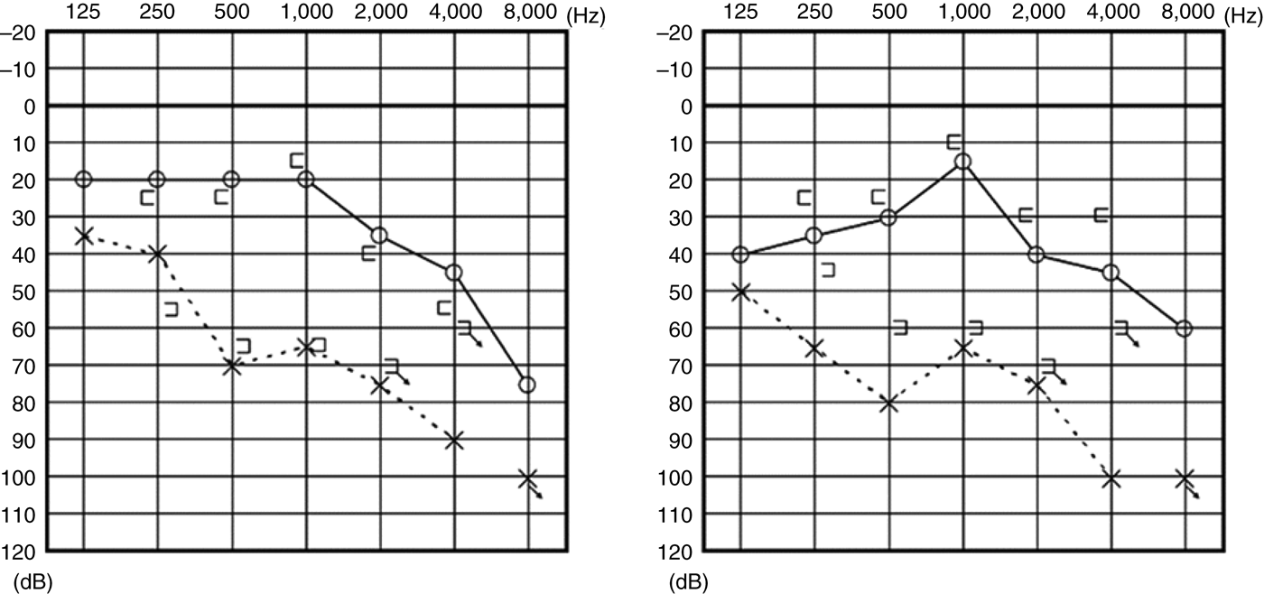
The patient attended our hospital because she wanted to know the cause of her hearing loss and vertigo. Audiography was performed, as shown in Figure 1. She had severe left hearing loss. Moreover, right hearing loss was occasionally present during our observation. Vestibular evoked myogenic potential testing showed that potentials were absent in the right ear but present in the left ear. Caloric response was present but poor at each ear (maximum slow phase velocity, <8°/second).
Two of the patient's audiograms, showing the best (right image) and worst (left image) hearing levels observed.

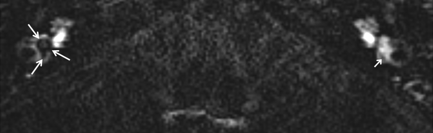
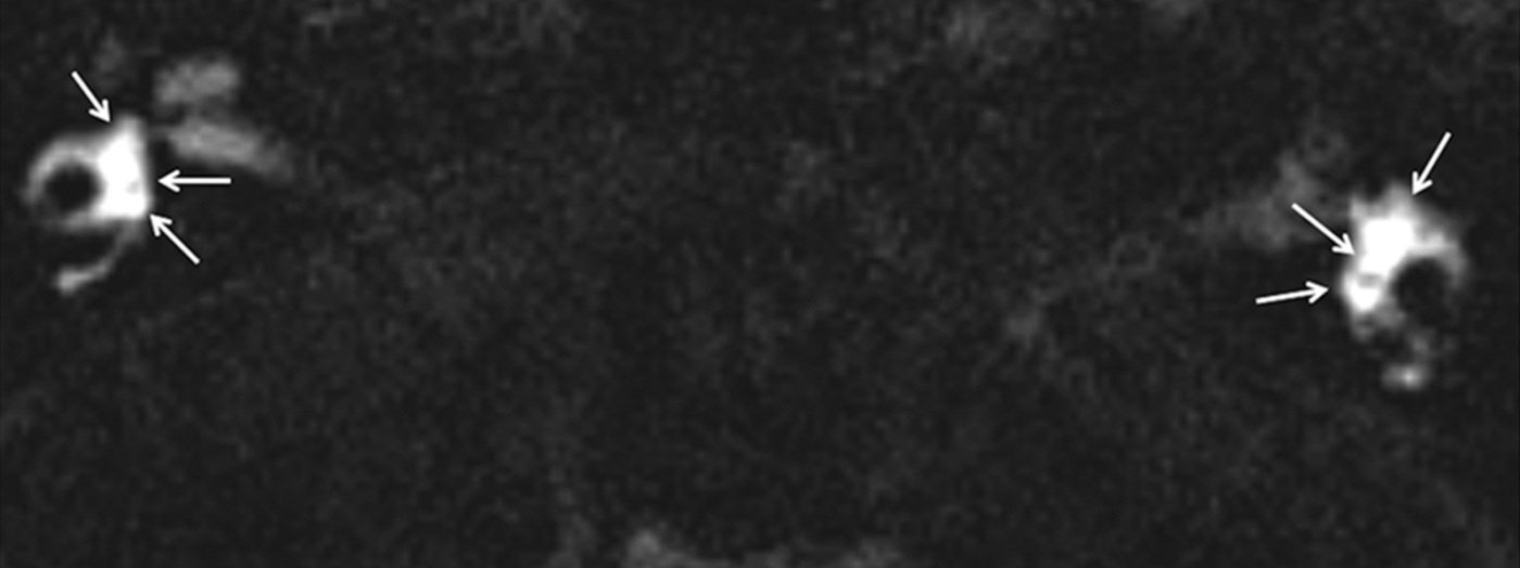
The patient underwent heavily T2-weighted 3D-FLAIR MRI, 4 hours after intravenous injection of a standard dose of gadolinium. The methods for gadolinium injection and 3D-FLAIR MRI have been reported previously.Reference Naganawa, Yamazaki, Kawai, Bokura, Sone and Nakashima6 In normal ears, intravenously administered gadolinium enters the perilymph but does not enter the endolymph. This difference facilitates visualisation of the endolymphatic space. The endolymphatic space consists of regions with low signal intensity which are partly surrounded by high signal intensity perilymphatic fluid when visualised using 3D-FLAIR MRI (Figure 2). However, in the present case the gadolinium enhancement was visible throughout the vestibule, and the endolymphatic space could not be visualised (Figure 3). Thus, we assumed that gadolinium had entered the endolymphatic space as well as the perilymphatic space.
Axial, heavily T2-weighted, three-dimensional, fluid-attenuated inversion recovery magnetic resonance imaging scan of a control subject. Short arrow shows the left vestibule, with a normal endolymphatic space. Long arrows show the right vestibule, with significant endolymphatic hydrops. Elsewhere, this same scan also demonstrated a normal left ear and right-sided Ménière's disease.
Axial, heavily T2-weighted, three-dimensional, fluid-attenuated inversion recovery magnetic resonance imaging scan of the presented patient. The endolymphatic space is not differentiated from the perilymphatic space, as gadolinium enhancement is visible throughout both vestibules (arrows).
Discussion
In this case, we could not differentiate the endolymphatic space from the perilymphatic space, using gadolinium contrast three-dimensional fluid-attenuated inversion recovery (3D-FLAIR) MRI. This was because gadolinium contrast was detected in the endolymph and the perilymph, which we suggest was due to disruption of the blood–labyrinth or blood–endolymph barrier. Tagaya et al.Reference Tagaya, Teranishi, Naganawa, Iwata, Yoshida and Otake7 reported disruption of the blood–labyrinth barrier in patients with sudden deafness. The present report gives the first radiological demonstration of breakdown of the blood–labyrinth or blood–endolymph barrier in a case of relapsing polychondritis with inner ear impairment. Vestibular evoked myogenic potentials were present only at the left ear, although the left hearing loss was more severe than the right hearing loss. We assumed that our patient's inner ear disorders were present in both ears, as the breakdown of the blood–labyrinth or blood–endolymph barrier was shown to be bilateral on 3D-FLAIR MRI.
In relapsing polychondritis, the developmental mechanism of inner ear disorders has yet to be elucidated. Cartilage is not present in the inner ear, so inner ear disorders may be due to circulatory problems caused by vasculitis (which increases the permeability of blood vessels) in the cochlear or vestibular branches of the internal auditory artery.Reference McAdam, O'Hanlan, Bluestone and Pearson1 Three-dimensional fluid-attenuated inversion recovery MRI provides a useful tool for the detection of minute abnormalities in the inner ear.
• Relapsing polychondritis is a rare autoimmune disease characterised by recurrent inflammation of cartilage at multiple sites
• Inner ear disorders, of unknown pathogenesis, appear in one-third of cases
• The reported case of relapsing polychondritis had hearing loss and vertigo
• Three-dimensional fluid-attenuated inversion recovery magnetic resonance imaging showed breakdown of the blood–labyrinth barrier
• This breakdown may be associated with inner ear disorders in relapsing polychondritis
We believe this report will expand understanding of the pathophysiology of inner ear impairments in patients with systemic diseases, including relapsing polychondritis.
Acknowledgements
This study was supported by research grants from the Ministry of Health, Labor and Welfare and from the Ministry of Education, Culture, Sports, Science and Technology of Japan.