Introduction
Paranasal sinus osteomas are benign tumours with an unpredictable growth rate.Reference Koivunen, Löppönen, Pfors and Jokinen1 Malignant transformation is unknown. Cases are often asymptomatic, with tumours detected incidentally during imaging. When symptoms occur, the most common are facial pain, deformity and orbit alteration. The commonest site is the frontal sinuses; this location accounts for between 80Reference Earwalker2 and 96 per centReference Zouloumis, Lazaridis, Papadaki and Epivatianos3 of cases. The second most common site is the anterior ethmoid sinus; when symptomatic, osteomas at this site usually also affect the frontal sinus (i.e. they are ethmoidofrontal). Postero-ethmoidal, maxillary and sphenoid osteomas are rare.
The incidence of osteomas has been established by reviewing radiological images. In a 1939 systematic review of 3510 sinus radiographs, osteomas were identified in 0.43 per cent.Reference Childrey4 A 1993 study of 1500 coronal computed tomography (CT) images identified osteomas in 3 per cent.Reference Earwalker2 Other authors have reported an incidence of only 0.01 per cent.Reference Mehta and Grewal5 The male/female ratio is 1.3/2.Reference Earwalker2
In histological terms, osteomas are divided into three categories: compact, mature and mixed.
Their aetiology is unknown, but three theories have been proposed: traumatic, infectious and embryological.
Complications may occur due to osteoma growth, obstruction of ostia drainage (at the infundibulum), and/or alteration of mucus and/or ciliary function in the sinus mucous lining. Reported complications include: sinusitis, mucocele,Reference Bourgeois, Gichten, Louis, Vincent, Pertuzon and Assaker6, Reference Nabeshima, Marutsuka, Shimao, Uehara and Kodama7 destruction of the anterior sinus wall, intracranial spread, palpebral abscess,Reference Dispenza, Saraniti, Ferrara, Martines, Caramanna and Salzano8 orbital penetration,Reference Namdar, Edelstein, Huo, Lazar, Kimmelman and Soletic9, Reference Karapantzos, Detorakis, Drakonaki, Ganasouli, Danielides and Kozobolis10 pneumocephalusReference Onal, Kaymaz, Arac and Dogulu11–Reference Huneidi and Afshar13 and brain abscess.Reference Roca, Casado, Borras and Gonzalez-Darder14, Reference Panagiotopoulos, Tzortzidis, Partheni, Iliadis and Fratzoglou15
Post-surgical recurrence is rare.Reference Zouloumis, Lazaridis, Papadaki and Epivatianos3
Symptomatic osteomas are conventionally treated by surgery,Reference Smith and Calcaterra16–Reference Savic and Djeric18 although controversy continues to exist over the timing of surgery for asymptomatic osteomas. Surgery is generally recommended if the frontal sinus is breached, if successive imaging reveals growth, or if the osteoma is located close to the fronto-nasal duct.Reference Savic and Djeric18
In this paper, we propose a revised classification of osteomas based on their radiological appearance (i.e. on axial, coronal and sagittal X-ray and/or CT imaging) and clinical symptoms (when present). We believe this staging classification will help clarify the indications for surgical treatment of osteomas.
Materials and methods
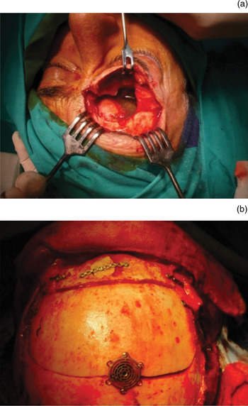
We reviewed 45 cases of frontal osteoma (1973–2007), 29 of which were treated surgically (64.44 per cent). Surgery generally employed the osteoplastic technique, with modified surgical approaches being used in nine cases (Figure 1). No patient underwent endoscopic resection, although in three cases the osteoplastic technique was combined with endoscopy to control the frontal recess.
(a) The osteoplastic technique, the usual surgical approach for osteoma treatment. More complicated techniques may occasionally be required: (b) shows craniotomy used in the case shown in Figure 5.

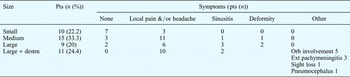
If classified by size only (i.e. using the classification system employed prior to our new system), these osteomas would have been categorised as follows: 10 small osteomas (22.22 per cent), 15 medium-sized ones (33.33 per cent), nine large ones without destruction (20 per cent), and 11 large ones with destruction of the osseous and/or bilateral walls (24.44 per cent) (Table I).
Conventional osteoma classification by size, with associated symptoms: current series

The proportion of asymptomatic cases decreases with increasing tumour size; patients with medium and large tumours have a high incidence of complaints. Pts = patients; destrn = wall destruction; orb = orbital; ext = external
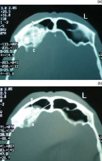
All cases were diagnosed by means of imaging, using conventional radiology for older cases, and simple skull X-ray plus CT (axial, coronal and sagittal views) for cases managed since the early 1980s (Figure 2). In surgical cases, diagnosis was confirmed by histology. Gardner's syndrome was ruled out in all cases.
Axial computed tomography scans showing digital measurement of (a) osteoma and (b) frontal sinus volume. L = left

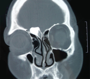
We believed that this classification system, based on osteoma size, was inadequate (Figure 3). Therefore, we set out to develop and implement a new method of classifying frontal osteomas, described below, with the aim of deriving a staging system which would better establish surgical indications.
Coronal computed tomography scan showing a fairly small osteoma in a slightly pneumatised sinus; thus, complications are more likely.

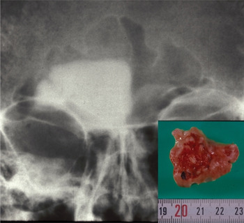
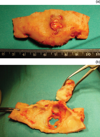
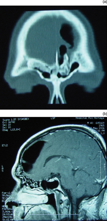
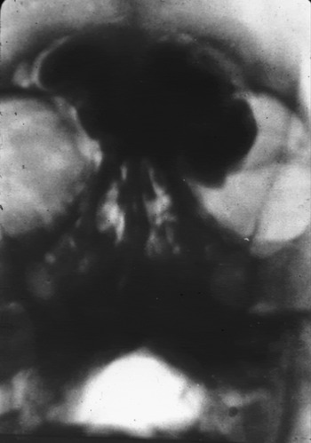
Our new staging system was based on the following four elements: (1) the relation between osteoma mass and total sinus size (i.e. the proportion of tumour mass to frontal sinus air cavity), calculated directly from CT images (using CT software; for older cases, the axes of the osteoma mass and the sinus were measured directly on the X-ray using a slide calliper) (Figure 2); (2) the distance between the osteoma and the infundibulum (i.e. whether the tumour is far away from the infundibulum or whether it partially or completely blocks it) (Figure 4); (3) the presence of destruction of the frontal sinus osseous walls, with osteoma penetration into surrounding areas (Figures 5 and 6); and finally, (4) the presence of complications (Figure 7).
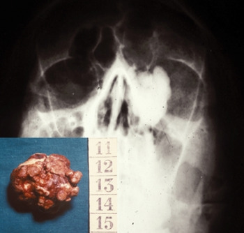
Coronal computed tomography scan showing an osteoma completely blocking the infundibulum. Inset shows the surgical specimen.

Surgical specimen of osteoma which had perforated the anterior wall of the frontal sinus (a). Resection and reconstruction forced a coronal approach with deep craniotomies (b).

(a) Coronal computed tomography scan and (b) sagittal MRI of an osteoma which has broken the posterior wall of the frontal sinus, causing pheumocephalus.

Conventional X-ray showing a large bilateral frontal sinus osteoma. The patient had lost vision in the left eye.

Using these four elements, we defined three discrete osteoma stages, as described below. We discuss the justification of our method in the Discussion section.
Results and analysis
Our 29 surgical cases had a favourable evolution. Our patients' orbital and central nervous system complications were resolved without adverse sequelae.
One patient had absence of unilateral vision post-operatively, but this problem had existed prior to surgery.
In cases with penetration of the orbit or frontal cranial cavity, reconstruction was carried out with a pedicled pericranial flap. Cranial reconstruction osteosynthesis was performed with silk sutures via drill holes in the bone, or with mini-plates (Figure 1).
No recurrence was detected after long-term follow-up (a minimum of two years).
We noted a wide range of sizes and morphological differences in the frontal sinuses encountered, with substantial differences between patients and, occasionally, between the right and left frontal sinuses of the same individual.
When making surgical treatment decisions, we encountered clinical ambiguity in 16 cases (35.56 per cent). As can be seen in Table I, we encountered small and medium-sized osteomas presenting with local pain and/or headache, together with large osteomas which were asymptomatic.
Of our 45 frontal osteoma patients, eight (17.78 per cent) had one or more complications, five had orbital involvement (Figures 7 and 8), five had sinusitis, three had destruction of the posterior sinus wall and external pachymeningitis, one had unilateral sight loss (Figure 7), one had pneumocephalus (Figure 6), and one had destruction of the anterior wall of the frontal sinus (Figure 5). These complications have been described previously,Reference Woldenberg, Nash and Bodner19–Reference Rodriguez Prado, Llorente Pendás, Del Campo Rodriguez, Puente Verez and Suarez Nieto22 although they occurred more frequently in our series.
Coronal computed tomography scan showing an osteoma originating in the infundibulum of the left frontal sinus, which had invaded the orbit leading to exophthalmia and impaired vision. Inset shows the surgical specimen.

In order to standardise the therapeutic indications for osteoma surgery, we established three discrete stages, which we used to categorise our 45 osteoma cases.
In stage I, the proportion of osteoma size to frontal sinus size was one-third or less. In addition, the tumour was well separated from the infundibulum, and there was no sinusitis and no complications. In our series, 18 cases (40 per cent) were categorised as stage I (Figure 9).
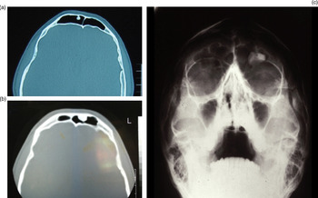
Coronal computed tomography scan (a & b) and conventional X-ray (c) showing three cases of stage I osteoma. The tumour tissue is clearly seen to occupy less than one-third of the frontal sinus cavity. L = left

In stage II, the proportion of osteoma size to frontal sinus size was between one-third and one-half. There was no obstruction of the infundibulum, although the osteoma might be located close to it. There was also no bone destruction, no sinusitis and no complications. Six of our cases (13.33 per cent) were categorised as stage II (Figure 2).
In stage III, the proportion of osteoma size to frontal sinus size was greater than one-half. There was also partial or complete obstruction of the infundibulum, sinusitis, bone destruction and/or complications. Twenty-one of our cases (46.67 per cent) were categorised as stage III (Figure 10).
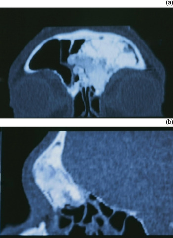
(a) Coronal and (b) sagittal computed tomography scans of an osteoma classified as stage III as it occupied more than half the sinus cavity. It had completely obstructed the infundibulum.

We believe that stage I osteomas do not require surgery, and that a patient follow-up protocol should instead be implemented. Stage II osteomas should be individually assessed to decide whether immediate surgery is indicated, or whether a ‘watchful waiting’ approach should be adopted. Stage III osteomas should always be treated surgically, once the diagnosis is confirmed.
The follow up of stage I osteomas should comprise anamnesis and simple antero-posterior and lateral skull X-rays, performed once a year. For stage II cases not requiring immediate surgery, follow up should comprise, in the first three years, a simple X-ray every six months, together with CT (axial, coronal and sagittal views) once a year. Thus, for those cases not requiring surgery, meticulous attention should be paid to the osteoma growth rate.
Of our 18 stage I cases (40 per cent), 10 required more than five years' follow up, while in the remaining eight cases follow up was interrupted earlier than five years after diagnosis (the minimum monitoring period was two years). Four patients initially categorised as stage I underwent surgery after they were recategorised as stage II, following verification of progressive osteoma growth.
Discussion
Most authors assume that all symptomatic osteomas should be treated surgically, and that the only controversy concerns the management of asymptomatic patients.Reference Smith and Calcaterra16–Reference Savic and Djeric18 However, whether patients are considered symptomatic or not is open to question, considering the fact that, in our experience, the most common symptoms (headache, facial pain and a feeling of localised pressure)Reference Namdar, Edelstein, Huo, Lazar, Kimmelman and Soletic9 are subjective and are often only reported after patients have received a diagnosis of osteoma. On the other hand, some asymptomatic patients who are diagnosed accidentally on skull X-ray have osteomas which extend intracranially.Reference Koivunen, Löppönen, Pfors and Jokinen1 These circumstances have forced us to implement a new classification approach in which subjective symptoms play a minor role.
After studying the characteristics of the cases presented above, we considered that the principal criterion for determining the need for surgery (undertaken to prevent the possibility of complications) was the relationship between osteoma size and frontal sinus air cavity size. We believe that this criterion is even more important than the proximity of the osteoma to the infundibulum. Voluminous osteomas which are situated well away from the infundibulum, but which occupy the majority of the sinus cavity, are highly likely to cause serious orbital and/or intracranial complications.
In our series, our new osteoma staging system accurately determined which cases should be carefully monitored and which should undergo surgery. However, this new staging system is not useful for determining which type of surgical intervention should be performed in individual cases, whether conventional (Figure 1a), modified (Figure 1b) or endoscopic.Reference Chiu, Schipor, Cohen, Kennedy and Palmer23, Reference Chen, Selva and Wormald24 The best surgical approach is that which achieves a safe and complete resection, while minimising any possible after-effects.
The staging system described by Kennedy's groupReference Chiu, Schipor, Cohen, Kennedy and Palmer23 principally refers to the indication for endoscopic resection. In these authors' series of nine patients, three underwent endoscopic resection, and the study assessed the variables which in each patient's case may have limited the use of this technique.
Destruction of one of the frontal sinus' osseous walls, with osteoma penetration into the surrounding areas, always indicates the need for surgery. This destruction is more likely to occur if the osteoma occupies most of the sinus cavity. Destruction of the inferior or naso-orbital wall, with the onset of ophthalmological complications (Figures 7 and 8), has frequently been reported. Destruction of the posterior wall is the usual cause of endocranial complications (Figure 6). Destruction of the anterior wall of the frontal sinus is less frequently described; this is important as it has a decisive influence on surgical planning (Figure 5).
Our proposed follow-up regime for the monitoring of stage I and non-surgical stage II osteomas may seem over-rigorous. It is accepted that osteomas grow slowly,Reference Namdar, Edelstein, Huo, Lazar, Kimmelman and Soletic9, Reference Dubin and Kuhn25, Reference Spencer and Mitchelli26 at a rate of 0 to 6.0 mm/year.Reference Koivunen, Löppönen, Pfors and Jokinen1 This wide variation in growth capacity leads us to believe that osteoma growth is unpredictable, mandating strict follow up.
Osteoma complications are extremely wide-ranging, and by no means rare. Those which occurred in our 45 cases are described above. There is no question that the existence of complications necessarily entails surgery, whatever the size of the osteoma and the sinus cavity which contains it.
Since the early publications on osteomas,Reference Gil-Carcedo, Alarcos, Alarcos and Izquierdo27–Reference Gil-Carcedo, Alarcos, Alarcos and Izquierdo29 no solid criteria have been established to determine the need for surgery. The proximity of the osteoma to the infundibulum is important but it cannot be applied as the single criterion; osteomas which are located away from the infundibulum but which occupy a large part of the sinus cavity (more than one-half) must be considered stage III, with a clear indication for surgery.
The validation and reproducibility of our osteoma staging system is based on its application in a series of 45 cases. The 29 patients treated surgically were operated upon by the same surgeon, allowing us to verify the correspondence between previous radiological images and the actual anatomical situation following surgical exposure of the tumour. We hope that our results will assist the tumour management decision-making process.
Our group works with a surgical navigation system and performs advanced endoscopic surgery. In osteomas considered to be stage I, and thus not requiring surgery, applications for endoscopic surgery are infrequent. Thus far in our practice, we have not identified the ideal circumstances in which to perform endoscopic osteoma resection, whether guided by a navigation system or not. However, the cases of endoscopic resection described by Chiu et al.,Reference Chiu, Schipor, Cohen, Kennedy and Palmer23 and the more numerous cases more recently described by Seiberling et al.,Reference Seiberling, Floreani, Robinson and Wormald30 encourage us to consider this surgical possibility in the future. CloseReference Close31 has published a clear definition of when the endoscopic Lothrop procedure can be used.
• Frontal sinus osteomas are frequently diagnosed as incidental findings on computed tomography (CT) scans
• No firm criteria have been established to determine the need for surgery
• This study proposes a revised osteoma classification based on CT appearance, together with clinical symptoms when present
We emphasise that our proposed osteoma staging system does not seek to advocate any particular surgical technique. Its usefulness is limited to clarifying, in the majority of cases, when surgery should be performed and when careful monitoring of osteoma growth should be implemented, a decision of considerable clinical importance.
Conclusion
The osteoma classification system we propose provides a simple and practical tool for determining the best course of therapeutic action when managing frontal sinus osteomas. The originality of the work lies in linking the size of the osteoma to the size of the frontal sinus cavity.
For stage I osteomas, no surgery is indicated. For stage II osteomas, treatment depends on clinical severity and the patient's condition. We have established a careful follow-up protocol for stage I and non-surgical stage II tumours. Stage III osteomas always require surgery.
Frontal osteoma complications may prove severe, and frequently appear in patients with stage III tumours who have not undergone surgery. In our series of 45 cases, eight (17.78 per cent) developed complications.