Introduction
The effects of resveratrol (3,5,4V-hydroxystilbene) on human and animal health have drawn attention because of its increased use. Resveratrol belongs to the stilbene class of the natural polyphenols, and is found in many common consumer products, including grapes, berries, nuts, red wine and skin care products (Baur & Sinclair, Reference Baur and Sinclair2006). Some in vitro studies have shown the involvement of resveratrol in oxidative stress of the oocytes by increasing reduced glutathione (GSH) intracellular levels (swine: Kwak et al., Reference Kwak, Cheong, Jeon, Lee, Choi, Jeung and Hyun2012; Li et al., Reference Li, Wang, Zhang, Yi, He, Wang, Tian, Yang, Song, He and Liu2016; caprine: Mukherjee et al., Reference Mukherjee, Malik, Saha, Dubey, Singhal, Boateng, Saugandhika, Kumar, De, Guha and Malakar2014). Moreover, Simsek et al. (Reference Simsek, Gurocak, Turkoz, Akpolat, Celik, Ozer, Yilmaz, Turhan and Ozyalin2012) demonstrated that pretreatment of irradiated rats with resveratrol (10–100 mg/kg) protected the ovarian follicle counts and increased the tissue levels of antioxidant enzymes. Resveratrol (2.0 μmol/l) protected porcine oocyte maturation in vitro from heat stress by significantly eliminating ROS, increasing GSH, and up-regulating the expression of SIRT1 (Li et al., Reference Li, Wang, Zhang, Yi, He, Wang, Tian, Yang, Song, He and Liu2016). An in vivo study in rats has shown that resveratrol restored the impaired mitochondrial activity induced by a high fat diet (Ku et al., Reference Ku, Cho, Hong, Lee, Lee, Hong and Lee2016).
However, the potential health benefits of resveratrol seem to be dose dependent. At lower doses, resveratrol can be very useful in maintaining the mammalian, including human, health. Whereas at higher doses it has proapoptotic actions on healthy cells and kills tumour cells (Mukherjee et al., 2010). After in vitro culture of rat theca-interstitial cells, resveratrol (70 or 100 µM) reduced cell viability and induced a dose-dependent proapoptotic effect, increasing caspase-3/7 activation and DNA fragmentation (Wong et al., Reference Wong, Villanueva, Cress and Duleba2010).
As women are able to absorb resveratrol through food, drink or skin contact and the effect of resveratrol on health benefits seems to be dependent on the concentration, it would be important to study the reproductive consequences of resveratrol exposure in vitro. It is important to carry out in vitro reproductive toxicology assays to measure changes in these compounds at the cellular level (Ducolomb et al., Reference Ducolomb, Casas, Valdez, González, Altamirano-Lozano and Betancourt2009). Alterations can, therefore, not only have negative effects on follicle development, oocyte maturation and ovulation, but also significantly impair fertility (Stefansdottir et al., Reference Stefansdottir, Fowler, Powles-Glover, Anderson and Spears2014).
Sheep have been chosen as animal model as, in addition to their economic importance, the ewe's ovary is similar to the human ovary in its architecture and physiology (Gosden et al., Reference Gosden, Baird, Wade and Webb1994; Demirci et al., Reference Demirci, Lornage, Salle, Poirela, Guerin and Franck2003; Fransolet et al., Reference Fransolet, Labied, Henry, Masereel, Rozet, Kirschvink, Nisolle and Munaut2014). Thus, this study was designed to test the hypothesis that resveratrol influences survival, growth, DNA damage and oxidative stress of ovine early-stage follicles cultured in vitro. Therefore, we evaluated the effect of resveratrol on secondary follicle morphology, DNA fragmentation, intracellular GSH levels and metabolically active mitochondria.
Materials and methods
Chemicals
Unless otherwise mentioned, culture medium, resveratrol, supplements and chemicals used in the present study were purchased from Sigma Chemical Co. (St. Louis, MO, USA).
Source of ovaries
Ovaries (n = 50) were collected at a local abattoir from 25 adult (1–3 years old) mixed-breed sheep. Immediately postmortem, pairs of ovaries were washed once in 70% alcohol and then twice in Minimum Essential Medium buffered with HEPES (MEM-HEPES) and supplemented with antibiotics (100 μg/ml penicillin and 100 μg/ml streptomycin). The ovaries were transported within 1 h to the laboratory in tubes containing MEM-HEPES supplemented with antibiotics (100 μg/ml penicillin and 100 μg/ml streptomycin) at 4°C (Chaves et al., Reference Chaves, Martins, Saraiva, Celestino, Lopes, Correia, Lima Verde, Matos, Báo, Name, Campello, Silva and Figueiredo2008).
Isolation and selection of secondary follicles
In the laboratory, the surrounding fatty tissues and ligaments were stripped from the ovaries. The ovarian cortical slices (1 to 2 mm thick) were cut from the ovarian surface using a surgical blade under sterile conditions and subsequently placed in fragmentation medium consisting of MEM-HEPES with antibiotics. Secondary follicles, approximately 250–400 μm in diameter without antral cavities, were visualized under a stereomicroscope (SMZ 645, Nikon, Tokyo, Japan) and mechanically isolated by microdissection using 26-gauge (26G) needles. These follicles were then transferred to 100-μl droplets containing base culture medium for evaluation of quality. Only secondary follicles that displayed the following characteristics were selected for culture: an intact basement membrane, two or more layers of granulosa cells and a visible and healthy oocyte that was round and centrally located within the follicle, without dark cytoplasm. Isolated follicles were pooled and then randomly allocated to the treatment groups, with approximately 45–50 follicles per group.
In vitro culture of secondary follicles
After selection, the follicles were divided randomly into four study groups and cultured individually (one follicle per droplet) in 100-μl droplets of culture medium under mineral oil in Petri dishes (60 × 15 mm, Corning, USA). The base control medium consisted of α-MEM (pH 7.2–7.4) supplemented with 3.0 mg/ml bovine serum albumin (BSA), 10 ng/ml insulin, 5.5 µg/ml transferrin, 5.0 ng/ml selenium, 2 mM glutamine, 2 mM hypoxanthine and 50 µg/ml ascorbic acid and then referred as α-MEM+. For the experimental conditions, the control medium was supplemented with resveratrol at different concentrations (2, 10 or 30 µM) (Ortega et al., Reference Ortega, Wong, Villanueva, Cress, Sokalska, Stanley and Duleba2012; Giaretta et al., Reference Giaretta, Spinaci, Bucci, Tamanini and Galeati2013). All follicles were cultured at 39°C under 5% CO2 for 18 days. Every 2 days, 60 μl of the culture medium were replaced with fresh medium in each droplet.
Evaluation of follicle morphology and development
During and after culture, follicles were classified according to their morphological characteristics, and those showing morphological signs, such as darkness of the oocytes and the surrounding granulosa cells, misshapen oocytes, rupture of the basement membrane and/or oocyte extrusion were classified as atretic. Analysis of follicular morphology was performed every 6 days of culture.
The following characteristics were analyzed in the morphologically normal follicles: (i) antral cavity formation, defined as the emergence of a visible translucent cavity within the granulosa cell layers; (ii) the diameter of healthy follicles, measured from the basement membrane, which included two perpendicular measures of each follicle; and (iii) the growth rate, calculated as the diameter variation during the culture period.
After 18 days of culture, all healthy follicles were carefully and mechanically opened with 26G needles under a stereomicroscope for oocyte recovery. The percentage of fully grown oocytes, i.e., oocyte ≥110 μm, was calculated as the number of acceptable quality oocytes (≥ 110 μm) recovered out of the total number of cultured follicles (× 100).
Assessment of DNA fragmentation by TUNEL assay
At the end of culture, parts of the oocytes were recovered from the follicles and subjected to the terminal deoxynucleotidyl transferase-mediated dUTP nick end labeling (TUNEL) assay as previously described (Li et al., Reference Li, Liu, Cang, Wang and Shorgan2009). Briefly, after in vitro culture, oocytes were fixed in 4% paraformaldehyde solution for 1 h at room temperature and incubated in droplets of 100 µl of permeabilizing solution [0.1% (v/v) Triton X-100 in 10 mM PBS] for 3 h at room temperature. Positive and negative controls were incubated in drops of 100 µl containing DNase-free RNase (Invitrogen Corporation, Carlsbad, CA, USA) at 37°C for 1 h and washed three times in 50-µl drops of PBS–polyvinylpyrrolidone (PVP). The TUNEL assay was prepared about 15 min prior to use and kept on as indicated by the manufacturer 4°C (In Situ Cell Detection Kit, Fluorescein: Boehringer Mannheim/Roche Diagnostics). To this end, 12.5 µl terminal deoxynucleotidyl transferase enzyme and 112.5 µl of marker solution of 2-deoxyuridine triphosphate 5-FITC were made to obtain 125 µl of TUNEL mixture for reaction. The experimental groups and the positive control were incubated with 15 µl of this solution for 1 h at 37°C in a moist chamber in the dark. The negative control was incubated at 15 µl with the marker solution. Oocytes were washed three times in 50-µl drops of PBS-PVP and incubated in drops of 50 µl containing 10 mM Hoechst 33342 for 15 min at room temperature in the dark. Oocytes were washed in PBS-PVP and slides were prepared for evaluation using an epifluorescence microscope (Nikon E200, Tokyo, Japan) at a magnification of ×400. DNA fragmentation was observed as green fluorescence chromatin.
Measurement of GSH levels and metabolically active mitochondria
After culture, other oocytes were recovered and intracellular GSH levels and mitochondrial activity were measured as previously described (Wang et al. 2014; Tanabe et al., Reference Tanabe, Tamura, Taketani, Okada, Lee, Tamura, Maekawa, Asada, Yamagata and Sugino2015), with minor modifications. Briefly, 4-chloromethyl-6.8-difluoro-7-hydroxycoumarin (CellTracker® Blue; CMF2HC; Invitrogen, Eugene, Oregon, USA) and MitoTracker Red (MitoTracker® Red, CMXRos, Molecular Probes, Melbourne, Victoria, Australia) were used to detect intracellular GSH and mitochondrial activity levels as blue and red fluorescence, respectively. Oocytes were incubated in the dark for 30 min in PBS supplemented with 10 mM of CellTracker Blue and 100 nM MitoTracker Red at 39°C. After incubation, the oocytes were washed with PBS and the fluorescence was observed under an epifluorescence microscope with UV filters (370 nm for GSH and 579 nm for active mitochondria). Fluorescence intensities of oocytes were analyzed by using the ImageJ software (National Institutes of Health, Bethesda, MD, USA).
Statistical analysis
Data from normal follicles, antrum formation and retrieval of grown oocytes after in vitro culture were expressed as percentages and compared by chi-squared test. Data from GSH, metabolic active mitochondria, follicular diameter and growth rates were submitted to the Shapiro–Wilk test to verify normal distribution of residues and homogeneity of variances. Thereafter, Kruskal–Wallis non-parametric test was used for comparisons. When main effects or interactions were significant, means were compared by Student–Newman–Keuls test. Data from TUNEL-positive cells were submitted to PLSD Fisher test and expressed as percentage. The results were expressed as the means ± standard error mean (SEM), and differences were considered significant when the P-value was < 0.05.
Results
Follicular morphology and development after in vitro culture
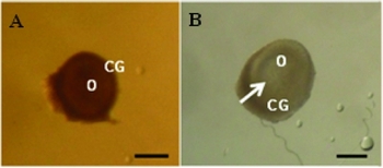
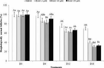
Morphologically normal follicles showed centrally located oocytes and normal granulosa cells, which were enclosed by an intact basement membrane (Fig. 1 A). As early as day 6 of culture, a small antral cavity appears in all treatments, filled with follicular fluid (Fig. 1 B). From day 0 to day 18, there was a reduction (P < 0.05) in the percentage of morphologically normal follicles in all treatments (Fig. 2). Considering the same culture period, the percentages of normal follicles were similar (P > 0.05) among control and resveratrol treatments at day 12 of culture. However, all resveratrol treatments significantly decreased the normal follicle rates at day 18 compared with the control group (α-MEM+).
Follicular development after in vitro culture. (A) Secondary follicle at day 0. (B) Antral follicle after 6 days of culture at 2 μM resveratrol. O: oocyte; GC: granulosa cell. Arrow: antral cavity. Scale bars: 200 µm.

Percentages of morphologically normal follicles cultured in α-MEM+ or different concentrations of resveratrol (RSV 2, 10 or 30 µM). a,bDifferent letters denote significant differences among culture periods in the same treatment (P < 0.05). A,BDifferent letters denote significant differences among treatments in the same period (P < 0.05).

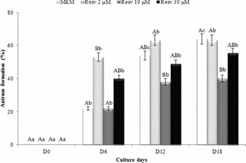
From day 6 of culture onward, antral cavity formation was observed in all treatments (Fig. 3). At the end of culture, antrum formation was higher in both α-MEM+ and 2 µM resveratrol treatments than 10 µM resveratrol (Fig. 3; P < 0.05). There were no significant differences (P > 0.05) in follicular diameter, in the daily growth rate or in the percentage of oocytes larger than 100 μm at the end of culture among treatments (data not shown).
Percentages of antral cavity formation in follicles cultured in α-MEM+ or different concentrations of resveratrol (RSV 2, 10 or 30 µM). a,bDifferent letters denote significant differences among culture periods in the same treatment (P < 0.05). A,BDifferent letters denote significant differences among treatments in the same period (P < 0.05).

DNA fragmentation after exposure to resveratrol
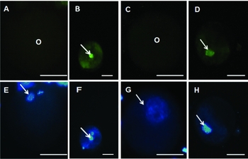
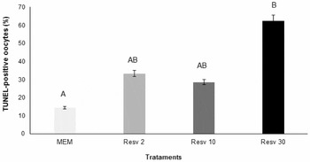
TUNEL assay illustrates healthy oocyte in the control group (α-MEM+) (Fig. 4 A) and oocytes with DNA fragmentation in medium containing 30 µM resveratrol (Fig. 4 B). The negative control did not show staining for TUNEL analysis (Fig. 4 C), while all oocytes showed DNA damage in the positive control (Fig. 4 D). Moreover, all oocytes showed stained chromatin with Hoechst 33342 in blue fluorescence (Fig. 4 E–H). Follicles cultured with 30 µM resveratrol showed a significantly higher percentage (62.5%) of oocytes with DNA fragmentation than the control group (14.3%; Fig. 5). However, no differences on TUNEL-positive oocytes were observed among α-MEM+, 2 or 10 µM resveratrol (P > 0.05).
Representative DNA fragmentation of ovine oocytes after 18 days of culture. Normal oocyte in the control group (A, E), TUNEL-positive oocytes in 30 µM resveratrol treatment group (B, F), normal oocyte in the negative control (C, G) and oocyte with DNA damage in the positive control (D, H). Oocytes stained with TUNEL (A–D) and Hoechst 33342 (E–H). Note the DNA fragmentation, as evidenced by green chromatin in (B) and (D). Scale bars: 50 µm.

Percentage (mean ± SEM) of TUNEL-positive oocytes after 18 days of culture in different concentrations of resveratrol (Resv 2; 10 or 30 µM). A,BDifferent letters denote significant differences between treatments (P < 0.05).

Effects of resveratrol on intracellular GSH levels and mitochondrial activity
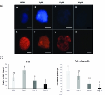
Oocytes from follicles treated with 10 and 30 µM resveratrol significantly decreased (P < 0.05) intracellular GSH levels compared with 2 µM resveratrol (Fig. 6). Moreover, the fluorescence intensity of metabolically active mitochondria measured after MitoTracker Red labeling was significantly decreased by 10 or 30 µM resveratrol, compared with the control group (α-MEM+) (Fig. 6).
Epifluorescence photomicrographic images of in vitro cultured ovine oocytes. (a) Oocytes were stained with CellTracker Blue (A–D) and MitoTracker Red (E–H) to detect intracellular levels of glutathione (GSH) and mitochondrial activity, respectively. Oocytes cultured in the control group (A, E) or with 2.0 (B, F), 10.0 (C, G) or 30 µM (D, H) resveratrol. Scale bars: 50 µm. (b) Effect of resveratrol on intracellular GSH and active mitochondria levels in sheep oocytes. Within each group (GSH and active mitochondria) of end point, bars with different letters (A–C) are significantly different (P < 0.05).

Discussion
Resveratrol is a therapeutic potential compound (cardioprotective, anticancer, anti-inflammatory and antioxidant effects) abundantly found in nuts, grapes and red wine (Baur & Sinclair Reference Baur and Sinclair2006; Pangeni et al., Reference Pangeni, Sahni, Ali, Sharma and Baboota2014). In vitro studies are a valuable tool to enable clear analysis of whether resveratrol acts positively or negatively on reproductive functions. Therefore, the present study evaluated the effects of resveratrol on sheep ovarian follicles in vitro by testing a concentration similar to that found in human serum (2 μM; Goldberg et al., Reference Goldberg, Yan and Soleas2003) and higher concentrations (10 μM and 30 μM). Under those conditions, resveratrol decreased the percentage of morphologically normal follicles compared with the control medium. In addition, almost 60% of the oocytes showed DNA fragmentation when exposed to the highest resveratrol concentration (30 µM), while only 14.2% of the oocytes with DNA damage could be detected in the control group (α-MEM+). Our data also showed that 10 or 30 µM resveratrol dramatically reduced mitochondrial activity compared with the control. These findings were also accompanied by decreased GSH levels compared with the lowest concentration of resveratrol tested (2 µM).
One of the important mechanisms leading to apoptotic cell death is oxidative stress, which is characterized by an inability of the cell to counteract an overwhelming production of ROS (Gupta et al., Reference Gupta, Miller, Babus and Flaws2006). Oxidative stress stimulates cellular damage and the mitochondria are the first organelles to degenerate because they are the site of oxygen radical production, which leads to mitochondrial dysfunction (Tanabe et al., Reference Tanabe, Tamura, Taketani, Okada, Lee, Tamura, Maekawa, Asada, Yamagata and Sugino2015). It was shown that resveratrol inhibits mouse skin tumour development through induction of apoptosis via mitochondrial cell death pathway, increasing the levels of cytochrome c, the adaptor Apaf-1, cleaved caspase 9, and caspase 3 (Kalra et al., Reference Kalra, Roy, Prasad and Shukla2008). Moreover, resveratrol inhibits ATP synthase, affecting the inter-organelle coupling of mitochondria and endoplasmic reticulum in cancer cells, yielding enhanced mitochondrial Ca2+ accumulation, the initiation of apoptotic pathways, and cell death (Madreiter-Sokolowski et al., Reference Madreiter-Sokolowski, Gottschalk, Parichatikanonda, Eroglu, Klec, Waldeck-Weiermair, Malli and Graier2016). Therefore, in the current study, a more reasonable explanation is that the greater DNA damage promoted by 30 µM resveratrol could be related to the reduced mitochondrial activity. After rat theca-interstitial cell culture, Wong et al., (Reference Wong, Villanueva, Cress and Duleba2010) also observed that resveratrol reduced cell viability at 70 or 100 µM as well as induced a dose-dependent increase in caspase-3/7 activation and DNA fragmentation at concentrations of 50 and 100 µM. In their study, 30 µM resveratrol did not increase cellular apoptosis.
Cells protect themselves from ROS through various defence mechanisms including antioxidant enzymes such as superoxide dismutase (SOD), catalase (CAT) (Gupta et al., Reference Gupta, Miller, Babus and Flaws2006) and GSH (Luberda, Reference Luberda2005). Compared with the highest concentration tested (30 µM), the increase in GSH levels and mitochondrial activity in response to 2 µM resveratrol might be an attempt to protect the follicles from oxidative damage as resveratrol may act as an antioxidant against oxidative stress in different types of cells (oocytes: Kwak et al., Reference Kwak, Cheong, Jeon, Lee, Choi, Jeung and Hyun2012; spermatozoa: Ünal et al., Reference Ünal, Take, Erdogan, Göktas and Sahin2013; kidney cells: Valentovic et al., Reference Valentovic, Ball, Brown, Terneus, McQuade, Van Meter, Hedrick, Roy and Williams2014). Therefore, as suggested previously, resveratrol can work as a pro-oxidant as well as an antioxidant agent depending on the concentration administered to the cells (Delmas et al., Reference Delmas, Aires, Dutartre, Mazué, Ghiringhelli and Latruffe2011). Moreover, although follicle structure may not be optimally preserved by culture in medium containing resveratrol, our findings appear to suggest that the remaining follicles in the 2 μM resveratrol treatment were in good condition and able to resume their development, as demonstrated by the presence of antral follicles and by oocyte quality (based on DNA damage, GSH and mitochondria activity levels), which were not different to that of the control medium.
In the current study, antral cavity formation was higher in α-MEM+ and 2 µM resveratrol treatments than 10 µM resveratrol, however, no effect on follicular growth was observed. After antrum formation, the follicle becomes dependent on FSH (Erickson & Shimasaki, Reference Erickson and Shimasaki2001), suggesting that the use of FSH in the base culture medium may be necessary to give a large support to antral follicle growth. Nevertheless, supplementation of resveratrol (0.24 and 0.5 µM) during in vitro maturation of goat oocytes improved embryonic developmental potential (Mukherjee et al., Reference Mukherjee, Malik, Saha, Dubey, Singhal, Boateng, Saugandhika, Kumar, De, Guha and Malakar2014), thus suggesting that at optimum concentration, resveratrol may be important for oocyte growth.
Overall, this study shows that ovine isolated secondary follicles are able to grow to the antral stage after in vitro culture in medium supplemented with 2 µM resveratrol, maintaining the same rates of DNA damage, GSH levels and mitochondrial function as the control medium. Conversely, at a concentration of 30 µM, resveratrol increased follicular DNA fragmentation and oxidative stress through decreasing mitochondrial activity. Our next goals are to extend the culture period in order to evaluate complete development of secondary follicles and their ability to produce normal oocytes with higher survival rates. Moreover, the potential reproductive risk associated with excessive levels of resveratrol exposure should be more studied.
Acknowledgements
This work was supported by FACEPE (Process APQ-1853–5.05/12). T.J.S. Macedo holds a scholarship from the FACEPE. M.H.T. Matos is supported by a grant from CNPq.
Conflict of interest
None of the authors has any conflict of interest to declare.