Introduction
There is a growing consensus that in order to understand the pathogenesis of Alzheimer's disease (AD), it is necessary to begin investigations in people who are at risk for the disease but are clinically healthy (Sperling et al., Reference Sperling2011). The recent growth in the development of putative neuroimaging, cerebro-spinal fluid (CSF), and blood biomarkers for AD pathology provides increasing impetus for studies of people at risk for AD, because biomarkers may allow models of AD risk to be tested without waiting for cognition and behavior to deteriorate to allow clinical classification of AD. Consequently, there is now substantial international effort to study prospectively relationships between clinical characteristics and putative AD biomarkers in groups who carry different risk factors for AD. Some of these studies focus on genetic risk factors (Reiman et al., Reference Reiman2011; Morris et al., Reference Morris2012); however, studies such as the Australian Imaging Biomarkers and Lifestyle Flagship Study of Ageing (AIBL) and Alzheimer's Disease Neuroimaging Initiative (ADNI) have examined cohorts of individuals aged over 60 years who possess varying risk factors for AD.
The AIBL study is a prospective study of a large group (1,112) of individuals aged over 60 years who are either classified as cognitively healthy, or meet clinical criteria for mild cognitive impairment (MCI) or AD, and who have agreed to reassessment every 18 months. Assessment comprises extensive study of cognitive function, neuroimaging, blood biomarkers, and lifestyle (diet and exercise) characteristics (Ellis et al., Reference Ellis2009). By combining these investigations in a prospective fashion, the AIBL study contributes to the understanding of the development and progression of AD through the prodromal, preclinical, and clinical stages of the disease. While data for clinical and pathological markers are continually being communicated in subsamples of the AIBL cohort, it is crucial that the clinical and cognitive characteristics of the entire AIBL cohort be reported as the study advances, especially as current clinical models of early AD are based almost entirely on the objective identification of subtle cognitive impairment.
Careful reporting of the clinical and cognitive status of the individuals at risk for AD who are enrolled in large prospective studies of relationships between biomarkers and clinical status is important because this will provide estimates of the rate and nature of progression of AD that have been derived from carefully characterized samples. Second, such reports provide important information about the nature and magnitude of cognitive impairment at different stages of disease progression and third, they set the context for any clinical–pathological relationships observed in sub-samples of the cohort. The first aim of this study was to describe the clinical status of the AIBL cohort at the 18-month reassessment, and in particular, the rate at which there was an emergence of cognitive impairment in the healthy cohort and progression of disease in the MCI cohort. The second aim of the study was to describe the nature and magnitude of change in cognitive function that accompanied change in clinical status from healthy to MCI and from MCI to AD.
Methods
Participants
We sought to reassess the baseline cohort of 1,112 individuals, 18 months after their enrollment and initial assessment in AIBL, from the following baseline groups: (1) 211 individuals with AD as defined by NINCDS-ADRDA criteria (McKhann et al., Reference McKhann, Drachman, Folstein, Katzman, Price and Stadlan1984); (2) 133 individuals with MCI – MCI is a clinical syndrome describing impaired cognitive performance (often involving memory), in the absence of frank dementia, which represents a high-risk state for the development of AD (Petersen et al., Reference Petersen, Smith, Waring, Ivnik, Tangalos and Kokmen1999; Winblad et al., Reference Winblad2004); (3) 768 healthy individuals without cognitive impairment (HC).
At the 18-month follow-up, individuals underwent cognitive and clinical assessment and the data collected were assessed by a clinical review panel (chaired by DA), before allocation of each participant into one of five diagnostic groups: AD by NINCDS-ADRDA criteria, MCI, HC reporting subjective memory concern (SMC), HC reporting no subjective memory concern (non-SMC), and other (e.g. non-AD dementia, psychiatric disorder other than dementia, etc.), details of which are outlined below. Subjective memory concern status was determined by the subject's “yes” or “no” response to the single question “Do you have difficulty with your memory,” and this same question was asked at both baseline and 18-month follow-up.
Ethics approval
The AIBL study, including the 18-month follow-up protocol, was approved by the institutional ethics committees of Austin Health, St Vincent's Health, Hollywood Private Hospital and Edith Cowan University, and all volunteers gave written informed consent before participating in the study.
Attendance for AIBL reassessment
Assessments took place at three locations in Melbourne and at two locations in Perth, depending on whether the participants were to undergo brain imaging and where they lived. A very small number of participants (particularly some affected by AD) were assessed by AIBL staff at their home.
All assessments were conducted in the mornings, after an overnight fast. Weight, height, abdominal girth, sitting blood pressure, and pulse were measured, and 80 ml of blood drawn. Participants were then provided with breakfast, following which their cognition and mood were assessed, as described below.
Cognitive and mood assessment
The AIBL cognitive and mood assessment has been described previously (Ellis et al., Reference Ellis2009). Cognitive and mood tests were performed by trained staff, most of whom were qualified neuropsychologists and all of whom had undergone extensive training in the assessment techniques. The full assessment battery comprised the Mini-Mental State Examination (MMSE; Folstein et al., Reference Folstein, Folstein and McHugh1975), California Verbal Learning Test – Second Edition (CVLT-II; Delis et al., Reference Delis, Kramer, Kaplan and Ober2000), Logical Memory I and II (WMS; Story 1 only; Wechsler, Reference Wechsler1945), D-KEFS verbal fluency (Delis et al., Reference Delis, Kaplan and Kramer2001), 30-item Boston Naming Test (BNT; Saxton et al., Reference Saxton2000), Wechsler Test of Adult Reading (WTAR; Wechsler, Reference Wechsler2001), Digit Span and Digit Symbol-Coding subtests of the Wechsler Adult Intelligence Scale – Third Edition (WAIS-III; Wechsler, Reference Wechsler1997), the Stroop task (Victoria version; Strauss et al., Reference Strauss, Sherman and Spreen2006), and the Rey Complex Figure Test (RCFT; Meyers and Meyers, Reference Meyers and Meyers1995). Participants also completed the computerized CogState battery (www.cogstate.com) and for all CogState tasks, speed (reaction time in milliseconds) and accuracy (number of correct responses made) of each performance were recorded. The length of a typical assessment was between one and two hours. For a more comprehensive account of the cognitive battery and the rationale behind the selection of individual tests, readers are referred to our baseline cohort paper (Ellis et al., Reference Ellis2009).
Both the 15-item version of the Geriatric Depression Scale (GDS-15; Sheik and Yesavage, Reference Sheikh and Yesavage1986) and the Hospital Anxiety and Depression Scale (HADS; Zigmond and Snaith, Reference Zigmond and Snaith1983; Snaith and Zigmond, Reference Snaith and Zigmond1986) were completed at the follow-up visit. For participants with a diagnosis of AD or MCI, and for some HC participants where decline was suspected (e.g. a decline of 1.5 SD over 18 months on one or more cognitive domains), we sought to contact an informant if such a person was available and if the participant consented to this contact. These informants were asked to provide additional information about the functional performance of the research participants, and to complete the Informant Questionnaire on Cognitive Decline (IQCODE – Short Form; Jorm and Jacomb, Reference Jorm and Jacomb1989).
Dementia severity was rated for all participants using the Clinical Dementia Rating (CDR) scale (Morris, Reference Morris1993) on the basis of information obtained from cognitive testing, direct questioning of the participant, and information from an informant and/or from the participant's treating clinician (for those diagnosed with AD or MCI). Both an overall CDR score (range 0–3) and a “sum of boxes” score (range 0–18) were generated. All CDR ratings were reviewed in detail by one person (DA) to ensure consistency.
Blood samples and lifestyle questionnaires
The 80 ml blood samples taken on arrival were processed and analyzed as detailed in our earlier baseline report (Ellis et al., Reference Ellis2009) and the findings of these 18-month repeat blood analyses will be reported in future publications. Basic pathology test results (e.g. full blood examination, electrolytes, thyroid function, etc.) were available to the diagnostic review panel if needed and results outside the normal range were flagged for immediate attention and reporting to the patient's regular doctor where indicated. All returning participants were asked to complete the International Physical Activity Questionnaire (IPAQ; Craig et al., Reference Craig2003).
Medical history and medication use
All participants completed a detailed questionnaire regarding personal medical history since baseline, medication use and smoking, and current and past alcohol and illicit drug use.
Brain imaging
At baseline, 288 participants (178 healthy controls, 57 MCI, and 53 AD) received neuroimaging using Positron Emission Tomography with Pittsburgh Compound B (C11-PiB PET imaging) and Magnetic Resonance Imaging (MRI) as previously reported (Rowe et al., Reference Rowe2010). Overall, 227 of 288 previously scanned participants received repeat scans at follow-up, 6 had died, 26 returned for AIBL follow-up but were not scanned, and 29 did not return for reassessment. These imaging results form the focus of other reports from our group, and will not be discussed here as the current paper reports on the entire cohort.
Clinical review and the diagnosis of AD or MCI
Monthly clinical review panel meetings were conducted to discuss the 18-month diagnostic classification for all participants with a baseline diagnosis of AD or MCI, and for those initially classified as HCs whose 18-month diagnostic status required further consideration. This latter group included participants who demonstrated any of the following: MMSE score <28/30, failure on the Logical Memory test (as per ADNI criteria, as described previously in Ellis et al., Reference Ellis2009), other evidence of possibly significant cognitive difficulty on neuropsychological testing (i.e. a score of 1.5 SD below the relevant normative mean on any neuropsychological measure), a CDR score of 0.5 or greater, medical history suggestive of the presence of illnesses likely to impair cognitive function, informant or personal history suggestive of impaired cognitive function, or the consumption of medications or other substances in quantities that could influence cognition. A consensus diagnosis was assigned for each such participant, using internationally agreed diagnostic criteria according to both DSM-IV (American Psychiatric Association, 1994) and ICD-10 (World Health Organization, 1992). Where appropriate, ICD-10 dementia severity rating (World Health Organization, 1992), NINCDS-ADRDA AD diagnosis (probable or possible), and MCI classifications were applied (see below). The clinical review panel comprised old age psychiatrists (DA, NL), neurologists (DD and CS), a geriatrician (MW) and psychologists (KE, GS), and other staff members who attended from time to time in order to provide information about individual subjects and their assessments. A quorum was formed by three members, and included at least one medically qualified member and at least one psychologist member. The panel conferred by telephone conference and five or more panel members attended all meetings, with all but one of the follow-up review conferences being chaired by DA (the other was chaired by NL). Diagnoses were made blind to the results of AIBL neuroimaging data, but were often informed by structural brain imaging that had been conducted for independent clinical reasons. Summary neuropsychological, mood, and informant data for both baseline and 18-month follow-up testing were available to the diagnostic panel.
As at baseline, MCI diagnoses were made according to a protocol based on the criteria of Winblad et al. (Reference Winblad2004), which are informed by the criteria of Petersen et al. (Reference Petersen, Smith, Waring, Ivnik, Tangalos and Kokmen1999). Consistent with Winblad criteria, all the participants classified with MCI had either personally, or through an informant, reported memory difficulties at follow-up. Participants with a baseline clinical diagnosis of MCI were required to demonstrate the following at reassessment in order to be retained in the MCI category: (1) a score 1.5 SDs or more below the relevant normative mean on at least one neuropsychological test, and (2) a current report of subjective memory concern either from the individual or from an informant. Participants who were classified as HCs at baseline had to fulfill the more stringent criterion of impairment on two or more cognitive tests at a level at least 1.5 SDs below the normative mean, in addition to reporting subjective memory concern, to be classified as MCI. The greater stringency applied to allocating individuals presenting as HCs to the MCI category was decided upon after extensive discussion, is justified by the acknowledged mutability of MCI diagnoses, and replicates our practice when diagnosing MCI at baseline. All participants with MCI manifested substantially intact activities of daily living, exhibited no clear evidence of significant impairment in their social or occupational functioning, and did not fulfill ICD-10 or DSM-IV diagnostic criteria for dementia.
Statistical analyses
For participants presenting for reassessment, the proportion of those transitioning from HC to MCI and MCI to AD over 18 months was calculated. Two-sample t-tests and tests of differences in proportions were used to investigate baseline differences between transitioning and non-transitioning groups. Analysis of variance (ANOVA) was undertaken to look for differences in cognition between HCs with and without a memory complaint. For both HC at baseline and MCI at baseline, a series of linear model (LM) analyses were conducted to examine cognitive change (18 months minus baseline assessment) between transition groups and stable groups (i.e. HC stable vs. HC to MCI transition; MCI stable vs. MCI to AD transition). All LM analyses were adjusted for age, sex, and APOE ε4 status. The magnitude of the difference in adjusted means between the transition group and the stable group for each performance measured at the 18-month assessment was expressed using Cohen's d.
Results
Cohort reassessment
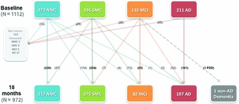
Figure 1 shows the total numbers of volunteers reassessed after 18 months and both the initial category to which each volunteer was assigned at baseline and the category of allocation after the 18-month reassessment and clinical review. Of the 1,112 participants in the inception cohort, follow-up data were collected for 89.9% (1,000) at 18 months (972 participants were reassessed and 28 had died) and a further 112 did not return for reassessment. The 18-month classifications were: 692 HCs (317 non-SMC and 375 SMC), 82 MCI cases, and 197 patients with AD dementia. One patient was classified as having non-AD dementia.
Summary of clinical classification and transitions over 18 months.
Note: SMC = Healthy control with subjective memory concern (answered “yes” to the question “Do you have difficulty with your memory?”); NMC = Healthy control with no subjective memory concern; MCI = Mild Cognitive Impairment; AD = dementia due to Alzheimer's disease by NINCDS/ADRDA criteria.

The number of deaths was greatest in the AD group (of 28 subjects who died, there were 17 AD, 5 MCI, and 6 HC). Of the additional 112 individuals who did not undergo 18-month reassessment (33 AD, 23 MCI, and 56 HC), the (sometimes multiple) reasons given for non-participation were as follows: not contactable (32), too ill for assessment (20), found the baseline cognitive assessment too difficult/challenging (20), too busy (17), AD now too severe (11), family illness (5), refused because of involvement in “too many” research projects (4), and no reason given (5).
Following detailed review by the clinical panel, 32 MCI cases had progressed to fulfill NINCDS-ADRDA diagnostic criteria for dementia due to AD. Accounting for baseline MCI cases lost to follow-up, a transition rate of 30.5% from MCI to AD was observed over 18 months. The rate of HCs progressing to MCI classification at 18 months was 2.5%, and 13 of these 17 incident MCI cases had been in the SMC category at baseline. There was considerable fluctuation in the status of reported subjective memory concern over 18 months. Thirty percent of baseline non-SMC (114 cases) reported memory difficulty at 18 months. In contrast, 25% of baseline SMC (97 cases) had no reported memory difficulty at 18-month reassessment.
Demographic characteristics
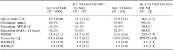
Table 1 shows the demographic characteristics of the following four groups; HC who remained stable over 18 months (stable HC), HC who transitioned to MCI (HC transition group), MCI who remained stable over 18 months (stable MCI), and MCI who transitioned to AD (MCI transition group).
Baseline demographic characteristics (presented as means with standard deviation in parentheses or percentages) for the following groups; HC stable, HC to MCI transition, MCI stable, and MCI to AD transition

*Indicates a significant difference using a test for the difference in proportions at the 1% level between MCI stable and MCI to AD transition groups.
Note: MMSE = Mini-Mental State Examination; HADS-D = Hospital Anxiety and Depression Scale, Depression Subscale; HADS-A = Hospital Anxiety and Depression Scale, Anxiety Subscale.
For the baseline HC group there were no significant differences between those who transitioned to MCI and those who did not in terms of age, proportion of females, proportion of APOE ε4 positive participants, baseline mood, premorbid IQ, and education levels or MMSE scores.
For the baseline MCI group, only the proportion of APOE ε4 groups differed between MCI stable (36.9% APOE ε4 positive) and MCI to AD transition (78.1% APOE ε4 positive) groups. Baseline age and mood measures, education, gender, premorbid IQ, and MMSE did not differ significantly between these groups.
Neuropsychological change
Healthy control at baseline
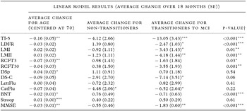
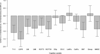
Linear models (LM), adjusted for age, sex, and APOE ε4 status, were conducted and the results presented in Table 2. The magnitudes of the differences in baseline-adjusted performance between the HC transition to MCI group and the stable HC group at 18-month assessment are shown in Figure 2 (HC data represent the baseline levels of performance).
Results of linear model analyses examining change in cognitive performance from baseline to 18 months for those who transitioned and did not transition to MCI in healthy older adults

*p < 0.05; **p < 0.01; ***p < 0.001; †p-value for the difference between transitioners and non-transitioners average change over 18 months after adjusting for other covariates.
Note: Each model additionally adjusted for sex, years of education, APOE ε4 status, baseline premorbid IQ (WTAR), depression (HADS-D), and anxiety levels (HADS-A). TI-5 = CVLT-II learning total correct over Trials 1 to 5; LDFR = CVLT-II total correct long delay free recall; LMI = Logical Memory I total correct; LMII = Logical Memory II total correct; RCFT3 = Rey Complex Figure Test 3 minute delayed recall; RCFT30 = Rey Complex Figure Test 30 minute delayed recall; DSp = WAIS-III Digit Span total correct; DS-C = WAIS-III Digit Symbol-Coding total correct; LettFlu = D-KEFS letter fluency total correct for F+A+S; CatFlu = D-KEFS category fluency total correct for animals+boys names; BNT = Boston Naming Test (30-item) total spontaneous correct; Stroop = congruency ratio; MMSE = Mini-Mental State Examination total correct.
Magnitude of change (Cohen's d effect size) for average change between transitioners and non-transitioners in the baseline HC participants.
Note: T-15 = CVLT-II total correct over Trials 1 to 5; LDFR = CVLT-II total correct long delay free recall; LMI = Logical Memory I total correct; LMII = Logical Memory II total correct; RCFT3 = Rey Complex Figure Test 3 minute delayed recall; RCFT30 = Rey Complex Figure Test 30 minute delayed recall; DSp = WAIS-III Digit Span total correct; DS-C = WAIS-III Digit Symbol-Coding total correct; LettFlu = D-KEFS letter fluency total correct for F+A+S; CatFlu = D-KEFS category fluency total correct for animals+boys’ names; BNT = Boston Naming Test (30-item) total spontaneous correct; Stroop = congruency ratio; MMSE = Mini-Mental State Examination.

Compared to the stable HC group, those who transitioned to MCI demonstrated significantly greater decline on all measures of episodic memory. The magnitude of impairment in episodic memory was, as defined by convention, moderate to large for all episodic memory measures (d > 0.6). The greatest difference between groups was observed on measures of verbal learning and delayed verbal recall where HC to MCI transitioners demonstrated decline relative to stable HC cases of very large magnitudes (d = 1.10 and 1.52, respectively). The transition group also demonstrated greater magnitude of decline than the stable group on confrontation naming (d = 0.91). However, these data demonstrated no difference between transitioners and stable HC on executive function, processing speed, and attention measures.
Mild cognitive impairment at baseline
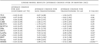
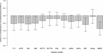
LM were also conducted for MCI at baseline. As above, the LM were adjusted for age, sex, and APOE ε4 status. Table 3 presents the results of the LM, and Figure 3 shows the magnitudes of the differences in baseline-adjusted performance between the MCI transitioners and stable MCI.
Results of linear model analyses examining change in cognitive performance from baseline to 18 months for those who transitioned and did not transition to AD in adults with MCI

*p < 0.05; **p < 0.01; ***p < 0.001; †p-value for the difference between transitioners and non-transitioners average change over 18 months after adjusting for other covariates.
Note: Each model additionally adjusted for sex, years of education, APOE ε4 status, baseline premorbid IQ (WTAR), depression (HADS-D), and anxiety levels (HADS-A). TI-5 = CVLT-II learning total correct over Trials 1 to 5; LDFR = CVLT-II total correct long delay free recall; LMI = Logical Memory I total correct; LMII = Logical Memory II total correct; RCFT3 = Rey Complex Figure Test 3 minute delayed recall; RCFT30 = Rey Complex Figure Test 30 minute delayed recall; DSp = WAIS-III Digit Span total correct; DS-C = WAIS-III Digit Symbol-Coding total correct; LettFlu = D-KEFS letter fluency total correct for F+A+S; CatFlu = D-KEFS category fluency total correct for animals+boys names; BNT = Boston Naming Test (30-item) total spontaneous correct; Stroop = congruency ratio; MMSE = Mini-Mental State Examination total correct.
Magnitude of change (Cohen's d effect size) for average change between transitioners and non-transitioners in the baseline MCI participants.
Note: T-15 = CVLT-II total correct over Trials 1 to 5; LDFR = CVLT-II total correct long delay free recall; LMI = Logical Memory I total correct; LMII = Logical Memory II total correct; RCFT3 = Rey Complex Figure Test 3 minute delayed recall; RCFT30 = Rey Complex Figure Test 30 minute delayed recall; DSp = WAIS-III Digit Span total correct; DS-C = WAIS-III Digit Symbol-Coding total correct; LettFlu = D-KEFS letter fluency total correct for F+A+S; CatFlu = D-KEFS category fluency total correct for animals+boys’ names; BNT = Boston Naming Test (30-item) total spontaneous correct; Stroop = congruency ratio; MMSE = Mini-Mental State Examination.

Inspection of Figure 3 demonstrates that there were small to moderate differences in the magnitude of decline between transitioners and stable MCI across most cognitive domains. However, as shown in Table 3 these differences only reached significance for Logical Memory I (d = 0.56, p = 0.02), and for letter and category fluency (d = 0.41 and 0.35 respectively, both p < 0.05).
Subjective memory concern
As shown in Figure 1, there are four different subgroups of HC volunteers: Stable non-SMC (N = 220), stable SMC (N = 254), non-SMC who became SMC (N = 114), and SMC who became non-SMC (N = 97). We examined whether these four subgroups differed either in baseline cognitive and clinical measures, and/or the magnitude of change over 18 months.
One-way ANOVA demonstrated no significant differences between the subjective memory concern subgroups in the magnitude of change over 18 months when controlling for multiple comparisons. However, examination of baseline clinical measures demonstrated small but significant differences in baseline anxiety (p < 0.0001) and depression scores (p < 0.0001), premorbid IQ (p < 0.01), and CDR sum of boxes (p < 0.01) between the groups. Stable non-SMC showed lower depression scores, lower anxiety scores, and lower CDR sums of boxes than stable SMC, and lower depression scores than the SMC cases that reverted to non-SMC status at 18 months.
Discussion
This is the first paper to report the diagnostic classification status of the entire AIBL cohort after the initial 18-month follow-up, and transition rates between classification categories over this initial review period. In terms of cohort retention, the AIBL investigators have been successful in reassessing the great majority of AIBL participants (89.9%). Approximately 30% of MCI participants in this cohort met criteria for AD dementia at follow-up, and this rate of transition to AD over 18 months is slightly higher than the reports of around 15% conversion per annum in carefully assessed clinically derived samples of MCI participants from North America and Europe (Kurz and Lautenschlager, Reference Kurz, Lautenschlager, Ames, Burns and O'Brien2010). It is likely that this higher transition rate reflects the fact that the AIBL baseline MCI cohort was recruited primarily from memory clinics and the private practices of geriatricians, neurologists, and psychiatrists specializing in the assessment and management of memory disorder, and reflected a group who were already close to transitioning to an AD diagnosis. That seven subjects initially classified as fulfilling criteria for MCI were reclassified as HCs after 18 months is also consistent with previous research experience and the mutability of MCI diagnoses.
At both baseline and 18 months, more than half of the HC volunteers reported a subjective memory complaint, which is within the range observed in Australian population samples (Slavin et al., Reference Slavin2010; Mewton et al., Reference Mewton, Sachdev, Anderson, Sunderland and Andrews2013). There was considerable instability in the report of subjective memory concern over the two assessments. This may reflect reassurance of some worried participants at their adequate performance on baseline testing, sensitization of some previously sanguine participants to subtle failures on complex and difficult tests, and the metric crudeness of assessing memory complaints on the basis of one single question. For this reason, a much more detailed assessment of memory concerns and perceptions of memory function has been undertaken at the 18-month and subsequent follow-ups, including administration of the Memory Complaint Questionnaire (MAC-Q), a brief measure of subjective memory complaint in people with normal cognitive function. Our current findings did suggest that affective factors play some role in the determination of memory complaint status, with lower depression and anxiety scores associated with a lack of memory complaint at baseline (Buckley et al., Reference Buckley2013), however these factors were not related to change in cognition over 18 months. This does not mean that individuals who have concerns about their memory should not be thoroughly assessed and then followed to detect actual or emergent cognitive impairment, but it does imply that they should also be assessed for the presence of any potentially treatable affective disorder.
There were a significantly higher proportion of participants with subjective memory concern in the group that converted to MCI over 18 months. However, the fact that 13/17 HCs who converted to MCI at follow-up reported difficulty with memory at baseline might merely reflect the diagnostic criterion of concern expressed about memory, which forms one essential element of the standard MCI diagnostic criteria (Winblad et al., Reference Winblad2004). Once we complete analysis of three-year follow-up data on the entire cohort, and more participants have undergone diagnostic transition, we intend to explore the effect of varying the criteria for MCI (e.g. adjusting the number of low cognitive performance measures required for classification and exploring the result of omitting the memory complaint criterion from the diagnostic algorithm) on transition rates and progression to AD.
Episodic memory deficits were greater both for those transitioning from HC to MCI than for those who remained classified as HC, as well as for those transitioning from MCI to AD compared to stable MCI volunteers. The salience of difficulties in retaining newly learned information as a very early indicator of cognitive decline has been inferred from previous research (for a review, see Savage, Reference Savage, Ames, Burns and O'Brien2010), and is reinforced by our findings. The magnitude of decline in episodic memory was larger in the HC to MCI transition than in the MCI to AD transition, and this likely reflects the fact that transition from MCI to AD is related more to changes on functional measures than episodic memory performance. Indeed, those MCI cases that transitioned to AD showed a significantly higher increase in CDR sum of box than stable MCI over 18 months. In terms of demographic characteristics, transitioners and stable cases did not differ significantly in age or gender (both for the HCs at baseline and MCI at baseline). However, the MCI to AD transition group included a significantly higher proportion of participants carrying an APOE ε4 allele, consistent with evidence that this allele is a strong risk factor for development of AD.
It is important to note that the AIBL sample is by design a convenience sample, drawn mainly from motivated volunteers and clinical populations, and is therefore unlikely to be truly representative of the Australian general population. Convenience sampling of cognitively healthy participants is vulnerable to self-selection bias, with convenience samples tending to be younger and better educated than population samples, while clinically referred samples (e.g. a vast majority of the MCI and AD cases within AIBL) are likely to include people who have better access to health care due to socioeconomic factors. Indeed, the AIBL population is observed to be highly educated, and with higher than average estimated IQ. As a result, there are limitations to the generalizability of the transition rates and cognitive effect size changes in this sample, with these data not directly comparable to population study rates, but rather forming an important comparison point for other large cohort studies (i.e. ADNI), which have established cohorts with varying risk factor profiles to examine AD pathology and early diagnosis.
No individual diagnosed with AD dementia at baseline and who was still alive and could be reassessed at 18 months failed to meet diagnostic criteria for AD at 18 months, indicating a high degree of rigor and accuracy in the baseline diagnostic process. The outcome for this group at 18 months has already been reported in detail (Sona et al., Reference Sona2012), with rapid decline (greater than 5 MMSE points lost in 18 months) being predicted by baseline CDR (both total score and sum of box score) and the prescription of a cholinesterase inhibitor at baseline. The relationship between prescription of cholinesterase inhibitors and rapid cognitive decline has also been observed within the ADNI study cohort (Schneider, Reference Schneider, Insel and Weiner2011).
Cognitive decline with age is not a rapid process for most people, and 18 months is a fairly short period over which to detect emergent decline or transition to clinically diagnosable MCI or AD among previously healthy individuals. Nevertheless, the detailed cognitive data collected in the AIBL study give us the best possible opportunity of detecting such changes that, after future waves of follow-up are complete, may allow us to make a major contribution to determining factors that may be predictive of future cognitive decline in older individuals who are cognitively healthy at present.
Conflict of interest
NL is editor and DA is a former editor of International Psychogeriatrics. For this reason the process of assessing this paper was conducted at arm's length under the supervision of one of the journal's deputy editors.
Description of authors’ roles
KE coordinated the baseline and 18-month data collection, supervised the data analyses, served on the diagnostic review panel and the AIBL management committee, assisted with devising the cognitive battery used for the AIBL study, wrote the initial draft of the paper, and contributed to its revision. CS served on the diagnostic review panel and AIBL management committee and contributed to the writing and revision of the paper. AIB served on the AIBL management committee, assisted with the initial conceptualization of the AIBL research plan, co-led the biomarkers arm of the study, and contributed to revising drafts of the paper. DD, NTL, GS, and MW assisted with the initial conceptualization of the AIBL research plan, served on the diagnostic review panel, assisted with devising the cognitive battery used for the AIBL study, and contributed to revising drafts of the paper. DD, NL, and MW also referred many subjects with AD and MCI to the AIBL study. PLG undertook data analyses and contributed to revising drafts of the paper. LM oversaw data analyses, led the CSIRO team working on the AIBL study, served on the management committee, and contributed to revising drafts of the paper. PM assisted with the initial conceptualization of the AIBL research plan, assisted with devising the cognitive battery used for the AIBL study, and contributed to revising drafts of the paper. RNM oversaw the Perth arm of the AIBL study, served on the AIBL management committee, assisted with the initial conceptualization of the AIBL research plan, co-led the biomarkers arm of the study, and helped revise drafts of the paper. SM manages the database and data integrity for the AIBL study and revised versions of this paper. CLM served on the AIBL management committee, assisted with the initial conceptualization of the AIBL research plan, co-led the biomarkers arm of the study, and contributed to revising drafts of the paper. SR-S oversaw data collection at the Perth site, served on the AIBL management committee and later on the diagnostic review panel, and contributed to revising drafts of the paper. AR coordinated the blood processing at the Melbourne site, served on the AIBL management committee, and contributed to revising drafts of the paper. JR and KP assessed many of the AIBL participants, attended most diagnostic review panel meetings, and contributed to revising drafts of the paper. CCR served on the AIBL management committee, assisted with the initial conceptualization of the AIBL research plan, led the imaging arm of the study, and contributed to revising drafts of the paper. VLV coordinated the neuroimaging stream, analyzed all PET images, and reviewed drafts of the paper. PZ conducted the initial data analysis and contributed to revising drafts of the paper. DA has led the AIBL study since its inception, was centrally involved in the conceptualization of the study program and the development of the study cognitive battery, chaired the diagnostic review panel and the AIBL management committee, wrote the second draft of the paper, and contributed to later revisions.
Acknowledgments
Funding for the study was provided in part by the study partners [Australian Commonwealth Scientific Industrial and Research Organization (CSIRO), Edith Cowan University (ECU), Mental Health Research Institute (MHRI), Alzheimer's Australia Vic (AA), National Ageing Research Institute (NARI), University of Melbourne, Austin Health, CogState Ltd., Hollywood Private Hospital, Sir Charles Gardner Hospital]. The study also received support from the National Health and Medical Research Council (NHMRC), the Dementia Collaborative Research Centres program (DCRC-EDP), and the McCusker Alzheimer's Research Foundation, Inc., as well as ongoing funding from the Science and Industry Endowment Fund (SIEF; www.SIEF.org.au). Pfizer International and GE Healthcare have contributed financial support to assist with analysis of blood samples and to further the AIBL research program. Ashley I. Bush is supported by a Federation Fellowship from the Australian Research Council. Cassandra Szoeke has received support from Alzheimer's Australia, the Ramiciotti Foundation, the Mason Foundation, and the NHMRC. Alzheimer's Australia (Victoria and Western Australia) assisted with promotion of the study and screening of telephone calls from volunteers.
The AIBL team wishes to thank the following clinicians who referred patients with AD and/or MCI to the study: Professor David Ames, Associate Professor Brian Chambers, Professor Edmond Chiu, Dr Roger Clarnette, Associate Professor David Darby, Dr Mary Davison, Dr John Drago, Dr Peter Drysdale, Dr Jacqui Gilbert, Dr Kwang Lim, Professor Nicola Lautenschlager, Dr Dina LoGiudice, Dr Peter McCardle, Dr Steve McFarlane, Dr Alastair Mander, Dr John Merory, Professor Daniel O'Connor, Professor Christopher Rowe, Dr Ron Scholes, Dr Mathew Samuel, Dr Darshan Trivedi, and Associate Professor Michael Woodward. We thank all those who took part as participants in the study for their commitment and dedication to helping advance research into the early detection and causation of AD.