Severe congenital cardiac disease occurs in approximately 6–8 of 1000 live births in North America.Reference Hoffman and Kaplan1, Reference Samanek2 Despite most forms of congenital cardiac disease being amenable to corrective surgical repair, survivors often have gross and fine motor delay, and may be left with behaviour difficulties and functional limitations in socialisation, communication, and daily living skills that persist into late childhood.Reference Majnemer, Limperopoulos, Shevell, Rohlicek, Rosenblatt and Tchervenkov3–Reference Miller, Tesman, Ramer, Baylen and Myers11 Historically, efforts to improve neurodevelopmental outcomes in newborns with congenital cardiac disease have focused on the surgical procedure itself, and specifically on the type and duration of cardiopulmonary bypass, lowest flow, and base deficit during bypass, which are known to be major risk factors for adverse neurodevelopmental outcome.Reference Bellinger, Jonas and Rappaport8, Reference Miller, Tesman, Ramer, Baylen and Myers11–Reference Hovels-Gurich, Seghaye and Schnitker13 However, increasing recognition that neurobehavioural abnormalities and brain injury may exist prior to the operative procedure has led to the examination of pre- and post-operative risk factors.Reference Limperopoulos, Majnemer, Shevell, Rosenblatt, Rohlicek and Tchervenkov14, Reference McQuillen, Barkovich and Hamrick15 Non-operative risk factors for adverse outcome or brain injury in this population include perioperative instability – for example, hypoxemia, hypotension, acidosis, or pre-operative need for balloon atrial septostomy – and cardiac lesion – that is, hypoplastic left heart syndrome.Reference Majnemer, Limperopoulos, Shevell, Rosenblatt, Rohlicek and Tchervenkov4, Reference McQuillen, Barkovich and Hamrick15–Reference Petit, Rome and Wernovsky19
Focal, non-cystic white matter injury, which is a characteristic pattern of brain injury in preterm newborns,Reference Miller, Ferriero and Leonard20, Reference Woodward, Anderson, Austin, Howard and Inder21 is now also recognised as the predominant pattern of injury in newborns with congenital cardiac disease. More than 50% of infants with congenital cardiac disease show some degree of white matter injury when imaged using magnetic resonance imaging or examined at autopsy in the post-operative period.Reference McQuillen, Barkovich and Hamrick15, Reference Petit, Rome and Wernovsky19, Reference Mahle, Tavani and Zimmerman22–Reference Kinney, Panigrahy, Newburger, Jonas and Sleeper24 The reasons why infants with congenital cardiac disease are susceptible to this classic “preterm” pattern of injury are not known, and may, in part, be related to the relative immaturity of the brain of newborns with congenital cardiac disease (reviewed by McQuillen and MillerReference McQuillen and Miller25).
Infection is an established risk factor for white matter injury in the preterm population.Reference Chau, Poskitt and McFadden26–Reference Leviton and Gilles31 The mechanism of white matter injury in the setting of infection is unclear, and current hypotheses have centred on direct injury due to inflammation, versus injury occurring as a consequence of hypoxia-ischaemia during septic shock. Chau et alReference Chau, Poskitt and McFadden26 recently found that both postnatal bloodstream infection and episodes of hypotension requiring treatment were risk factors for white matter injury, whereas chorioamnionitis confirmed by histopathology was not. These findings suggest that the pathogenesis of white matter injury may be multifactorial, with contributions to the underlying pathogenesis from both inflammation and haemodynamic shock. Acquired infection is also common in newborns with congenital cardiac disease, whose critical illness results in prolonged need for mechanical ventilation, intravenous therapy, and intensive care. Whether infection is also associated with white matter injury in infants with congenital cardiac disease, over and above established risk factors relating to impaired oxygen delivery, is not known.
We analysed infants enrolled in a cohort study examining magnetic resonance imaging predictors of neurodevelopmental outcome in term newborns with congenital cardiac disease in order to examine the hypothesis that infection is an independent risk factor for white matter injury in this population.
Methods
The study subjects were infants greater than 36 weeks’ gestation at birth who were admitted to the intensive care units at the University of California, San Francisco Medical Center or the Children's and Women's Health Centre of British Columbia within the first month of life requiring surgery for congenital cardiac disease. Neonates with a diagnosis of congenital infection were excluded. The University of California, San Francisco and University of British Columbia Committees on Human Research approved the study protocol.
Clinical data collection
From August, 2001 to March, 2009, 127 of the enrolled newborns were examined with magnetic resonance imaging pre-operatively, and 108 (85%) had repeat studies post-operatively. Clinical data were prospectively collected from the medical records by a team of trained neonatal research nurses, and reviewed by a paediatric intensivist who was blinded to the neuroimaging findings. Perinatal variables included gestational age at birth and birth weight. Perioperative clinical variables included cardiac lesion – transposition of the great arteries, single ventricle anatomy or “other”; lowest oxygen saturation, SaO2, bypass type; blood pressure on the first post-operative day; days intubated; and days of inotrope therapy. The lowest SaO2 was recorded from pulse oximetry and extracted from the bedside patient record for each 24-hour period.
Data for infections – bloodstream infection, pneumonia, and surgical site infection – were extracted from clinical charts and microbiology reports, and reviewed by a specialist in paediatric infectious diseases according to the following definitions: (1) Bloodstream infection was diagnosed when blood cultures grew pathogenic species, or Staphylococcus epidermidis in the setting of a central venous catheter, and clinical signs or symptoms suggesting infection – temperature instability, low (less than 4000 cells per cubic millimetres) or elevated (greater than 12,000 cells per cubic millimetres) white blood cell count, greater than 10% band forms, or stated clinical suspicion by a treating physician; (2) pneumonia, viral or bacterial, was diagnosed if an infiltrate was identified on chest X-ray and “clinical signs of respiratory symptoms were documented”; (3) surgical site infection was diagnosed in children who had clinical signs of cellulites, erythema with purulent discharge or wound dehiscence, within 30 days of operation, and received antibiotic therapy. None of the infants had meningitis. With the exception of the patient diagnosed with respiratory syncytial virus pneumonia, in order to further enhance our certainty of a clinically significant infection, we only considered infections that were treated with antibiotic therapy for at least 5 days.
Magnetic resonance imaging
Pre-operatively, magnetic resonance imaging studies were performed as soon as the baby could be transported safely from the cardiac intensive care unit to the imaging suite. Post-operative studies were performed after temporary pacemaker wires were removed. If necessary, infants were sedated according to institution guidelines.
Magnetic resonance was acquired at the University of California, San Francisco using a 1.5 Tesla scanner, General Electric Signa, and a specialised, high-sensitivity neonatal head coil, built into the magnetic resonance imaging compatible incubator, General Electric or Lammers Medical Technologies. Image sequences included: (1) T1-weighted sagittal spin echo images, 4-millimetre thickness, using a repetition time (TR) of 500 milliseconds, echo time of 11 milliseconds, 1 excitation, and 192 × 256 acquisition matrix; (2) dual-echo T2-weighted spin echo, 4-millimetre thickness, with a repetition time of 3000 milliseconds, echo time of 60 and 120 milliseconds, and 192 × 256 acquisition matrix; and (3) coronal volumetric three-dimensional gradient echo images with radiofrequency spoiling, spoiled gradient recalled images (1.5-millimetre thickness) with a repetition time of 36 milliseconds, a echo time of 9 milliseconds, a flip angle of 35°, and 1 excitation. At the University of British Columbia, magnetic resonance imaging studies were carried out without pharmacological sedation on a Siemens 1.5 Tesla Avanto using VB 13A software and included comparable imaging to that obtained at the University of California, San Francisco (repetition time/echo time/field of view/Slice thickness/Gap): (1) three-dimensional coronal volumetric T1-weighted images (36/9.2/200/1/0 millimetres); (2) axial fast spin echo T2-weighted images (4610/107/160/4/0.2 millimetres). Average diffusivity (Dav) maps were generated from diffusion tensor imaging acquired with a multi-repetition, single-shot echo planar sequence with 12 gradient directions (4900/104/160/3/0 millimetres), b = 0, 600, and 700 seconds per square millimetre, and an in-plane resolution of 1.3 millimetres.
A neuroradiologist blinded to all clinical information beyond corrected gestational age and cardiac anatomic diagnosis scored each magnetic resonance imaging scan for acquired focal, multi-focal, or global changes, as reported previously.Reference McQuillen, Hamrick and Perez32 Acquired focal and multi-focal lesions included focal infarct, germinal matrix and intraventricular hemorrhage, and white matter injury. Focal infarct referred to discrete areas of cortical and/or white matter reduced intensity on Dav maps, or focal hyperintensity on T2-weighted images. Single white matter lesions measuring less than 3 millimetres were classified as white matter injury, whereas larger lesions were considered as stroke.Reference McQuillen, Hamrick and Perez32, Reference Block, McQuillen and Chau33 White matter injury was considered “minimal” if there were three or fewer areas of T1 signal abnormality, each less than two millimetres. Injury was considered “moderate” if there were more than three areas of T1 signal abnormality or if these areas measured more than two millimetres but less than 5% of the hemisphere was involved. “Severe” injury was defined as involvement of more than 5% of the hemisphere.Reference Miller, Ferriero and Leonard20 The presence of newly acquired injury on the post-operative study was noted.
Statistical analysis
Statistical analysis was performed using Stata 10.0 software (Stata Corporation, College Station, Texas, United States of America). Differences between clinical predictors were assessed using two-tailed Student's t-test and Wilcoxon rank sum for continuous variables and chi-square, or Fisher's exact test for categorical variables. We performed stratified analysis by cardiac lesion and presence of stroke to look at the relative risk of white matter injury in these sub-populations. We also performed multivariable logistic regression analysis to look at the adjusted odds of white matter injury. We included in the initial model any potential confounders associated with both the predictor – infection – and the outcome – white matter injury – at a significance of p less than or equal to 0.1.
Results
During the study period, 135 newborns were enrolled. Of these, eight were excluded from the study for the following reasons: never imaged, four infants, congenital cytomegalovirus infection, one infant, gestational age less than 36 weeks, one infant, and no surgery, one infant. Of the remaining 127 infants, 89 (70%) constituted the University of California, San Francisco cohort.
Infection
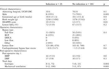
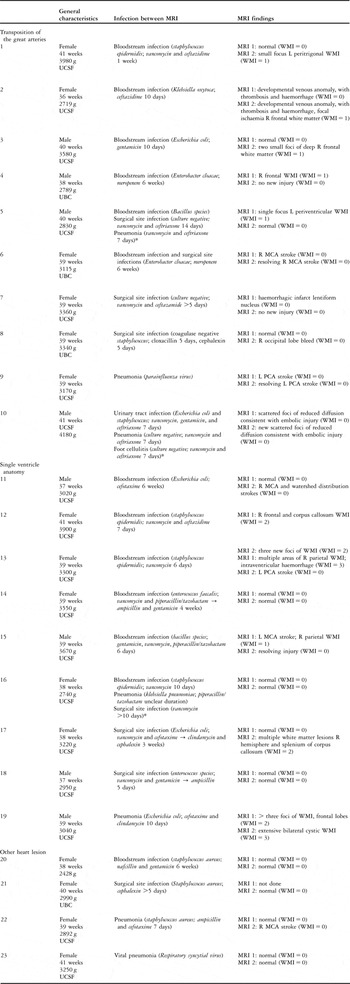
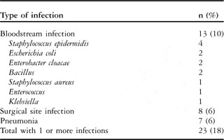
The clinical characteristics of study subjects appear in Table 1. Infection was common, with 23 of 127 infants (18%) sustaining at least one of bloodstream infection, pneumonia, or surgical site infection during the admission, and prior to the second scan. The clinical and imaging characteristics of the 23 newborns with infection appear in Table 2. Bloodstream infection was most common, 13 infants (Table 3). Organisms cultured from the blood included: Staphylococcus epidermidis – four cases – Escherichia coli, Enterobacter cloacae, and Bacillus species – two cases each – Staphylococcus aureus, Enterococcus, and Klebsiella – one case each. Of the 23 children with infection, surgical site infection was present in eight (6%) and pneumonia in seven (6%) infants. There were five infants (5%) with recurrent infections.
Clinical characteristics for 127 infants with congenital cardiac disease with and without nosocomial infection.

DBP = diastolic arterial blood pressure; DHCA = deep hypothermic circulatory arrest; MBP = mean arterial blood pressure; RCP = regional cerebral perfusion; SaO2 = oxygen saturation; UCB = University of British Columbia; UCSF = University of California, San Francisco
Values are listed as n (%), mean (±standard deviation) or median (range)
Characteristics of 23 infants with congenital cardiac disease and infection.

MCA = middle cerebral artery; MRI = magnetic resonance imaging; PCA = posterior cerebral artery; SVA = hypoplastic left heart syndrome; TGA = transposition of the great arteries; UBC = University of British Columbia; UCSF = University of California, San Francisco; VSD = ventricular septal defect; WMI = white matter injury
*Overlapping antibiotic regimens
Nosocomial infection in 127 infants with congenital cardiac disease.

Infection and white matter injury
Pre-operative magnetic resonance imaging was performed at a median age of 5 days of life (range 1, 43 days). A total of 108 children were also imaged post-operatively, at a median of 21 days of life, (range 9, 65 days) which was a median of 10 days after the operation day (range 4, 61 days post-operative). Infection occurred before the first magnetic resonance imaging in four children (3%, two with bloodstream infection) whereas the remainder of infections occurred after the time of the first magnetic resonance imaging but prior to the second scan.
Out of 127 infants, 30 (24%) had white matter injury on the pre-operative magnetic resonance imaging. None of these newborns were diagnosed with nosocomial infection prior to the time of imaging, and therefore we considered only new white matter injury acquired on the second magnetic resonance imaging for the remainder of the analysis. Newborns with white matter injury on the first scan were not at increased risk for developing infection (p greater than 0.6).
Of the 108 infants reimaged post-operatively, new white matter injury, not seen on the first scan, was present in 33 (31%). There was no difference in the timing of magnetic resonance imaging, post-operative day or day of life, in newborns with and without injury (p = 0.7). Overall, there was no significant difference in the frequency of newly acquired post-operative white matter injury in infants with infection (30%), compared to those without (31%). When restricting the analysis to bloodstream infection, there was also no significant association with white matter injury (relative risk 1.3, 95% confidence interval 0.6–2.9, p = 0.5) pneumonia, (relative risk 0.9; 95% confidence interval 0.3–3.1, p = 0.9), and surgical site infection, (relative risk 0.8; 95% confidence interval 0.2–2.7, p = 0.7) were also not associated with white matter injury.
Infection and white matter injury: subgroup analyses
Given the hypothesised association of infection with white matter injury, we examined the infants without stroke to avoid the potential overlapping impact of infection on both stroke and white matter injury. When analysing only the 73 newborns without stroke, the estimated association between infection and white matter injury was higher, though the results were not significant for bloodstream infection (relative risk 1.9, 95% confidence interval 0.8–4.2, p = 0.1) pneumonia (relative risk 1.6, 0.5–5.1, p = 0.4), and surgical site infection (relative risk 1.0, 95% confidence interval 0.3–3.4, p = 1.0).
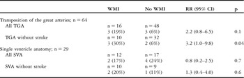
Since post-operative hypotension is an independent risk factor for white matter injuryReference Galli, Zimmerman and Jarvik23, Reference Petit, Rome and Wernovsky34 and post-operative haemodynamics vary by cardiac diagnosis and procedure, we performed subgroup analyses by cardiac anatomy. When considering cardiac anatomy, white matter injury was more common in the 29 infants with single ventricle anatomy (41%) as compared to the 64 with transposition of the great arteries (25%). Despite the overall association between infection and white matter injury among newborns with single ventricle anatomy and transposition of the great arteries being the same (relative risk 1.2; 95% confidence interval 0.4–3.7 versus relative risk 1.1; 95% confidence interval 0.4–2.8), infants with transposition of the great arteries and bloodstream infection had an estimated risk of white matter injury that was twice as high as those with single ventricle anatomy and bloodstream infection (relative risk 2.2; 95% confidence interval 0.8–6.5 versus relative risk 0.8; 95% confidence interval 0.2–2.5) though the difference was not significant. When considering only those without stroke, the risk of white matter injury in newborns with bloodstream infection was higher and significant for those with transposition of the great arteries (relative risk 3.2; 95% confidence interval 1.0–9.8, p = 0.04), but not single ventricle anatomy (relative risk 1.3; 95% confidence interval 0.4–4.0, p = 0.6; Table 4).
Bloodstream infection and white matter injury among 64 infants with TGA and 29 infants with SVA, and stratified by presence of stroke.

SVA = hypoplastic left heart syndrome; TGA = transposition of the great arteries; WMI = white matter injury
Adjusted analysis
Of the possible confounding covariates listed in Table 1, only duration of inotrope therapy was associated with white matter injury. Again, there was no overall association between infection and white matter injury. However, the risk of bloodstream infection among the 64 newborns with transposition of the great arteries, after adjusting for days inotropes and mean post-operative blood pressure, persisted and was higher (odds ratio 7.3; 95% confidence interval 0.9–61.0, p = 0.06), especially when considering only the 42 newborns without strokes (odds ratio 182.3; 95% confidence interval 1.8 to greater than 100, p = 0.003).
Discussion
In this cohort of newborns with congenital cardiac disease admitted to one of two university hospitals for surgery, nosocomial infection was present in almost 20% of the population. Overall, infection had no effect on white matter injury. Bloodstream infection was associated with a trend towards higher frequency of white matter injury in those infants with transposition of the great arteries, but not those with single ventricle anatomy. After adjusting for markers of illness severity, and when considering only the children without stroke, the effect was stronger and significant. Since the pneumonias, bloodstream, and surgical site infections that we identified were hospital acquired, nearly all were potentially preventable.
Newborns with congenital cardiac disease may be critically ill with unstable haemodynamics before, during, and following surgical repair. Multiple risk factors for stroke or white matter injury, including cyanosis, invasive catheterisation, hypotension, and cardiac arrest,Reference Hoffman and Kaplan1, Reference Galli, Zimmerman and Jarvik23, Reference Petit, Rome and Wernovsky34, Reference Dent, Spaeth and Jones35 have been identified in this population. Each type of congenital cardiac disease may have variable exposure to specific risk factors such as intracardiac shunting. Given the complex interplay between infection, inflammation, and adverse haemodynamics, perhaps it is not surprising that we were not able to uncover a strong association between infection and brain injury in this heterogeneous sample of newborns with congenital cardiac disease. Nonetheless, these results suggest that the association between bloodstream infection and white matter injury in children with congenital cardiac disease is not as clear as for preterm children.Reference Chau, Poskitt and McFadden26, Reference Glass, Bonifacio and Chau27, Reference Shah, Doyle and Anderson29 The larger effect in children with transposition of the great arteries when compared with those with single ventricle anatomy may be related to differences in perioperative haemodynamic stability. The Norwood operation for palliation of single ventricle anatomy is associated with higher risk of mortality and hospital length of stay compared with the arterial switch procedure for correction of transposed great arteries.Reference Qamar, Goldberg, Devaney, Bove and Ohye36, Reference Wernovsky, Kuijpers and Van Rossem37 For children with single ventricle anatomy, the presence of multiple potential risk factors for brain injury over a more prolonged admission may make an effect of infection more difficult to detect. The increased association after excluding those newborns with strokes highlights the fact that multiple potential risk factors, including infection, may have overlapping effects on the brain.
The reasons why children with congenital cardiac disease are susceptible to white matter injury, a pattern of injury more typically associated with premature birth, is not known but may be related to the fact that children with congenital cardiac disease appear to have delayed brain growth and maturation, making their brains resemble those of younger gestation children.Reference Miller, McQuillen and Hamrick16, Reference Limperopoulos, Tworetzky and McElhinney17 Brain immaturity is the substrate upon which other insults such as hypotension and hypoxemia provide the proximate injury mechanism. Furthermore, like their preterm counterparts, children with congenital cardiac disease often have prolonged hospitalisations, extending the period of risk for multiple potential factors including nosocomial infection and haemodynamic instability. White matter injury in preterm newborns appears to be related to damage to pre-oligodendroglial cells, which are important for myelination. These oligodendrocyte precursors are extremely vulnerable to glutamate-receptor mediated injury and cell death following exposure to free radicals, reactive oxygen, and nitrogen species, as well as inflammatory cytokines (reviewed by BackReference Back38). The causative “upstream mechanisms” – that is, the events leading to production of these harmful factors – are poorly understood, and may include ischaemia and/or infection/inflammation (reviewed by Khwaja and VolpeReference Khwaja and Volpe39). There is increasing evidence from clinical studies in preterm newborns that nosocomial infection is an important risk factor for white matter injury.Reference Chau, Poskitt and McFadden26, Reference Glass, Bonifacio and Chau27, Reference Shah, Doyle and Anderson29 It is not known whether the injury is a direct result of inflammation to the central nervous system or whether it is due to ischaemia suffered as a result of impaired cerebral blood flow during periods of hypotension.
We acknowledge several limitations of this study. Despite this being the largest reported cohort of newborns with congenital cardiac disease studied with pre- and post-operative imaging, the relatively small numbers of infants with infection require our conclusions to be viewed with caution. Moreover, the cohort includes patients with multiple forms of congenital cardiac disease including transposition of the great arteries and single ventricle anatomy. In this critically ill population, there may be multiple causes for brain injury, and the small cohort size may have limited the ability to find an association between our variables of interest. Finally, the optimal timing for detecting white matter injury in this cohort has not been determined; it is therefore possible that we missed white matter injury in some affected newborns who were imaged too early or too late to see changes related to infection, which would further restrict our ability to observe an effect.
Conclusions
The strong association of infection with white matter injury in premature newborns, combined with observations of delayed brain development in term newborns with congenital cardiac disease, provides clear rationale for this study. However, heterogeneity inherent in the congenital cardiac disease population, along with multiple interacting risk factors, may have obfuscated a relationship between infection and brain injury in this population. The fact remains that newborns with congenital cardiac disease suffer acquired brain injury at unacceptably high rates. In this study, we have provided a first measurement of the association of nosocomial infection with brain injury in infants with congenital cardiac disease. Interventions to reduce nosocomial infection may decrease brain injury, but a larger sample in more uniform populations will be necessary to definitively prove this concept. An alternative possibility is that previously identified risk factors for brain injury that directly impair brain oxygen delivery are relatively more potent, and thus more important, determinants of brain injury in newborns with congenital cardiac disease.
In order to improve developmental outcomes in this population, each of the risk factors for brain injury must be addressed individually. The infections that we identified in this study were hospital acquired, and therefore many may have been preventable. The suggestion that infection prevention has the potential to mitigate long-term neurologic impairment as a consequence of white matter injury underscores the importance of vigilant attention to infection control practices for paediatric patients undergoing cardiac surgery.
Acknowledgements
We are grateful to Drs Sarah Tabbutt and David Teitel for their careful evaluation of the paper. This project was supported by NIH/NCRR/OD UCSF-CTSI Grant Numbers KL2 RR024130, NIH/NINDS, K23NS066137, 1R01NS063876 and 5M01RR01271; Canadian Institutes of Health Research, MOP 93780; March of Dimes Birth Defects Foundation, 6-FY2009-303, 0365018Y, and 5-FY05-1231; the American Heart Association; and a grant from the Larry L. Hillblom Foundation.