Introduction
In vitro production (IVP) of bovine embryos has been improved immensely throughout the last decades. Nevertheless, embryos generated in vitro still differ from their in vivo produced counterparts (Greve et al., Reference Greve, Callesen, Hyttel, Avery, Greppi and Enne1994; Wrenzycki et al., Reference Wrenzycki, Herrmann, Carnwath and Niemann1998, Reference Wrenzycki, Herrmann and Niemann2007). It is possible to achieve blastocyst rates of up to 70% if in vivo matured oocytes are used. In contrast, if oocytes are matured in vitro, blastocyst rates are only half that of those matured in vivo (35%; Rizos et al., Reference Rizos, Ward, Duffy, Boland and Lonergan2002). In vitro culture (IVC) conditions have been enhanced in the last few years, mainly by adjustment of media formulations, whereas the in vitro maturation (IVM) seems to be invariable (for review see Sutton et al., Reference Sutton, Gilchrist and Thompson2003). Therefore, improvement of cumulus–oocyte complex (COC) selection as well as IVM might enhance the outcome of IVP.
Physiologically, the oxygen concentration in follicular fluid ranges between 3–13% (McNatty, Reference McNatty and Jones1978), whereas the oxygen concentration employed in most IVM protocols is around 20% (atmospheric oxygen concentration; Landau et al., Reference Landau, Braw-Tal, Kaim, Bor and Bruckental2000). In fact, no cells in intact mammals are exposed to an atmospheric oxygen concentration (Bavister, Reference Bavister1995). Thus, the gaseous environment to which the oocyte is exposed varies considerably between in vitro and in vivo conditions. The effect of employing a reduced oxygen concentration during IVM has been investigated in various species (hamster, Gwatkin & Haidri, Reference Gwatkin and Haidri1974; pig, Park et al., Reference Park, Hong, Yong, Hwang, Lim and Lee2005; mouse, Preis et al., Reference Preis, Seidel and Gardner2007) with contradictory results. In the bovine, a concentration of 5% O2 during IVM was shown to have detrimental effects on maturation rates (Pinyopummintr & Bavister, Reference Pinyopummintr and Bavister1995). The reduced oxygen concentration also seems to have an impact on total cell numbers of blastocysts, increasing them in murine (Banwell et al., Reference Banwell, Lane, Russell, Kind and Thompson2007; Preis et al., Reference Preis, Seidel and Gardner2007) and bovine (Jiang et al., Reference Jiang, Wang, Lu, Gordon and Polge1992) blastocysts. Nevertheless, using a reduced oxygen concentration while simultaneously supplementing the maturation medium with glucose led to increased consecutive blastocyst rates (Hashimoto et al., Reference Hashimoto, Minami, Takakura, Yamada, Imai and Kashima2000a). This situation indicates a close connection between glucose and oxygen metabolism.
It is reported that the intrafollicular glucose concentration changes in dependency of the expansion and growth of the follicle (Wise, Reference Wise1987). The glucose concentration also increases with increasing size of the follicle (Landau et al., Reference Landau, Braw-Tal, Kaim, Bor and Bruckental2000; Leroy et al., Reference Leroy, Vanholder, Delanghe, Opsomer, Van Soom, Bols and A. de Kruif2004). Regardless of this factor, other authors state that the intrafollicular glucose content does not vary in dominant or non-dominant bovine follicles throughout the estrous cycle (Orsi et al., Reference Orsi, Gopichandran, Leese, Picton and Harris2005).
Matching these diverse results, the measured millimolar glucose concentrations vary vastly, for example ranging from 1.4 –2.01 mM in follicles with a diameter < 4 mm (Leroy et al., Reference Leroy, Vanholder, Delanghe, Opsomer, Van Soom, Bols and A. de Kruif2004; Sutton-McDowall et al., Reference Sutton-McDowall, Gilchrist and Thompson2005). These discrepancies may be due partly to post-mortem effects that occur during transportation of ovaries from the abattoir to the laboratory. Despite the diverse reports on the intrafollicular glucose concentration, this concentration is always significantly lower than the blood glucose concentration (Leroy et al., Reference Leroy, Vanholder, Delanghe, Opsomer, Van Soom, Bols and A. de Kruif2004; Orsi et al., Reference Orsi, Gopichandran, Leese, Picton and Harris2005). It has been suggested that an optimal glucose concentration in the maturation medium – similar to glucose concentrations in vivo – is elementary for a successful development up to the blastocyst stage. Insufficient or excessive glucose concentrations can diminish embryonic development (Hashimoto et al., Reference Hashimoto, Minami, Yamada and Imai2000b).
The environmental conditions to which oocytes and early embryos are subjected in vitro – especially the glucose concentration – seem to be crucial for the sex ratio. Gender distribution is reported to be deferred towards the male by supplementing glucose to the medium. For IVC, a shift in the sex ratio towards the male could be detected if glucose was added to synthetic oviduct fluid (SOF) medium to a concentration of 5.56 mM (Larson et al., Reference Larson, Kimura, Kubisch and Roberts2001; Kimura et al., Reference Kimura, Spate, Green and Roberts2005). Several research groups have examined glucose as a possible physiological marker for sex distribution in mammalian offspring. A shift in the sex ratio could also be detected in embryos that had been cultured in SOF medium with 1.5 mM glucose in contrast with a group of embryos cultured in SOF medium when the glucose had been substituted with citrate and myo-inositol. Here, significantly, more male embryos developed in the SOF–glucose group (Rubessa et al., Reference Rubessa, Boccia, Campanile, Longobardi, Albarella, Tateo, Zicarelli and Gasparrini2011).
Therefore, the present study was conducted to investigate the effects of an oxygen tension of 5% during IVM (Experiment 1). Secondly, it was examined whether measuring the follicular glucose concentration (Experiment 2) prior to IVM allows conclusions regarding development, morphological quality and sex distribution of bovine embryos generated from these cumulus–oocyte complexes, thus possibly presenting a new selection parameter for COC used in IVP.
Materials and methods
Unless otherwise stated all chemicals were obtained from Sigma (Steinheim, Germany).
Cumulus–oocyte complex collection
Bovine ovaries were obtained from a local abattoir and transported into the laboratory in Dulbecco's phosphate-buffered saline (PBS). Complete (PBS supplemented with 11.2 mg/l heparin and 1.0 g/l bovine serum albumin (BSA)) at 37°C within 4 h of slaughter. The ovaries were washed three times with 0.9% sodium chloride (NaCl) solution that contained 0.06 g l−1 penicillin and 0.01 g l−1 streptomycin. Cumulus–oocyte complexes (COC) were isolated using the slicing method described previously by Eckert & Niemann (Reference Eckert and Niemann1995). The selection of COC was performed under a stereomicroscope according to morphological criteria. Only those complexes deemed suitable (Kastrop et al., Reference Kastrop, Bevers, Destree and Kruip1990) were pooled in tissue culture media (TCM) air (1.51 mg TCM199, 5 mg gentamycin, 2.2 mg sodium pyruvate, 35 mg NaHCO3, 100 ml sterile water [Ampuwa®, Fresenius, Bad Homburg, Germany], 100 mg BSA) and used for IVM.
Follicle dissection and measurement of follicular glucose concentration
A total of 240 individual follicles with a diameter of >3 mm were isolated individually from ovaries of the same origin as stated above. The procedure was performed on a hot plate at 37°C using a scalpel and surgical forceps. During the process of follicle dissection, the follicle surface was kept moist with PBS Complete medium. The outer diameter of each follicle was measured and ruptured carefully to release the follicular fluid and the COC. Follicular glucose concentration was determined using a blood glucose metre (Freestyle Freedom Lite, Abbott, Wiesbaden, Germany). Afterwards, COC were allocated to two groups according to their individual follicular glucose concentration: G<1.1 for <1.1 mM and G≥1.1 for ≥1.1 mm. COC were then treated as described above.
In vitro production
Embryos were generated as described recently (Stinshoff et al., Reference Stinshoff, Wilkening, Hanstedt, Bruning and Wrenzycki2011) according to a standard protocol. Briefly, COC (n = 20/drop) were placed in 100-μl droplets of maturation medium (0.453 g TCM199 supplemented with 1.5 g gentamicin sulphate; 0.66 μg sodium pyruvate; 0.066 mg NaHCO3; 0.3 g BSA fatty acid free (FAF), dissolved in 30 ml sterile water [Ampuwa®, Fresenius, Bad Homburg, Germany]) and 25 μl suigonan (10 IU ml−1 eCG [equine chorionic gonadotropin] and 5 IU ml−1 hCG [human chorionic gonadotropin]) under silicon oil for in vitro maturation (IVM). Maturation was carried out for 24 h at 39°C in an humidified atmosphere under 5% O2, 5% CO2 or under atmospheric (~20%) O2 conditions and 5% CO2 (control group, G<1.1, G≥1.1).
For IVF (in vitro fertilization), matured COC were rinsed in fertilization medium (Fert-TALP Tyrode's albumin lactate pyruvate supplemented with 6 mg ml−1 BSA) and placed in 100-μl droplets of Fert-TALP (supplemented with 10 μM hypotaurine, 1 μM epinephrine, 0.1 IU/ml heparin and 6 mg ml−1 BSA) in groups of 20. Bull semen with proven IVF fertility was thawed in a water bath at 30°C for 10 s and then centrifuged for 16 min at 380 g with a 90% gradient of Sperm Filter® (Gynemed, Lensahn, Germany). The supernatant was discarded, the remaining semen pellet was resuspended in 750 μl Fert-TALP-solution and centrifuged (380 g for 3 min). The supernatant was discarded leaving 50 μl. The pellet was resuspended in 750 μl HHE (hypotaurine heparin epinephrine) and centrifuged for 3 min at 380 g. In the last step, the supernatant was removed leaving 100 μl of sperm solution. A final concentration of 100,000 spermatozoa per 100 μl Fert-TALP/HHE was co-incubated in vitro with the COC for 19 h at 39°C in a humidified atmosphere and 5% CO2 in air.
Subsequently, for IVC, presumptive zygotes were transferred into TCM air. Adherent cumulus cells were removed completely by repeated pipetting using a micropipette (diameter: 125 μm or 135 μm, respectively). SOFaa (Holm et al., Reference Holm, Booth, Schmidt, Greve and Callesen1999) supplemented with 4 g l−1 BSA (FAF) was used as medium for IVC. Presumptive zygotes were placed in drops of culture medium in groups of six and cultivated at 39°C under 5% O2, 5% CO2. Cleavage and development rates of the embryos were assessed on days 3, 7 and 8 post insemination (pi; day 0 = IVF) as means of morphological quality evaluation. Embryos that had reached either the stage of blastocysts, expanded blastocysts or hatched blastocysts on day 8 p.i. were washed in PVA and stored individually at –80°C until used for sex determination.
Total cell number and live–dead ratio
Only embryos that had reached the stage of an expanded blastocyst on day 8 p.i. were used for the cell count. The embryos were washed three times in a PBS + 0.1% polyvinylalcohol (PVA) solution and incubated for 15 min in 0.01% ethidium homodimer (Invitrogen, Karlsruhe, Germany) under exclusion of light. Ethidium homodimer is not able to pass intact cell membranes and thus only stains dead cells. Subsequently, blastocysts were incubated for 3 min in PBS + 0.002% Hoechst bisbenzimide 33342 (Invitrogen, Germany). In contrast to ethidium homodimer, the Hoechst stain is able to pass intact cell membranes and stains live cells blue. Stained expanded blastocysts were pipetted on an object slide and covered with a coverslip. The total cell number was counted using a fluorescence microscope (Olympus, Hamburg, Germany) at ×20 magnification. In addition the live–dead cell ratio was determined.
Sex determination of the embryos
Embryos (5% O2: n = 21; 20% O2: n = 20; G<1.1: n = 20; G ≥1.1: n = 22) were chosen randomly for sexing via PCR. For PCR, as described previously by Morton et al. (Reference Morton, Herrmann, Sieg, Struckmann, Maxwell, Rath, Evans, Lucas-Hahn, Niemann and Wrenzycki2007), embryos were diluted in 11 μl sterile water (Ampuwa, Germany). Preparation for PCR employed three steps: 5 min at 95°C followed by 2 min on ice and again 5 min at 95°C. Samples were centrifuged (16100 g; Biofuge Fresco, Haereus, Hanau, Germany) and kept on ice. PCR was performed in a final volume of 50 μl (1× PCR buffer, 25 mM MgCl2, 20 mM of each dNTP, 10 μM of the bovine-specific primer [see Table 1], 10 μM of the Y-specific primer [see Table 1] and 5 U/μl Taq polymerase). The PCR program employed an initial step of 97°C for 2 min followed by 35 cycles each of 30 s at 95°C for denaturation, 30 s at 60°C for annealing of primers and 30 s at 72°C for primer extension. The last cycle was followed by a 5 min extension at 72°C and cooling to 4°C. PCR products were subjected to electrophoresis on a 4% agarose gel and visualized under a UV lamp.
Specific primers used for polymerase chain reaction (PCR)

Experimental design
Experiment 1
In the first experiment COC were divided randomly into two groups. Those assigned to the first group were matured in an oxygen deprived atmosphere (5% O2 IVM group). The COC of the control group were matured under atmospheric conditions. IVF and IVC were performed as described above. Cleavage rates on day 3 pi, developmental rates on days 7 and 8 pi, total cell number and live–dead cell ratio, as well as the sex on day 8 p.i. of embryos that had reached the stage of an expanded blastocyst were determined.
Experiment 2
In the second experiment follicles were isolated from ovaries and COC pooled according to the glucose content of the originating follicle and employed in IVP. Morphological quality was assessed by determining cleavage rates on day 3 p.i. and development rates on days 7 and 8 pi. Total cell numbers, live–dead cell ratio and sex ratio were analysed in embryos that had reached the stage of an expanded blastocyst on day 8 p.i. according to the above mentioned protocols.
Statistical analysis
Evaluation of cell numbers and ratio of live and dead cells was performed via variance analysis followed by multiple pair wise comparisons using the Tukey test. Distribution of embryonic sex was determined using chi-squared analyses. A P-value ≤ 0.05 was considered to be statistically significant.
Results
Experiment 1
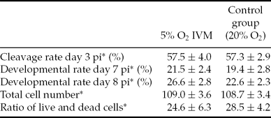
In total, 540 COC were matured under an oxygen concentration of 5% and 519 COC under atmospheric oxygen concentration. The subsequent cleavage (day 3 p.i.) and development rates (day 7 p.i. and day 8 p.i.) did not differ significantly between the two groups (Table 2).
Effect of oxygen tension during in vitro maturation on bovine embryo development, total cell number and ratio of live and dead cells in expanded blastocysts matured under different oxygen concentrations
*Mean ± standard deviation (SD); IVM, in vitro maturation; pi, post insemination.
Neither the total cell number nor the live–dead cell ratio of expanded blastocysts differed significantly (Table 2).
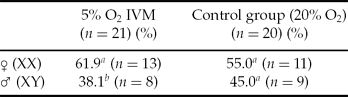
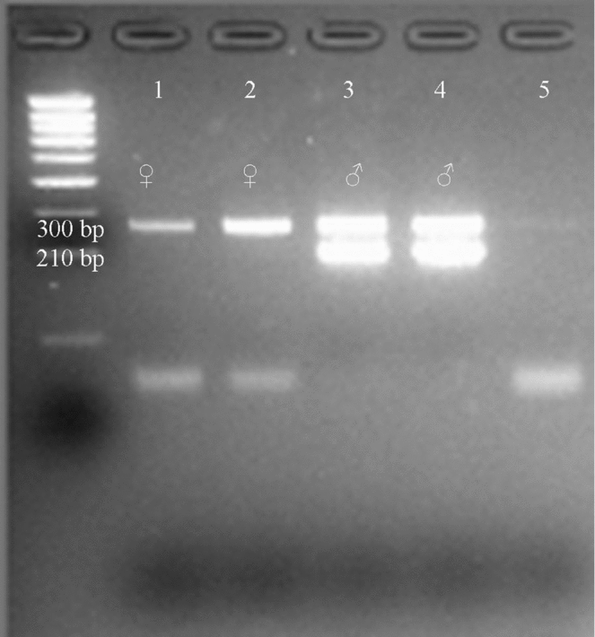
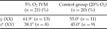
The distribution of sexes among embryos showed a significant shift towards the female in those embryos that developed of COC matured under restricted oxygen conditions (Table 3) in comparison with the expected ratio of 50:50%. A representative gel photo of PCR results is shown in Fig. 1.
Sex distribution of bovine embryos matured under 5% O2 or atmospheric oxygen concentration
a,bp ≤ 0.05 within column. IVM, in vitro maturation.
Representative gel photo (4% agarose gel) of analysis of sex distribution of bovine embryos: (1) female embryo, (2) control for ‘female,’ (3) male embryo, (4) control for ‘male’.
Experiment 2
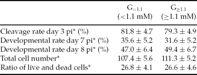
In total, 117 COC were isolated from follicles with an intrafollicular glucose concentration of <1.1 mM. Employing the same method, 123 COC were obtained from follicles with a glucose content of ≥ 1.1 mM. Cleavage (day 3 p.i.) and development rates on day 7 and on day 8 p.i. did not differ significantly between the two groups (Table 4).
Effect of follicular glucose content on bovine embryo development and total cell number and ratio of live and dead cells of expanded blastocysts from groups G<1.1 and G≥1.1
*Mean ± standard deviation (SD); pi = post insemination.
Total cell number (107.4 ± 5.6 vs. 111.3 ± 5.2) and live–dead cell ratio (26.8 ± 4.1 vs. 26.6 ± 4.6) of expanded blastocysts did not differ significantly between the two groups (Table 4).
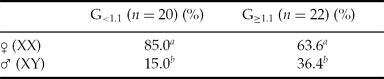
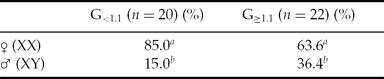
The sex distribution of embryos of both groups differed significantly from the expected distribution of 50:50. In both groups more female embryos (G<1.1: 85.0% female vs. 15% male embryos and G≥1.1: 63.6% female vs. 26.4% male) could be observed (Table 5).
Sex distribution of bovine embryos from groups G<1.1 and G≥1.1
a,bp ≤ 0.05 within column.
Discussion
The aim of maturation systems in vitro is to supply the oocyte with a suitable environment to resume and complete meiosis, finally reaching the second metaphase stage. There is a wide range of available protocols for this specific step in IVP. van de Sandt et al. (Reference van de Sandt, Schroeder and Eppig1990) showed that the use of different maturation media resulted in differences in preimplantation development in murine embryos. This observation has been verified in the bovine (Rose & Bavister, Reference Rose and Bavister1992). Initially, TCM199 proved to be a suitable basis for a maturation medium, resulting in good quality embryos. Nevertheless, it was not originally designed for IVM but for somatic cell culture (Morgan et al., Reference Morgan, Morton and Parker1950, Reference Morgan, Campbell and Morton1955). Other media employed in IVM include a modified SOF medium (de Castro & Hansen, Reference de Castro and Hansen2007). Alterations in protocols have gone as far as the use of a two-step maturation protocol with a period of meiosis inhibition prior to the final maturational step (Pavlok et al., Reference Pavlok, Lapathitis, Cech, Kubelka, Lopatarova, Holy, Klima, Motlik and Havlicek2005).
Subsequent embryo development rates are known to differ vastly with regards to the employed media in each step of IVP. At present, the final culture step is mostly conducted in oxygen-reduced conditions, which are considered to be beneficial to successive development rates (Thompson et al., Reference Thompson, Simpson, Pugh, Donnelly and Tervit1990). Interestingly, there is no consent about the influence of a reduced oxygen concentration during maturation on the subsequent development of embryos. The obtained results differ vastly, possibly due to the varying media employed. Apart from offering an explanation for the controversial results this fact also inhibits a direct comparison.
Interactions between oxygen concentration and the employed media have been shown. Especially in TCM199-based medium, the oxygen concentration is a determinant of development to the blastocyst stage (de Castro & Hansen, Reference de Castro and Hansen2007). Furthermore, it was possible to link the glucose concentration in the employed media in combination with the oxygen concentration to alterations in subsequent embryo development. It has been suggested that in a static in vitro system, as used for maturation, employed media should contain a supra-physiological glucose concentration in order to supply enough glucose at all times during maturation (Sutton-McDowall et al., Reference Sutton-McDowall, Gilchrist and Thompson2010). A glucose concentration similar to that of the follicular glucose concentrations in vivo in the maturation medium is needed to show effects induced by a reduced oxygen tension – visible in increased developmental rates (Hashimoto et al., Reference Hashimoto, Minami, Yamada and Imai2000b). The employed glucose concentration in the present work was 5.6 mM. The lack of distinctions regarding cleavage and development rates may be because this concentration could be too low to evoke effects in combination with a reduced oxygen concentration during IVM.
Staining of expanded blastocysts revealed no significant difference in total cell numbers between the 5% O2 IVM group and the control group. There are also controversial results in total cell numbers of blastocysts among studies, reflecting the diverse effects of different oxygen concentrations on embryo development. Total cell numbers of bovine (Oyamada & Fukui, Reference Oyamada and Fukui2004), porcine (Kikuchi et al., Reference Kikuchi, Onishi, Kashiwazaki, Iwamoto, Noguchi, Kaneko, Akita and Nagai2002; Iwamoto et al., Reference Iwamoto, Onishi, Fuchimoto, Somfai, Takeda, Tagami, Hanada, Noguchi, Kaneko, Nagai and Kikuchi2005; Karja et al., Reference Karja, Kikuchi, Fahrudin, Ozawa, Somfai, Ohnuma, Noguchi, Kaneko and Nagai2006) and murine blastocysts (Preis et al., Reference Preis, Seidel and Gardner2007) were increased if oocytes were matured previously under reduced oxygen tension. Nevertheless, a different study conducted in the porcine also detected no effect on total cell numbers (Park et al., Reference Park, Hong, Yong, Hwang, Lim and Lee2005). Additionally, staining allowed differentiation between live and dead cells, and allowed evaluation of the injuriousness of a method, as determined by the live–dead cell ratio. No differences could be detected in the present study regarding this parameter, reinforcing the assumption that, under the present conditions, a reduction of the oxygen concentration during IVM neither impairs nor improves subsequent embryonic development.
Differences in development between female and male embryos, such as in speed of development, arise in the disproportionate expression of sex-specific genes or genes that are regulated by X-chromosomal transcripts (reviewed by Gutierrez-Adan et al., Reference Gutierrez-Adan, Perez-Crespo, Fernandez-Gonzalez, Ramirez, Moreira, Pintado, Lonergan and Rizos2006). For the first time, we were able to describe the effect of a reduced oxygen concentration during IVM on subsequent gender distribution of the embryos. In the present study, significantly more female embryos were generated from oocytes that had been matured previously under O2-reduced conditions. The sex shift among embryos of the control group did not differ significantly from the expected 1:1 ratio (Gamarra et al., Reference Gamarra, Le Guienne, Lacaze and Ponsart2011; Table 2). These results seem to be controversial when compared with studies conducted earlier. Here it was discussed that oocytes that were matured under a higher oxygen concentration contained an increased amount of reactive oxygen species (ROS; Hashimoto Reference Hashimoto2009). The amount is regulated by glucose-6-phosphate dehydrogenase (G6PD; Filosa et al., Reference Filosa, Fico, Paglialunga, Balestrieri, Crooke, Verde, Abrescia, Bautista and Martini2003) – the gene of which is located on the X chromosome (Gutierrez-Adan et al., Reference Gutierrez-Adan, Oter, Martinez-Madrid, Pintado and De La Fuente2000). Accordingly, it would seem to be more likely to expect that a larger number of female embryos originated from oocytes that had been matured under atmospheric oxygen conditions. This proposal is supported by the results of Iwata et al., (Reference Iwata, Kimura, Hashimoto, Ohta, Tominaga and Minami2002), which showed a higher developmental competence for embryos with higher G6PD activity under stress conditions, e.g. high oxygen concentrations. However, these results were obtained by the analysis of early embryos and with regards to the early embryos’ environment and not to that of the oocyte. An analysis of obtained embryos at the molecular level will be necessary to clarify the arisen discrepancies.
Cleavage and development rates between embryos generated with oocytes from follicles with defined glucose levels (< 1.1 mM and ≥1.1 mM) did not differ significantly.
Interestingly, cleavage and development rates obtained in this experiment are higher than those obtained in Experiment 1, even though IVP was performed in the same laboratory. A possible explanation may lie in the different handling of ovaries at the time of COC collection. As follicle dissection is more time consuming than the slicing method a so-called ‘post-mortem’ effect may have occurred. The duration of storage after slaughter has a subsequent effect on the development of embryos. A storage duration of 4 h after slaughter resulted in the highest development rates on day 7 pi, compared with either a shorter or a longer storage time (Blondin et al., Reference Blondin, Coenen, Guilbault and Sirard1997). Additionally, the time for handling of oocytes and zygotes during IVF or IVC steps was shortened on account of the restricted number of oocytes isolated individually from their follicles.
So far, the effects of intrafollicular glucose concentrations in premature follicles on subsequent embryo development have not been studied, whereas the effects of differing glucose concentrations during IVM have been studied in varying species, including the bovine (Rose-Hellekant et al., Reference Rose-Hellekant, Libersky-Williamson and Bavister1998; Khurana & Niemann, Reference Khurana and Niemann2000; Sutton-McDowall et al., Reference Sutton-McDowall, Gilchrist and Thompson2005; Herrick et al., Reference Herrick, Lane, Gardner, Behboodi, Memili, Blash, Echelard and Krisher2006). In conjunction with other results, it is possible to link glucose supplementation during IVM to an increase in total cell numbers in caprine blastocysts (Sato et al., Reference Sato, Iwata, Hayashi, Kimura, Kuwayama and Monji2007).
Staining of expanded or hatched blastocysts of groups G<1.1 and G≥1.1 showed no significant differences in the total cell number and the live–dead cell ratio between the two groups in the present study, a finding that indicated that embryos of both groups were of similar quality.
There is growing evidence for a maternal influence on sex distribution of offspring mediated through testosterone concentration and glucose availability (reviewed by Grant & Chamley, Reference Grant and Chamley2010). Sex analysis of embryos of the groups G<1.1 and G≥1.1 showed a significant alteration from the expected distribution of 50:50 (Gamarra et al., Reference Gamarra, Le Guienne, Lacaze and Ponsart2011; Table 4). It is suggested that glucose supplementation produces a preferential loss of female embryos during culture (Gutierrez-Adan et al., Reference Gutierrez-Adan, Lonergan, Rizos, Ward, Boland, Pintado and de la Fuente2001) whereas the presence of glucosamines during maturation did not affect the sex ratio of bovine IVP-derived embryos (Kimura et al., Reference Kimura, Iwata and Thompson2008). In the present study, the oocytes were pooled according to the glucose content of their originating follicles. Although a shift towards the female could be detected in both groups, an increase in male embryos generated from the oocytes of the G≥1.1 group was seen. In other species it has been proposed that a high maternal blood glucose concentration is related to male-biased litters (Mus musculus and Microtus agrestis: Rosenfeld et al., Reference Rosenfeld, Grimm, Livingston, Brokman, Lamberson and Roberts2003; Helle et al., Reference Helle, Laakson, Adamsson, Paranko and Huitu2008).
It has been suggested that glucose plays an important role in the final maturational phase of the oocytes. An excessive glucose concentration (20 mmol) during IVP has detrimental effects on the embryos development just as well as COC matured in a glucose-deprived medium (Hashimoto et al., Reference Hashimoto, Minami, Yamada and Imai2000b). Here, fewer oocytes were able to reach metaphase II than oocytes cultured in media supplemented with physiological glucose concentrations.
The results of this work indicate that, under the present conditions, a reduced oxygen concentration during IVM – although not affecting in vitro development or morphological quality – shifts the sex ratio of bovine embryos towards the female.
Additionally, we were able to show that with high(er) follicular glucose concentrations, an increase in the number of male embryos may be obtained. This result shows that the glucose concentration of the oocyte's environment plays a crucial role in gender distribution even before the final maturational stage is reached.
Acknowledgements
The authors would like to thank Doris Mueller for her assistance in the IVP laboratory.