INTRODUCTION
The short-bodied onuphids of the genus Nothria Malmgren, Reference Malmgren1867, inhabiting distinctive dorsoventrally flattened tubes, adorned with large shell fragments and foraminiferans, are well known as epibenthic crawlers and have been reported worldwide from shallow subtidal waters to abyssal depths (Kucheruk Reference Kucheruk and Kuznetsov1980, Reference Kucheruk1985; Paxton Reference Paxton1986). Less well known is their sister genus Anchinothria Paxton, Reference Paxton1986, also widely distributed, with most species occurring deeper than 500 m, constructing flattened soft muddy tubes, often encrusted with elongated foreign objects (Paxton, Reference Paxton1986; Orensanz, Reference Orensanz1990). The generic name Nothria was widely used in the older onuphid literature for species having only single branchial filaments (Hartman, Reference Hartman1944; Day, Reference Day1967; Fauchald, Reference Fauchald1968) until Pettibone (Reference Pettibone1970) redefined the genus. She recognized the close relationship between Nothria and Paradiopatra Ehlers, Reference Ehlers1887, in both having enlarged anterior parapodia, although modified in different ways. Pettibone (Reference Pettibone1970) suggested the lobes of the anterior parapodia as diagnostic characters, and recognized auricular prechaetal lobes in Nothria and bi- to trilobed prechaetal lobes in Paradiopatra. The first attempt to revise both genera was undertaken by Fauchald (Reference Fauchald1982a) who re-examined the type material, referred eight species to Nothria and 11 species to Paradiopatra. Paxton (Reference Paxton1986) revised the whole family Onuphidae, uncovered that the name ‘Paradiopatra’ was already in use, and proposed the new name Anchinothria for species previously placed in Paradiopatra sensu Pettibone (Reference Pettibone1970). Paxton's phylogenetic analysis of the generic relationships within onuphids based on morphological characters confirmed Pettibone's earlier observation in revealing two sister genera: Nothria and Anchinothria, sharing a number of synapomorphies. Both genera comprise short-bodied onuphid worms with enlarged anterior parapodia, distally wide so-called ‘scoop-shaped’ pectinate chaetae with rolled lateral margins, and subacicular hooks inserted medially in the posterior parapodia between two bundles of limbate chaetae.
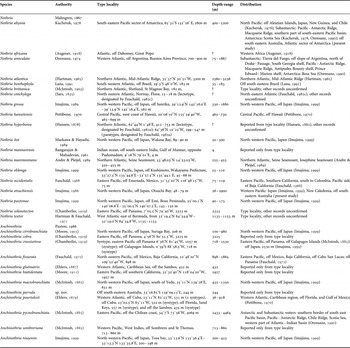
Paxton (Reference Paxton1986) re-evaluated the morphology of the anterior parapodia of species revised by Fauchald (Reference Fauchald1982a), re-assigned them among the two genera, and recognized 13 species of Nothria and 12 species of Anchinothria. Seven more species of Nothria have been described during the following two decades, and two were later synonymized by Orensanz (Reference Orensanz1990) increasing the total number of species in the genus to 19. Of the 12 species referred to Anchinothria, three were later synonymized by Orensanz (Reference Orensanz1990), and Imajima (Reference Imajima1999) described a new Anchinothria species from Japan, bringing the total number of species in the genus up to 11 (Table 1).
Species composition and distribution of Nothria and Anchinothria.

Nothria demonstrates a significant variation in the majority of morphological characters which was underestimated in the early species descriptions and revisions of the genus (Fauchald, Reference Fauchald1982a; Kucheruk, Reference Kucheruk1985). Several attempts to estimate the morphological variation based on the examination of large material were performed. Kucheruk (Reference Kucheruk and Kuznetsov1980) analysed 1200 specimens of Nothria collected worldwide at depths from 45 to 2930 m. He referred all the examined specimens to Nothria conchylega (Sars, Reference Sars1835) despite the significant variability found in the studied material. Kucheruk (Reference Kucheruk and Kuznetsov1980) reported that the chaetal composition (presence or absence of falcigers, limbate, and pectinate chaetae) in the anterior three pairs of parapodia was invariable in all specimens studied while the shape of falcigers in three anterior chaetigers was size-related. Jirkov & Yermolaev (Reference Jirkov and Yermolaev1989) examined extensive collections of Nothria specimens from the northern hemisphere and identified two distinct morphotypes (A and B) differing from each other in the presence/absence of limbate and pectinate chaetae on the second pair of parapodia. They have analysed 12 morphological characters in 191 specimens belonging to morphotype A and 62 specimens assigned to morphotype B. Three characters were found to be size-dependent while eight characters, including chaetal composition in anterior pairs of parapodia, lacked significant correlation with the size (expressed by body width) of studied worms. Orensanz (Reference Orensanz1990) discussed the ontogenetic variation in Nothria anoculata Orensanz, Reference Orensanz1974 and demonstrated the gradual replacement of pseudocompound falcigers by simple falcigers in the first two pairs of parapodia with increasing size of specimens. The segment number of the first appearance of subacicular hooks and branchiae was also variable in the examined material, with positive correlation with the size of the worms. In contrast, the morphology of N. abyssia was shown to be less variable and became stabilized earlier in the development in comparison with N. anoculata (Orensanz, Reference Orensanz1990).
Intertidal and shallow water onuphids of Australia have been studied by Paxton (Reference Paxton1979, Reference Paxton1993, Reference Paxton1996). The present study focuses on Nothria and Anchinothria which are better represented in low subtidal and slope depths. The genus Nothria has been reported from New Zealand for N. conchylega (Knox & Hicks, Reference Knox and Hicks1973), was known to occur in Australia, but had not been identified to species (Paxton, Reference Paxton, Beesley, Ross and Glasby2000), whilst Anchinothria has never been reported from Australian waters.
The present study aims to revise the species of Nothria and Anchinothria collected from shelf and slope depths off southeastern Australia. The detailed description of intraspecific variability in at least four different species of Nothria (Kucheruk, Reference Kucheruk and Kuznetsov1980; Jirkov & Yermolaev, Reference Jirkov and Yermolaev1989; Orensanz, Reference Orensanz1990) showed several different patterns of size-dependency of major diagnostic characters. We also aim to analyse the ontogenetic variation of the major diagnostic morphological characters in Australian Nothria such as chaetal morphology and composition in the anterior three pairs of parapodia, morphology and distribution of branchiae, prechaetal and postchaetal parapodial lobes, and appearance of the subacicular hooks. The description of size-dependent morphological variation in Nothria from Australian waters will become a basis for future revision of diagnostic characters in other species of the genus with subsequent generic revision of Nothria.
MATERIALS AND METHODS
Specimens were stained with methylene blue solution in 30% ethanol and examined under a dissecting stereomicroscope. methylene blue staining increased the contrast of some morphological structures, such as ceratophores, branchiae, and parapodial lobes and cirri. After examination stained specimens were transferred to 70% ethanol, which led to the complete loss of the stain from the specimens. Unstained specimens were examined under a compound light microscope. Temporary glycerol slides of specimens smaller than 1.3 mm wide were used for the detailed examination of chaetal distribution and the morphology of parapodia along the body. Permanent slides of anterior parapodia embedded into Hydro-Matrix® mounting medium were made for the examination of chaetae in worms larger than 1.3 mm wide. Line drawings were made with the aid of a camera lucida and digital photography.
Specimens used for scanning electron microscopy (SEM) analysis were initially fixed with 4% formalin and then transferred into 70% ethanol. They were run through the series of ethanol: 70%, 80%, 90%, 100%, 100%, 100%, 30 minutes at each step, critically point dried with a Bal-Tec CPD 030 Critical Point Dryer, mounted on stubs, coated with gold using Emitec K550 Gold Sputter Coater and imaged with a Zeiss EVO LS15 scanning electron microscope with a Robinson Backscatter Detector.
Kucheruk (Reference Kucheruk and Kuznetsov1980) demonstrated strong positive correlation between the width and the length of the body and the number of chaetigers in Nothria. He suggested that the width of a worm can be used for size estimation since posterior regions were frequently missing or in poor shape in the studied samples. Following Kucheruk (Reference Kucheruk and Kuznetsov1980) the width of the sixth chaetiger measured as a distance between the inner parts of dorsal cirri was used as an estimation of the size of the worms.
We followed chaetal terminology suggested by Budaeva & Fauchald (Reference Budaeva and Fauchald2010). The term ‘falciger’ was used to describe simple or pseudocompound hooded dentate chaetae present on anterior modified parapodia. The term ‘hook’ was used to define simple bidentate chaetae appearing in median position in posterior unmodified parapodia. The terminology for prostomial appendages followed that of Paxton (Reference Paxton1998).
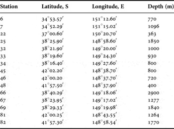
Specimens borrowed from the following institutions have been examined: Australian Museum, Sydney, New South Wales, Australia (AM); Museum Victoria, Melbourne, Victoria, Australia (MV); and Senckenberg Museum, Frankfurt-am-Main, Germany (SMF). The holotype and paratypes of the new species were deposited in the Marine Invertebrate Collection of the Australian Museum, Sydney (AM). Station details of the material from the RV ‘Franklin’ SLOPE expedition (MV) are given in Table 2.
Details of RV ‘Franklin’ epibenthic sled stations (Poore et al., Reference Poore, Just and Cohen1994).

SYSTEMATICS
Order EUNICIDA Dales, 1962
Family ONUPHIDAE Kinberg, 1865
Subfamily HYALINOECIINAE Paxton, Reference Paxton1986
Genus Anchinothria Paxton, Reference Paxton1986
DIAGNOSIS
Body short, up to 100 segments. Median antenna longer and thicker than lateral antennae. Ceratophores short, consisting of 2–5 rings. Nuchal grooves straight. Anterior 2–3 pairs of parapodia enlarged, directed anteroventrally with bi- to trilobed prechaetal lobes. Uni- or bidentate simple or pseudocompound falcigers on first 2–4 pairs of anterior parapodia, in one species on first seven pairs of parapodia. Subacicular hooks in median position from chaetigers 4–16. Pectinate chaetae wide with rolled margins, so-called ‘scoop-shaped’, from chaetigers 2–3, in one species from chaetiger 14. Branchiae present or absent, single to up to 10 filaments. Tubes dorsoventrally flattened with parchment-like inner layer covered with mud and often incrusted with scattered large elongated foraminiferans, glass sponge spicules or echinoid spines attached along longitudinal margins of tubes.
Anchinothria parvula sp. nov.
(Figures 1–3)
TYPE MATERIAL
AM: W.41478, Tasman Sea, Taupo Seamount, RV ‘Franklin’ Station 8, 33°16.85′S 159°09.15′E, 244 m, 2 October 1989 (holotype); W.41475.001, the same location as holotype (1 paratype gold-coated for SEM); W.41476, the same location as holotype (1 paratype with dissected maxillae); W.41476.001, the same location as holotype (microscopic slide with dissected parapodia 1–3, 7, and 13 from paratype W.41476); W.41477, the same location as holotype (10 paratypes).
DIAGNOSIS
First pair of parapodia with pseudocompound uni- or bidentate falcigers. Second and third pairs of parapodia with pseudocompound bidentate falcigers and limbate chaetae. Pectinate chaetae flat, from chaetiger 14. Branchiae absent. Subacicular hooks from chaetigers 6–8. Prechaetal lobes as low rounded ridges on all chaetigers. Postchaetal lobes present on first three chaetigers. Two pairs of eyes.
TYPE LOCALITY
Off south-eastern Australia, 33°16.85′S 159°09.15′E, 244 m.
DESCRIPTION
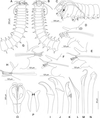
All specimens examined represented by anterior fragments. Holotype, largest specimen, consisting of 17 chaetigers, 0.73 mm wide and 5.9 mm long (Figure 1A–C). Paratypes consisting of 9–18 chaetigers with width varying from 0.4 to 0.73 mm. Specimens pale yellowish in colour with dark brown strap along lateral posterior margin of prostomium and anterior margin of peristomium; light brown spots located dorsally at bases of first pair of parapodia (Figure 1C). Some specimens without coloration.
Prostomium with rounded anterior margin, globular frontal lips not visible from dorsal side, projecting anteroventrally (Figures 1A–C; 2A). Palps reaching chaetiger 1 (1–2); lateral antennae reaching chaetiger 5 (3–7); median antenna longer and thicker than lateral antennae reaching chaetiger 9 (6–9). Ceratophores consisting of 3 (2–4) rings (Figure 1C). Nuchal grooves straight with narrow middorsal separation. Two pairs of eyes present, large dark eyes located laterally near bases of lateral antennae (Figure 1A); pair of small eyespots located dorsally at anterior margin of peristomium (Figure 1C). Peristomium half as long as first chaetiger, bearing short, tapering peristomial cirri (Figure 1A, C).
Anchinothria parvula, new species: (A) anterior part of body, dorsal view; (B) same, ventral view; (C) same, lateral view; (D) parapodium of chaetiger 1, anterior view; (E) parapodium of chaetiger 2, anterior view; (F) parapodium of chaetiger 3, anterior view; (G) parapodium of chaetiger 7, posterior view; (H) parapodium of chaetiger 13, anterior view; (I–J) pseudocompound falcigers from chaetiger 1; (K) same from chaetiger 2; (L) limbate chaeta from chaetiger 3; (M) same from chaetiger 13; (N) subacicular hook from chaetiger 13; (O) maxillae; (P) mandibles.

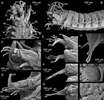
First pair of parapodia slightly larger than subsequent parapodia projecting lateroventrally and directed anteriorly (Figures 1C; 2A, B). Anterior pair of parapodia with short rounded prechaetal lobe, subulate dorsal cirri, digitiform postchaetal lobes and ventral cirri (Figures 1D; 2C–D). Second pair of parapodia similar in size with posterior parapodia bearing short, rounded prechaetal lobes, subulate dorsal cirri, digitiform postchaetal lobes and conical ventral cirri (Figures 1E; 2E). Third pair of parapodia with very short rounded prechaetal lobes, bearing subulate dorsal cirri, digitiform postchaetal lobes and slightly conical ventral cirri with distal glandular pads (Figures 1F; 2E). Prechaetal lobes becoming invisible after chaetiger 3 (Figures 1F; 2E); postchaetal lobes abruptly disappearing from chaetiger 4 (Figure 2F). Ventral cirri subulate on first two chaetigers (Figures 1D, E; 2C–E), slightly conical with round glandular tips on chaetiger 3 (Figures 2E) transforming into flat, round glandular pads from chaetiger 4 (Figure 2F, I, J).
Anchinothria parvula, new species: (A) anterior part of body, ventral view; (B) same, lateral view; (C) parapodium of chaetiger 1, posterior view; (D) same, anteroventral view; (E) parapodia of chaetigers 2–3, ventral view; (F) parapodium of chaetiger 4, ventral view; (G) parapodium of chaetiger 7, ventral view; (H) parapodium of chaetiger 15, anterior view; (I–J) ventral glandular pad of chaetiger 5; dc, dorsal cirrus; fl, frontal lip; la, lateral antenna; ma, median antenna; p, palp; psl, postchaetal lobe; ul, upper lip; vc, ventral cirrus; vgp, ventral glandular pad.

First pair of parapodia of holotype and seven paratypes with 3 (4) bidentate clearly pseudocompound falcigers (Figures 1D, J; 3A); six paratypes with unidentate falcigers (Figure 1I). Falcigers with wide, short distal parts, covered with short blunt hoods. Second tooth, when present, relatively small, widely separated from distal tooth (Figure 1J). Shafts of anterior falcigers smooth with scattered small spines. Second and third pairs of parapodia bearing single dorsal limbate chaetae (Figure 1E, F, L) and three pseudocompound clearly bidentate falcigers with short blunt hoods and group of small spines located beyond pseudoarticulation (Figures 1E, F, K; 2E; 3B). Single bundle of bilimbate chaetae with wide distal parts from chaetiger 4 (Figures 2F; 3C). Subacicular hook starting from chaetiger 8 (6–8) in median position with single limbate chaeta located dorsally and ventrally (Figures 1G, N, M; 3D, E). Subacicular hooks first present singly in parapodium (Figures 1G; 2G), becoming paired posteriorly (Figures 1H; 2H). Pectinate chaetae first present on chaetiger 14, flat, slightly bent with approximately 18–20 denticles, one per parapodium (Figures 2H; 3F, G). Neuroaciculae pale yellow with pointed tips (Figure 3G), 2–3 per parapodium (Figure 1D–H).
Anchinothria parvula, new species: (A) pseudocompound falcigers from chaetiger 1; (B) pseudocompound falcigers from chaetiger 3; (C) limbate chaetae from chaetiger 4; (D) subacicular hooks from chaetiger 13; (E) limbate chaeta from chaetiger 13; (F) pectinate chaeta from chaetiger 15; (G) pectinate chaeta and emerging acicula from chaetiger 14.

Mandibles white with short shafts (Figure 1P). Maxillae very delicate almost transparent with approximately 10 teeth in each plate of MxII–MxIV. Mx V not observed (Figure 10). Branchiae absent. Pygidium and tubes unknown.
ETYMOLOGY
The species name ‘parvula’ meaning ‘little’, ‘pretty’, ‘young’ in Latin refers to the small size and dainty appearance of individuals of the new species.
REMARKS
Anchinothria parvula belongs to a group of species with falcigers on the first three pairs of parapodia. This group comprises the following species: Anchinothria crassisetosa (Chamberlin, 1919), A. hiatidentata (Moore, Reference Moore1911), A. macrobranchiata (McIntosh, Reference McIntosh1885), and A. pycnobranchiata (McIntosh, Reference McIntosh1885). The new species can be distinguished from A. crassisetosa, A. pycnobranchiata and A. hiatidentata in the origin of the subacicular hook. They are present from chaetiger 4 in the two former species and from chaetiger 12 in the latter, while A. parvula sp. nov. has subacicular hooks starting from chaetigers 6–8. Anchinothria parvula sp. nov. differs from A. macrobranchiata in lacking rather than having long branchiae from chaetiger 8 and being much smaller in size. The largest specimen of A. parvula is 0.73 mm wide without parapodia while A. macrobranchiata reaches 6 mm in width including parapodia (McIntosh, Reference McIntosh1885).
DISTRIBUTION
Anchinothria parvula is known from a single locality, off south-eastern Australia, at a depth of 244 m.
Genus Nothria Malmgren, Reference Malmgren1867
DIAGNOSIS
Body short, up to 100 segments. Median antenna longer and thicker than lateral antennae. Ceratophores short, consisting of 2–5 rings. Nuchal grooves straight. Anterior 2–3 pairs of parapodia enlarged, directed anteroventrally with large auricular prechaetal lobes. Uni-, bi- or tridentate simple or pseudocompound falcigers on first 2–3 pairs of anterior parapodia. Subacicular hooks in median position from chaetigers 7–15. Pectinate chaetae wide with rolled margins, so-called ‘scoop-shaped’, from chaetigers 2–3, in one species from chaetiger 9. Branchiae present or absent, single to up to five filaments. Tubes dorsoventrally flattened with thin inner parchment-like layer covered with large shell fragments, small stones and foraminiferans.
Nothria abyssia Kucheruk, Reference Kucheruk1978
(Figures 4–10)
Nothria abyssia Kucheruk, Reference Kucheruk1978: 101–103, figures 4, 5.—Orensanz Reference Orensanz1990: 48–50, pl. 10, chart 5B.
Nothria abranchiata – Hartman Reference Hartman1964: 112, pl. 24, figures 1–3.—non Nothria abranchiata McIntosh, Reference McIntosh1885: 314–317, pl. 21A, figure 27; pl. 22A, figures 1–3, pl. 40, figures 1–12.
Onuphis (Nothria) abranchiata—Averincev Reference Averincev1972: 178.—Averincev Reference Averincev1974: 219.
Onuphis (Nothria) australatlantica Fauchald, Reference Fauchald1982b: 238, figure 1, table 1.
Paradiopatra abyssia—Fauchald Reference Fauchald1982a: 83.
MATERIAL EXAMINED
AM: W42762, RV ‘Franklin’ SLOPE Station 69 (2), W42763.001, RV ‘Franklin’ SLOPE Station 69 (2 specimens gold-coated for SEM); W42764.001, RV ‘Franklin’ SLOPE Station 69 (1 specimen gold-coated for SEM).
MV: F183731, RV ‘Franklin’ SLOPE Station 82 (2); F183732, RV ‘Franklin’ SLOPE Station 67 (1); F183733, RV ‘Franklin’ SLOPE Station 25 (1); F183734, RV ‘Franklin’ SLOPE Station 69 (35); F183735, RV ‘Franklin’ SLOPE Station 48 (3); F183736, RV ‘Franklin’ SLOPE Station 66 (1); F192362, RV ‘Franklin’ SLOPE Station 69 (one slide with dissected parapodia).
ZMH: P.26039, ANDEEP III, RV ‘Polarstern’ ANT XXII-3 Station 59-5, 67°29.81′S 0°00.23′ W, 4651 m, 14 February 2005, epibenthic sledge, supranet (2); P.26040, ANDEEP III, RV ‘Polarstern’ ANT XXII-3 Station 59-5, 67°29.81′S 0°00.23′W, 4651 m, 14 Feb 2005, epibenthic sledge, epinet (2); P.26041, ANDEEP III, RV ‘Polarstern’ ANT XXII-5 Station 102-13, 65°33.18′S 036°31.05′W, 4817 m, 6 March 2005, epibenthic sledge, epinet (1); P.26042, ANDEEP III, RV ‘Polarstern’ ANT XXII-5 Station 102-13, 65°33.18′S 036°31.05′W, 4817 m, 6 March 2005, epibenthic sledge, supranet (1); P.260043, ANDEEP III, RV ‘Polarstern’ ANT XXII-4 Station 81-8, 70°31.08′S 14°34.82′W, 4419 m, 24 February 2005, epibenthic sledge, supranet (1); P.26044, ANDEEP II, RV ‘Polarstern’ ANT XIX-4 Station 141-10, 58°25.08′S 25°00.77′W, 2313 m, 23 March 2002, epibenthic sledge, epinet (1).
DIAGNOSIS
Based on specimens wider than 1.0 mm at chaetiger 6 excluding parapodia: first pair of parapodia extending beyond frontal margin of prostomium, with simple and pseudocompound bidentate falcigers. Second pair of parapodia with bidentate pseudocompound falcigers and limbate chaetae. Third pair of parapodia with limbate and pectinate chaetae. Pectinate chaetae flat. Branchiae absent. Subacicular hooks from chaetigers 7–9. Auricular prechaetal lobes on first chaetiger only, chaetigers 2–3 with conical prechaetal lobes, reduced posteriorly. Postchaetal lobes present on first 2–3 chaetigers. Paired eyespots may be present on dorsal side of prostomium.
TYPE LOCALITY
Antarctic Ocean, 65°31′S 152°26′E, 2800 m.
DESCRIPTION
All examined specimens represented by anterior fragments varying in width from 0.4 to 1.53 mm. Specimens light brownish, lacking colour pattern. Following description based on specimens wider than 1.0 mm at chaetiger 6 excluding parapodia.
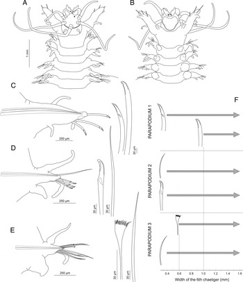
Prostomium anteriorly rounded with paired ovoid to oval frontal lips, palps reaching chaetiger 1, lateral antennae reaching chaetiger 4 and median antenna reaching chaetigers 3–6 (Figure 4A). Ceratophores short, consisting of 2–3 indistinct rings. Two little eyespots present on dorsal side of prostomium (Figure 4A). Some specimens without eyes. Nuchal grooves wide and straight, with narrow mid-dorsal separation. Upper lips oval; ventral lip wide and massive. Peristomium half as long as first chaetiger (Figure 4A). Peristomial cirri as long as peristomium.
Nothria abyssia Kucheruk, Reference Kucheruk1978, morphology and chaetal composition: (A) anterior region, dorsal view; (B) same, ventral view; (C) parapodium and chaetae from chaetiger 1; (D) parapodium and chaetae from chaetiger 2; (E) parapodium and chaetae from chaetiger 3; (F) diagram of chaetal replacement in relation to body width of worms.

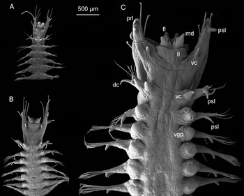
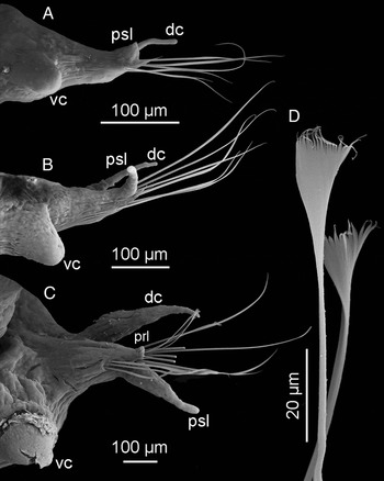
First pair of parapodia enlarged, prolonged significantly extending beyond anterior margin of prostomium, bearing large truncate auricular prechaetal lobes, short digitiform postchaetal lobes, subulate dorsal cirri and short digitiform ventral cirri (Figures 4C; 5C; 6C). Second pair of parapodia similar in size to subsequent parapodia, bearing very short foliose prechaetal lobes, well developed digitiform postchaetal lobes, dorsal cirri and conical ventral cirri (Figures 4D; 7C). Third pair of parapodia with smaller but distinct prechaetal lobes, digitiform postchaetal lobes, dorsal cirri and transitional slightly conical ventral cirri (Figures 4E; 8C). Dorsal cirri present on all chaetigers of specimens examined, gradually decreasing in size and becoming subulate. Prechaetal lobes decreasing in size considerably from chaetigers 3–4, becoming short and rounded, almost invisible from chaetigers 4–5. Postchaetal lobes gradually decreasing posteriorly, visible on up to 24 chaetigers in longest specimens examined. Ventral cirri replaced by round glandular pads from chaetiger 4 (Figure 4B).
Nothria abyssia Kucheruk, Reference Kucheruk1978, ventral view: (A) specimen of 0.4 mm width; (B) specimen of 0.6 mm width; (C) specimen of 1.57 mm width; dc, dorsal cirrus; fl, frontal lip; ll, lower lip; md, mandibles; prl, prechaetal lobe; psl, postchaetal lobe; vc, ventral cirrus; vgp, ventral glandular pad.

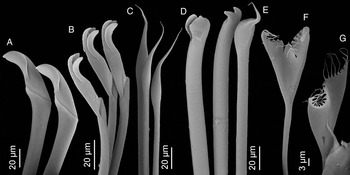
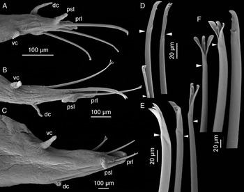
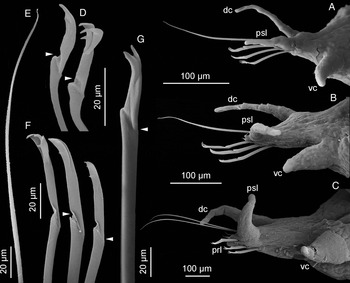
Nothria abyssia Kucheruk, Reference Kucheruk1978, parapodia and chaetae from chaetiger 1 of specimens of different sizes: (A) parapodium from chaetiger 1 of specimen of 0.4 mm width, posterior view; (B) same of specimen of 0.6 mm width; (C) same of specimen of 1.57 mm width; (D) pseudocompound falcigers of specimen of 0.4 mm width; (E) pseudocompound falcigers of specimen of 0.6 mm width; (F) simple and pseudocompound falcigers of specimen of 1.57 mm width. Arrow heads indicate pseudoarticulation of falcigers; dc, dorsal cirrus; prl, prechaetal lobe; psl, postchaetal lobe; vc, ventral cirrus.

First pair of parapodia with one simple and 2–3 indistinctly pseudocompound bidentate falcigers (Figures 4C; 6F). Subdistal tooth on anterior falcigers small and often broken (Figure 6F). Anterior falcigers bearing paired short hood with blunt tips, in some falcigers hoods missing, presumably broken (Figures 4C, 6F). Second pair of parapodia with 3–4 clearly bidentate pseudocompound falcigers with short, paired hoods and 1–4 simple, thin limbate chaetae (Figures 4D; 7G). Third pair of parapodia with 5–8 thin limbate chaetae and 2–3 flat and wide pectinate chaetae with 17–20 denticles (Figures 4E; 8D). Subsequent parapodia with numerous limbate and pectinate chaetae. Subacicular hooks starting from chaetigers 7–9, two per parapodium. Neuroaciculae yellow with pointed tips, 2–3 per parapodium.
Nothria abyssia Kucheruk, Reference Kucheruk1978, parapodia and chaetae from chaetiger 2 of specimens of different sizes: (A) parapodium from chaetiger 2 of specimen 0.4 mm width, posterior view; (B) same of specimen of 0.6 mm width; (C) same of specimen of 1.57 mm width; (D) pseudocompound falcigers of specimen of 0.4 mm width; (E) limbate chaeta of specimen of 0.6 mm width; (F) pseudocompound falcigers of specimen of 0.6 mm width; (G) pseudocompound falciger of specimen of 1.57 mm width; dc, dorsal cirrus; prl, prechaetal lobe; psl, postchaetal lobe; vc, ventral cirrus.

Nothria abyssia Kucheruk, Reference Kucheruk1978, parapodia and chaetae from chaetiger 3 of specimens of different sizes: (A) parapodium from chaetiger 3 of specimen of 0.4 mm width, ventral view; (B) same of specimen of 0.6 mm width, posterior view; (C) same of specimen of 1.57 mm width, anterior view; (D) pectinate chaetae of specimen of 1.57 mm width; dc, dorsal cirrus; prl, prechaetal lobe; psl, postchaetal lobe; vc, ventral cirrus.

Branchiae absent. Tubes flattened, covered by numerous foraminiferan tests; no large pieces of shell fragments were observed.
ONTOGENETIC VARIATION
Thirty-two specimens of Nothria abyssia ranging in width from 0.4 to 1.53 mm (Figure 5) were examined (SLOPE Station 69).
Chaetal composition of the first pair of parapodia
Specimens smaller than 1.0 mm wide had 3–4 (rarely up to 5) pseudocompound falcigers in the parapodia of the first chaetiger (Figure 4F; 6D, E). In specimens larger that 1.0 mm wide, a single pseudocompound falciger was replaced by a simple falciger (Figures 4F; 6F). Both simple and preudocompound falcigers frequently were with partly broken or worn subdistal tooth and lacked hoods in the largest specimens examined (Figure 6F). No truly unidentate falcigers were observed in the studied material.
Chaetal composition of the second pair of parapodia
Parapodia of all specimens examined were with the same set of chaetae consisting of 3–4 pseudocompound bidentate falcigers (Figures 4D, F; 7D, F, G) and 1–2 limbate chaetae (Figure 7E). Pectinate chaetae were absent in all studied specimens. No variation was observed.
Chaetal composition of the third pair of parapodia
Parapodia of all specimens examined lacked falcigers (Figure 4E, F). Specimens smaller than 0.6 mm wide were with limbate chaetae only (Figure 8A), the larger specimens had both limbate and pectinate chaetae, 2–4 per parapodium (Figure 8C, D). Pectinate chaetae were flat with approximately 20 long teeth.
Presence of subacicular hooks
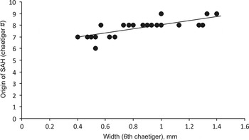
The segment of the first presence of subacicular hooks varied from six to nine with positive correlation with the body size (Figure 9).
Nothria abyssia Kucheruk, Reference Kucheruk1978: relationship between body width of specimens and origin of subacicular hooks (SAH), R 2 = 0.556.

Distribution of prechaetal and postchaetal parapodial lobes
Well-developed auricular prechaetal lobes were present only on the first pair of parapodia in all specimens examined, independently of their size (Figure 6A–C). Short foliose prechaetal lobes were distinguishable on the second and the third pairs of parapodia in specimens larger than 1.5 mm wide (Figures 7C; 8C). In smaller specimens they were present as short rounded ridges (Figures 7B; 8A, B). All subsequent parapodia were with very short prechaetal lobes in all material examined, independently of the size of the worms. Postchaetal lobes were digitiform on the first two pairs of parapodia in all the specimens. In specimens smaller that 0.5 mm wide they became short conical or knob-shaped from chaetiger 3 (Figure 8A), gradually decreasing in size posteriorly. In larger specimens the anterior 5–6 chaetigers were with digitiform postchaetal lobes, becoming short and conical posteriorly, present at least till chaetiger 24 in the longest specimen examined.
REMARKS
Nothria abyssia is similar to N. paxtonae Imajima, Reference Imajima1999 in lacking branchiae, having falcigers present on the first two pairs of parapodia and limbate chaetae starting from chaetiger 2. Both species share the presence of flat pectinate chaetae rather than scoop-shaped pectinate chaetae characteristic of other species of Nothria. In N. abyssia pectinate chaetae were reported on chaetiger 2 by Kucheruk (Reference Kucheruk1978) however were found only on chaetiger 3 in the examined material. Imajima (Reference Imajima1999) reported pectinate chaetae starting from chaetiger 9 in N. paxtonae. In addition, N. paxtonae has eight papillae surrounding the anus. Anal papillae were never reported in N. abyssia.
Three specimens from SLOPE Station 48 (400 m) were with two pairs of eyes, one large pair located laterally and a pair of small dorsal eyespots. This record represents the shallowest finding of N. abyssia and the presence of eyes could have an adaptive value. Other specimens examined lacked eyes or had a single pair of small eyespots on the dorsal side of the prostomium.
DISTRIBUTION
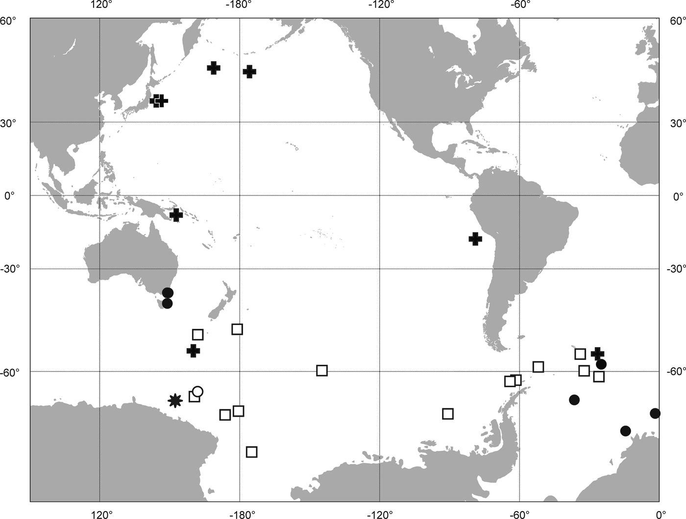
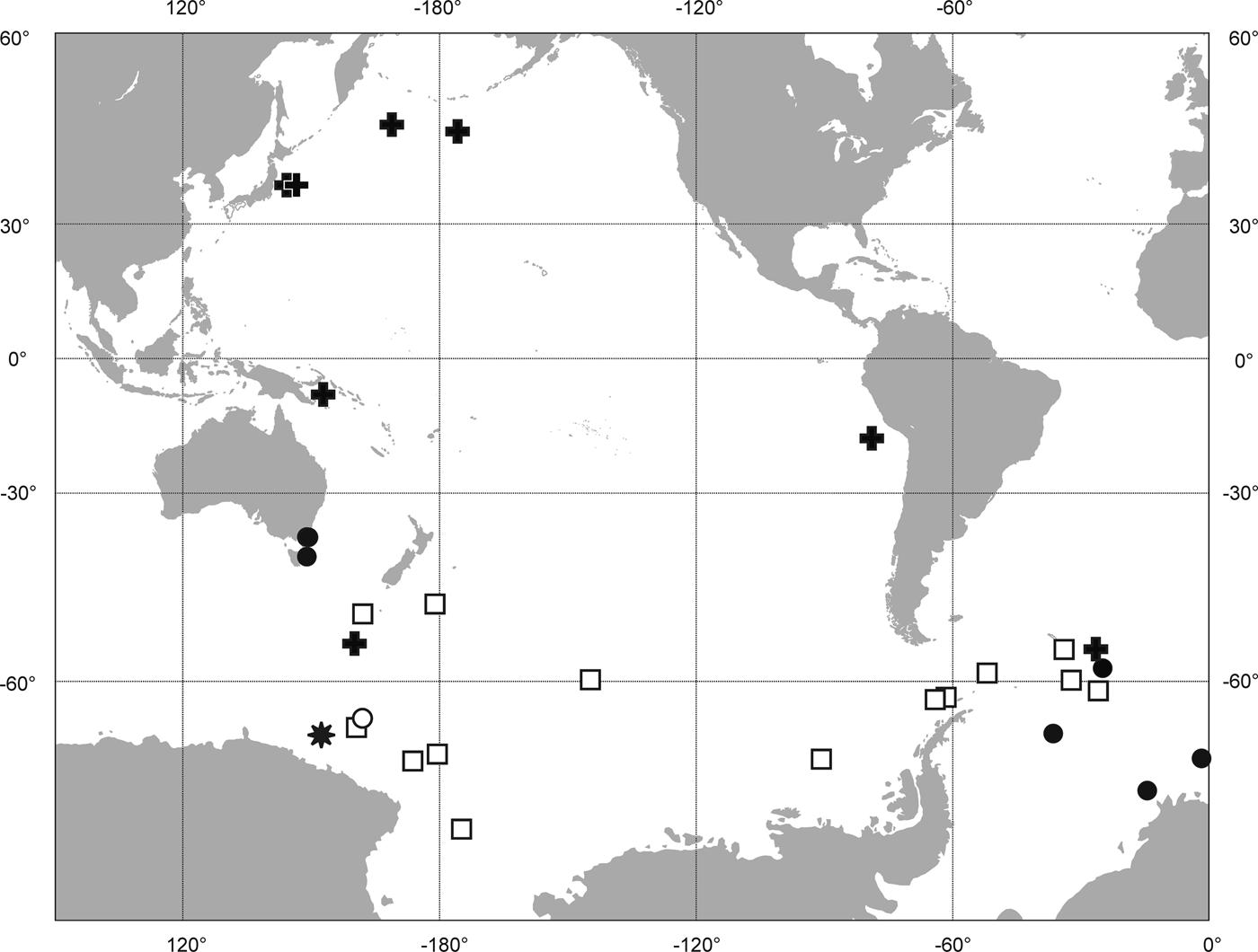
Nothria abyssia has been reported worldwide at bathyal and abyssal depths. It has been reported by Kucheruk (Reference Kucheruk1978) from the Antarctic Ocean, off the Aleutian Islands, Japan, New Guinea, and Chile at depths from 2700 to 5200 m. Orensanz (Reference Orensanz1990) reported N. abyssia from the Pacific–Antarctic Ridge, the Macquarie Ridge, southern part of the South-east Pacific Basin, the Scotia Sea, depth-range 609–4370 m. In the present study Nothria abyssia is reported from slope depths (400–2900 m) south-east of Australia and from bathyal–abyssal depths (2313–4817 m) from the Atlantic sector of Antarctica (Figure 10).
Distribution of Nothria abyssia Kucheruk, Reference Kucheruk1978: black asterisk – type locality; black circles – examined material; black crosses – records from Kucheruk (Reference Kucheruk1978); white squares – records from Orensanz (Reference Orensanz1990); white circles – record from Averincev (Reference Averincev1972).
Nothria otsuchiensis Imajima, Reference Imajima1986
(Figures 11–17)
Nothria otsuchiensis Imajima, Reference Imajima1986: 108–110, figure 8.—Imajima Reference Imajima1999: 46–51, figure 26.
Nothria otsuchiensis binoculata Maekawa & Hayashi, Reference Maekawa and Hayashi1989: 66–68, figure 4a–m.
Nothria conchylega—Imajima Reference Imajima1970: 116.—Imajima Reference Imajima1982: 157.—Imajima Reference Imajima1997: 177–178.
MATERIAL EXAMINED
AM: W.24760, RV ‘Tangaroa’, NZOI, East of Newcastle, NSW, 32°49′18′S 152°49′6″E, 9 October 1982 (11); W.24762, RV ‘Kapala’, Sydney–Newcastle, NSW, 33°37′S 152°4′E – 33°39′S 152°2′E, 896–924 m, 10 December 1980, benthic dredge (17); W.16567, RV ‘Kapala’, east of Port Stephens, NSW, K78-26-09, 32°50′S 152°40′E, 274 m, 6 December 1978 (1); W.198982, RV Kimbla, 32 km north of Fraser Island., QLD, Station 25, 24°22.3′S 153°17.1 E, 192–229.5 m, 15 December 1977. Coll.: W. Ponder, I. Loch, P. Terrill (3); W.41761, RV ‘Kimbola’, south-east of Percy Island, QLD, 21°47′S 150°34′E, 59 m, 13 December 1977, coarse shell and bryozoa substrate, Coll.: W. Ponder, I. Loch, P. Terrill (6); W.41764, RV ‘Kimbola’, east of Broad Sound, QLD, 22°6′S 150°49′E, 53 m, coarse shell, 13 December 1977, Coll.: W. Ponder, I. Loch, P. Terrill (6); W.41762, Off Cape Bowling Green Light, QLD, 19°17′S 147°32E, 24 m, coarse shells, foraminiferans, 20 November 1977, Coll.: P. Colman, F. Lowe (1); W.41763, 96 km-north-north-east of Port Headland, WA, 20°35′S 118°34′E, 36–37 m, sand, 24 October 1988, Coll.: B.W. Jenkins (1); W.41766, RV ‘Franklin’ Station 22, 18°11.01′S 147°25.5.′E, 472–490 m, 26 August 1988, sledge, (1); W.41484, BIOCAL Station DW44, New Caledonia, 22°47′S 167°14′E, 440–450 m, 30 August 1985 (25, one slide with dissected parapodia); W.41481, off Japan: Shimoda, Station 12 34°38.875′N 138°57.123′E–34°38.625′N 138°56.750′E, 45–40 m, dredge, 20 October 1999. Coll.: R. Ueshima (3); W.41482, off Japan: Shimoda, Station 43 34°36.380′N 138°57.722′E–34°36.375′N 138°58.265′E, 93–109 m, 14 July 2000, Coll.: R. Ueshima (1); W.41483.001, off Japan: Shimoda, Station 43 34°36.380′N 138°57.722′E–34°36.375′N 138°58.265′E, 93–109 m, 14 July 2000, Coll.: R. Ueshima (1 specimen gold-coated for SEM); W.42765.001, RV ‘Franklin’ SLOPE Station 32 (1 specimen gold-coated for SEM); W.42766.001, RV ‘Franklin’ SLOPE Station 32 (1 specimen gold-coated for SEM); W.42767.001, RV ‘Franklin’ SLOPE Station 32 (4 specimens gold-coated for SEM); W.42768.001, RV ‘Franklin’ SLOPE Station 32 (2 specimens gold-coated for SEM); W.42769.001, RV ‘Franklin’ SLOPE Station 32 (2 specimens gold-coated for SEM).
MV: F183649, Bass Trait, RV ‘Tangaroa’ Cruise 81-T-1 Station 177, 38°53.7′S 147°06.5′E, 59 m, 18 November 1981, coarse shell, Agassiz trawl (1); F43639, Bass Strait, RV ‘Tangaroa’ Cruise 81-T-1 Station 177, 38°53.7′S 147°06.5′E, 59 m, 18 November 1981, sled (2); F43648, Bass Trait, RV ‘Tangaroa’ Cruise 81-T-1 Station 193, 39°16.7′S 143°06.7′E, 95 m, 21 November 1981, shelly sand, Zock dredge (1); F43613, Bass Strait, FV ‘Sadra’ GCB Poore Cruise 80-Sa-1 Station 117, 40°38.0′S 145°23.0′E, 4 November 1980, muddy shell, grab (1); F183647, Bass Strait, RV ‘Tangaroa’ Cruise 81-T-1 Station 170, 31°51.8′S 148°26.5′E, 130 m, 15 November 1981, sled (3); F183648, Bass Strait, RV ‘Tangaroa’ Cruise 81-T-1 Station. 171, 38°53.7′S 147°55.2′E, 71 m, 17 November 1981, sled (3); F183646, Bass Strait, FRV ‘Hai Kung’ Cruise 81-HK-1 Station 120, 39°01.0′S 143°22.1′E, 84 m, 31 January 1981 (1); F183737, RV ‘Franklin’ SLOPE Station 46 (19); F183738, RV ‘Franklin’ SLOPE Station 67 (5); F183739, RV ‘Franklin’ SLOPE Station 33 (99); F183740, RV ‘Franklin’ SLOPE Station 22 (2); F192351, RV ‘Franklin’ SLOPE Station 66 (4); F192352, RV ‘Franklin’ SLOPE Station 82 (4); F192353, RV ‘Franklin’ SLOPE Station 34 (15); F192354, RV ‘Franklin’ SLOPE Station. 81 (18); F192355, RV ‘Franklin’ SLOPE Station 69 (4); F192356, RV ‘Franklin’ SLOPE Station 32 (398); F192357, RV ‘Franklin’ SLOPE Station 7 (46); F192358, RV ‘Franklin’ SLOPE Station. 6 (2); F192359, RV ‘Franklin’ SLOPE Station 45 (91); F192363–F192375, RV ‘Franklin’ SLOPE Station 32 (13 slides with dissected parapodia); F192376, RV ‘Franklin’ SLOPE Station 7 (one slide with dissected parapodia); F192377, RV ‘Franklin’ SLOPE Station 45 (one slide with dissected parapodia).
DIAGNOSIS
Based on specimens wider than 1.4 mm at chaetiger 6 excluding parapodia: first pair of parapodia reaching frontal margin of prostomium. First and second pair of parapodia with simple and pseudocompound bidentate falcigers. Third pair of parapodia with pseudocompound bidentate falcigers, limbate and pectinate chaetae. Pectinate chaetae with rolled lateral margins. Branchiae from chaetiger 9. Subacicular hooks from chaetigers 10–13. Auricular prechaetal lobes on first chaetigers, becoming foliose on chaetiger 2, gradually reduced posteriorly, present till end of body. Postchaetal lobes present on first 14–16 chaetigers. One or two pairs of eyes.
TYPE LOCALITY
Japan, Otsuchi Bay, 49–79 m.
DESCRIPTION
All specimens in samples from Australian waters incomplete anterior fragments from 0.35 to 2.1 mm wide (Figure 11A–D). Largest complete specimen from BIOCAL Station DW44 (New Caledonia) 2.2 mm wide, 28 mm long consisting of 52 chaetigers. All specimens light brownish lacking distinct colour pattern. The following description based on specimens wider than 1.4 mm at chaetiger 6 excluding parapodia.
Prostomium distally slightly tapering, forming triangle with ovoid frontal lips projecting anteroventrally (Figure IIC). Palps reaching chaetiger 1, lateral antennae reaching chaetigers 4–9, median antenna thicker and longer than lateral antennae, reaching chaetigers 6–12. Ceratophores with 2–4 rings. One pair of large dark eyes present laterally on prostomium near bases of lateral antennae. One of specimens examined (SLOPE Station 22) with second pair of small eyes dorsally on prostomium. Nuchal grooves straight with narrow mid-dorsal separation. Peristomium half as long as first chaetiger. Peristomial cirri thin and slender, longer than peristomium. All specimens examined light beige lacking distinct colour pattern.
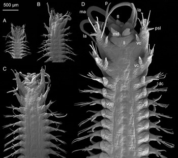
First pair of parapodia enlarged projecting lateroventrally, directed anteriorly with large auricular prechaetal lobes, digitiform postchaetal lobes and dorsal and ventral cirri (Figures 11D; 12D; 15A). Second pair of parapodia slightly enlarged projecting anteriorly with foliose distally tapering prechaetal lobes, digitiform postchaetal lobes and subulate dorsal and ventral cirri (Figures 11D; 13D; 15C). Third pair of parapodia similar in size with subsequent parapodia, directed laterally with foliose distally tapering prechaetal lobes, well developed digitiform postchaetal lobes, subulate dorsal cirri and conical ventral cirri (Figures 11D; 14D; 15D). Prechaetal lobes present on all parapodia of longest anterior fragment (up to chaetiger 28) gradually reduced posteriorly. Postchaetal lobes reduced in size towards posterior region, completely disappearing from chaetigers 15–18. Dorsal cirri present on all chaetigers, becoming shorter and thinner posteriorly. Transitional ventral cirri present on chaetigers 3 and 4, replaced by oval glandular pads from chaetiger 5 (Figure 11D).
Nothria otsuchiensis Imajima, Reference Imajima1986, ventral view: (A) specimen of 0.35 mm width; (B) specimen of 0.54 mm width; (C) specimen of 0.97 mm width; (D) specimen of 1.49 mm width; dc, dorsal cirrus; fl, frontal lip; la, lateral antenna; ll, lower lip; ma, median antenna; md, mandibles; p, palp; psl, postchaetal lobe; vc, ventral cirrus; vgp, ventral glandular pad.

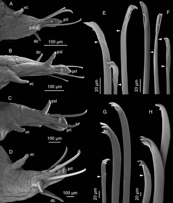
Nothria otsuchiensis Imajima, Reference Imajima1986, parapodia and chaetae from chaetiger 1 of specimens of different sizes: (A) parapodium from chaetiger 1 of specimen of 0.35 mm width, posterior view; (B) same of specimen of 0.54 mm width; (C) same of specimen of 0.97 mm width; (D) same of specimen of 1.49 mm width; (E) pseudocompound falcigers of specimen of 0.35 mm width; (F) same of specimen of 0.54 mm width; (G) simple and pseudocompound falcigers of specimen of 0.97 mm width; (H) same of specimen of 1.49 mm width; dc, dorsal cirrus; prl, prechaetal lobe; psl, postchaetal lobe; vc, ventral cirrus.

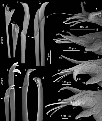
Nothria otsuchiensis Imajima, Reference Imajima1986, parapodia and chaetae from chaetiger 2 of specimens of different sizes: (A) parapodium from chaetiger 2 of specimen of 0.35 mm width, posterior view; (B) same of specimen of 0.54 mm width; (C) same of specimen of 0.97 mm width; (D) same of specimen of 1.49 mm width; (E) pectinate chaetae of specimen 0.35 mm width; (F) pseudocompound falciger of specimen of 0.35 mm width; (G) same of specimen of 0.54 mm width; (H) same of specimen of 0.97 mm width; (I) same of specimen of 1.49 mm width; (J) simple falciger of specimen of 1.49 mm width; dc, dorsal cirrus; prl, prechaetal lobe; psl, postchaetal lobe; vc, ventral cirrus.

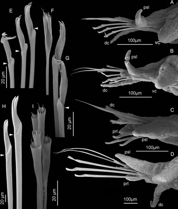
Nothria otsuchiensis Imajima, Reference Imajima1986, parapodia and chaetae from chaetiger 3 of specimens of different sizes: (A) parapodium from chaetiger 3 of specimen of 0.35 mm width, posterior view; (B) same of specimen of 0.54 mm width; (C) same of specimen of 0.97 mm width; (D) same of specimen of 1.49 mm width; (E) pseudocompound falcigers of specimen of 0.35 mm width; (F) pectinate chaetae of specimen of 0.54 mm width; (G) pseudocompound falcigers of specimen of 0.54 mm width; (H) same of specimen of 1.49 mm width; (I) pectinate chaetae of specimen 0.97 mm width; dc, dorsal cirrus; prl, prechaetal lobe; psl, postchaetal lobe; vc, ventral cirrus.

Nothria otsuchiensis Imajima, Reference Imajima1986, parapodia and chaetal composition: (A) parapodium and chaetae from chaetiger 1 of specimen of 1.6 mm width; (B) parapodium and chaetae from chaetiger 2 of specimen of 0.5 mm width; (C) same of specimen of 1.6 mm width; (D) parapodium and chaetae from chaetiger 3 of specimen of 1.6 mm width; (E) diagram of chaetal replacement in relation to body width of worms.

First pair of parapodia with 1–2 simple bidentate falcigers and 1–2 bidentate pseudocompound falcigers bearing paired short pointed hoods. Frequently hoods on anterior falcigers broken (Figures 12D, H; 15A). Second pair of parapodia with one simple and two pseudocompound bidentate falcigers with paired hoods (Figures 13D, I, J; 15C). Third pair of parapodia with two bundles of chaetae: dorsal bundle of 1–2 slender limbate chaetae and 5–10 pectinate chaetae with 18–22 denticles and rolled lateral margins; ventral bundle of 3–4 bidentate pseudocompound falcigers with paired short and pointed hoods (Figures 14D, H; 15D). Only limbate and pectinate chaetae present from chaetiger 4. Paired bidentate subacicular hooks appearing in median position within ventral bundle of limbate chaetae from chaetigers 10–13. Neuroaciculae pale yellow with pointed tips, 2–3 per parapodium.
Branchiae starting from chaetiger 9 in all specimens examined from Australian waters, single, strap-like, becoming longer and wider towards median body section and reduced in size posteriorly. Specimens from BIOCAL Station DW44 with branchiae starting from chaetigers 9–12. Pygidium with dorsal anus and one pair of anal cirri. Tubes dorsoventrally flattened, covered with shell fragments of various size, foraminiferans and large sand particles.
ONTOGENETIC VARIATION
Seventy-six specimens of Nothria otsuchiensis ranging in width from 0.35 to 2.1 mm (Figure 11A–D) were examined (SLOPE Stations 7(5), 32(49), 33(8), 45(14)). A number of morphological characters were size related.
Chaetal composition of the first pair of parapodia
Specimens smaller than 0.6 mm wide had 3–4 pseudocompound falcigers in the parapodia on the first chaetiger (Figures 12A, B, E, F; 15E). Left and right parapodia were asymmetrical in chaetal composition in 33% of specimens examined (N = 21). In specimens larger that 0.6 mm wide 1–2 pseudocompound falcigers were replaced by simple falcigers (Figures 12C, D, G, H; 15A, E). The most frequent composition of anterior chaetae was two simple falcigers and one pseudocompound falciger which was found in both parapodia in 50% of specimens examined (N = 45). In other specimens the chaetal composition was asymmetrical in the left and right parapodia with various combinations of simple and pseudocompound falcigers, but not exceeding five chaetae in total per parapodium. In nine of the largest specimens (wider than 1.33 mm) only left parapodia were observed, and the combination of two simple and one pseudocompound falcigers was found in eight of them. Simple falcigers of the largest specimens often lacked their hoods, which presumably were broken (Figure 12H).
Chaetal composition of the second pair of parapodia
Specimens smaller than 0.6 mm wide had limbate and pectinate chaetae starting from chaetiger 2 (Figures 13A, E; 15B, E). Occasionally limbate and pectinate chaetae were found in the second parapodia of larger specimens (up to 0.77 mm wide) but never in both right and left parapodia and rarely together. No limbate and/or pectinate chaetae were observed in specimens larger than 0.8 mm wide. Specimens of all sizes had three (up to five) pseudocompound falcigers in the parapodia of the second chaetiger (Figures 13A–D, F–I; 15A–E). One or two simple falcigers replaced pseudocompound falcigers in specimens larger that 1.4 mm wide (Figures 13D, J; 15C, E). The total number of falcigers in the largest specimens was always three per parapodium. Neither simple nor pseudocompound falcigers in the second pair of parapodia lost their hoods (Figure 13F–J).
Chaetal composition of the third pair of parapodia
Parapodia of all specimens examined were with the same set of chaetae consisting of 1–5 pseudocompound bidentate falcigers (Figures 14A–H; 15D, E), 1–3 limbate chaetae and pectinate chaetae (Figures 14A–D, I; 15D, E). No variation was observed.
Distribution of prechaetal and postchaetal parapodial lobes
All specimens examined consisted of anterior fragments. Therefore, we estimated the variation in distribution of the parapodial lobes in the anterior body region. All specimens, independently of their size, had enlarged auricular prechaetal lobes on the first pair of parapodia (Figure 12 A–D) becoming foliose and slightly tapering on the second and the third pairs of parapodia (Figures 13A–D; 14A–D). In the specimens smaller than 0.6 mm wide prechaetal lobes were visible on the anterior 7–9 chaetigers and almost untraceable posteriorly. All the larger specimens had prechaetal lobes on all segments, gradually reduced posteriorly, but distinguishable.
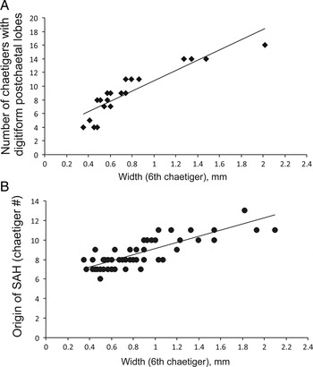
Twenty-six specimens examined were anterior fragments with a sufficient number of segments allowing estimation of the variation of the distribution of postchaetal lobes. The number of chaetigers with digitiform postchaetal lobes was a size-dependent character varying from four chaetigers in specimens of 0.37–0.5 mm wide to 16 chaetigers in specimen of 2.1 mm wide (Figure 16A).
Nothria otsuchiensis Imajima, Reference Imajima1986: (A) relationship between body width of specimens and number of chaetigers with digitiform postchaetal lobes, R 2 = 0.812; (B) relationship between body width of specimens and origin of subacicular hooks (SAH), R 2 = 0.677.

Presence of subacicular hooks
The first presence of subacicular hooks was a size-dependent character. The variation of this character ranged from hooks starting from chaetiger 6 to chaetiger 13 (Figure 16B).
Presence of branchiae
Branchiae were totally absent in five specimens smaller than 0.5 mm wide. In specimens from Australian waters, branchiae started strictly from chaetiger 9 with no variation observed. Specimens from New Caledonia had branchiae starting from chaetigers 9–12 independent of size of worms.
REMARKS
Nothria otsuchiensis is similar to Nothria oblonga Imajima, Reference Imajima1999 in having at least one pair of large eyes and sharing the presence of falcigers in the first three pairs of parapodia and the presence of limbate and pectinate chaetae from the third pair of parapodia. The former species can be distinguished from the latter in having simple and pseudocompound falcigers rather than exclusively pseudocompound falcigers on the first two pairs of parapodia. Imajima (Reference Imajima1999) reported that N. oblonga resembles the juveniles of N. otsuchiensis bearing pseudocompound and compound falcigers on the anterior parapodia. However, he stated that the holotype of N. oblonga was a mature specimen with oocytes in the body cavity. Consequently, he considered it as a valid species. Imajima reported the holotype of N. oblonga being the largest specimen at 2 mm wide (presumably with parapodia). The body width of the holotype at the level of the sixth chaetiger without parapodia estimated from the original illustration (Imajima, Reference Imajima1999, p. 45, figure 24a) was 1.5 mm. Simple falcigers were found in the first pair of parapodia of the examined specimens of N. otsuchiensis larger than 0.6 mm wide. Therefore, N. oblonga does not fall within the described pattern of the ontogenetic variation of N. otsuchiensis and is considered to be a valid species.
Imajima (Reference Imajima1986) described two pairs of eyes in N. otsuchiensis, one large pair of eyes located laterally near the bases of the lateral antennae and a pair of small eyespots located dorsally on the prostomium near the bases of the palps. Brown patches on the dorsal side of the anterior parapodia were also observed by Imajima (Reference Imajima1986) in N. otsuchiensis. Nothria otsuchiensis binoculata Maekawa & Hayashi (Reference Maekawa and Hayashi1989) lacking the dorsal pair of eyes and brown pigmentation on the parapodia was reported from Wakasa Bay, Japan. Imajima (1999) re-examined the type specimens of the new subspecies and reported two pairs of eyes, stating the pigmentation can disappear in the preserved specimens. All the specimens examined in the present study from Australia and New Caledonia lacked the dorsal pair of eyespots. The only specimen with two pairs of eyes was found at Slope Station 22. All specimens from Japan examined in the present study had two pairs of eyes. No specimens with brown pigmented patches on the dorsal side of parapodia were found in the present material.
DISTRIBUTION
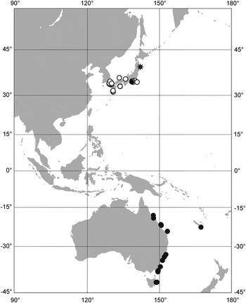
Nothria otsuchiensis was widely reported from Japan from Otsuchi to Kagoshima Bay at water depth from 26 to 1070 m (Imajima, Reference Imajima1986, Reference Imajima1999). In Australian waters it was found along the western and south-western coast, depth range 24–2900 m. A single record was reported from New Caledonia, 440–450 m (Figure 17).
Distribution of Nothria otsuchiensis Imajima, Reference Imajima1986: black asterisk,–type locality; black circles,–examined material; white circles,–records from Imajima (Reference Imajima1999).

DISCUSSION
Ontogenetic variation of morphological characters involving changes in chaetal and parapodial morphology has been widely reported for onuphid worms (Paxton, Reference Paxton1986, Reference Paxton1993, Reference Paxton1996, Reference Paxton1998; Orensanz, Reference Orensanz1990; Budaeva & Fauchald, Reference Budaeva and Fauchald2010, Reference Budaeva and Fauchald2011). Chaetae of onuphids undergo replacement during the worm's growth and maturation. Onuphid larvae and early juveniles bear various provisional chaetae that are subsequently replaced by definitive chaetae in later juveniles and adults. Provisional larval chaetae differ significantly from adult chaetae and can be clearly distinguished from the latter (Budaeva & Fauchald, Reference Budaeva and Fauchald2010). Chaetal replacement continues in later development of onuphids: anterior falcigers replace each other, increasing in size and slightly changing in morphology, limbate and pectinate chaetae increase in numbers, subacicular hooks frequently shift posteriorly (Paxton, Reference Paxton1986, Reference Paxton1993, Reference Paxton1996, Reference Paxton2005; Hsieh & Simon Reference Hsieh and Simon1987; Budaeva & Fauchald, Reference Budaeva and Fauchald2010). The general pattern of growth related changes in parapodial morphology is represented by the increasing number of modified parapodia and spreading of digitiform postchaetal lobes towards the posterior end of the body (Jirkov & Yermolaev, Reference Jirkov and Yermolaev1989; Orensanz, Reference Orensanz1990). In a number of onuphid genera (e.g. Paradiopatra, Diopatra, Onuphis, Kinbergonuphis and Mooreonuphis) branchiae tend to appear in the anterior part of the body and spread more anteriorly and/or posteriorly with increasing worm size (Blake, Reference Blake1975; Fauchald, Reference Fauchald1982c; Hsieh & Simon, Reference Hsieh and Simon1987; Fadlaoui et al., Reference Fadlaoui, Lechapt and Retière1995; Budaeva & Fauchald, Reference Budaeva and Fauchald2010). However, in Nothria the segment of the first appearance of branchiae remains variable but not size-dependent (Kucheruk, Reference Kucheruk and Kuznetsov1980; Jirkov & Yermolaev, Reference Jirkov and Yermolaev1989; Orensanz, Reference Orensanz1990).
Intraspecific variability has never been studied thoroughly in Anchinothria, presumably due to insufficient material, as was also the case during the present study. Nevertheless, the investigation of morphological variation in this genus is desirable to re-evaluate the taxonomic status of a number of species. Examination of 95 specimens of Anchinothria including non-type material and types of Anchinothria pycnobranchiata (McIntosh, Reference McIntosh1885), A. abranchiata (McIntosh, Reference McIntosh1885) and A. antarctica (Hartman, Reference Hartman1967) revealed significant variation in the presence/absence of branchiae, a character used to distinguish these three species (Orenzanz, 1990). The diameter of subacicular hooks was shown to be strongly size-dependent in A. pycnobranchiata (Orensanz, Reference Orensanz1990) but an analysis of variation of other diagnostic characters was never performed.
We followed Kucheruk (Reference Kucheruk and Kuznetsov1980) and Jirkov & Yermolaev (Reference Jirkov and Yermolaev1989) in distinguishing variation in two parameters of chaetal distribution in the anterior parapodia of Nothria. The first parameter was chaetal composition, i.e. presence or absence of different types of chaetae: falcigers, limbate and pectinate chaetae. The second parameter was morphology of falcigers, e.g. dentition, presence or absence of pseudoarticulation and hoods. Chaetal composition was previously reported independent of the size of specimens. Only morphology of falcigers was shown to be size-dependent (Kucheruk, Reference Kucheruk and Kuznetsov1980; Jirkov & Yermolaev, Reference Jirkov and Yermolaev1989; Orensanz, Reference Orensanz1990). Consequently, chaetal composition of the anterior parapodia was used as a major diagnostic character in species of Nothria. Kucheruk (Reference Kucheruk1985) recognized four different types of chaetal composition in the anterior three pairs of parapodia and presented them as four formulas with different letters indicating several types of chaetae. He assigned nine Nothria species to the four types based on literature data. Imajima (Reference Imajima1999) distinguished five species of Nothria reported from Japanese waters based on the presence/absence of different types of chaetae in the anterior parapodia.
The cases of invariance of chaetal composition in Nothria were presumably reported due to unavailability of small specimens for examination. The smallest specimens studied by Jirkov & Yermolaev (Reference Jirkov and Yermolaev1989) were 0.7 mm wide, at the level of chaetiger 10. Kucheruk (Reference Kucheruk and Kuznetsov1980) included specimens 1.0–3.3 mm wide into his analysis. The size of the smallest specimens examined by Orensanz (Reference Orensanz1990) was not reported. Examination of the wide size-range (0.35–2.1 mm) in two Nothria species in the present study demonstrated that chaetal composition can vary significantly in specimens of different sizes. No provisional chaetae were found in the studied material, demonstrating that all the specimens examined were juveniles and adults. Hence, ontogenetic changes in morphology associated with the transition from larval stages to juveniles could not be considered in the present study.
The general pattern of falciger morphology in Nothria specimens of various sizes represents three growth related chaetal replacement tendencies: replacement of bidentate falcigers of smaller specimens by unidentate falcigers in larger specimens, replacement of pseudocompound falcigers by simple falcigers, and loss of paired hood in the largest specimens examined (Kucheruk, Reference Kucheruk and Kuznetsov1980; Jirkov & Yermolaev, Reference Jirkov and Yermolaev1989; Orensanz, Reference Orensanz1990). A similar pattern was found in the two species examined in the present study. Pseudocompound falcigers were gradually replaced by simple falcigers in the larger specimens of both species. Falcigers on the first pair of parapodia frequently lacked hoods in both species and the second tooth in N. abyssia.
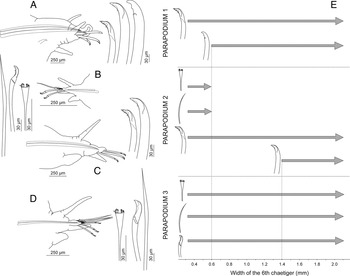
The patterns of chaetal morphology and composition elucidated during the present study have been summarized in Figure 4 for Nothria abyssia and Figure 15 for N. otsuchiensis, and compared to each other in Figure 18. In N. abyssia the third pair of parapodia lacked pectinate chaetae in specimens smaller than 0.5 mm wide, having only limbate chaetae on the third chaetiger. Pectinate chaetae appeared from chaetiger 3 in N. abyssia larger than 0.5 mm wide. Specimens of N. otsuchiensis smaller than 0.6 mm wide had limbate and pectinate chaetae in addition to falcigers on the second pair of parapodia, while in adults only pseudocompound falcigers were found on the second chaetiger. Kucheruk (Reference Kucheruk1978) reported pectinate chaetae starting from chaetiger 2 in N. abyssia. His largest specimen examined was 4 mm wide and could potentially represent the next stage of chaetal replacement with pectinate chaetae emerging on chaetiger 2.
Chaetal composition and morphology in anterior parapodia of Nothria abyssia Kucheruk, Reference Kucheruk1978 and N. otsuchiensis Imajima, Reference Imajima1986: (L) limbate chaetae; (P) pectinate chaetae; (pF) pseudocompound falcigers; (sF) simple falcigers.

As a result, three size-related morphotypes can be recognized in Nothria otsuchiensis and N. abyssia based on both chaetal composition and morphology of falcigers (Figure 18). The patterns of chaetal replacement in the two examined species differed from each other. In N. abyssia new types of chaetae (pectinate chaetae and simple falcigers) appeared in the anterior parapodia with the growth of a worm. In N. otsuchiensis emergence of simple falcigers was combined with disappearance of limbate and pectinate chaetae. Assessment of ontogenetic variation in other species of Nothria may reveal patterns of chaetal replacement different from the patterns described here. Therefore, examination of every species within the genus is required for accurate evaluation of their validity.
Origin of subacicular hooks in Nothria was shown to be size-dependent in several species. Generally subacicular hooks appear in more posterior segments in larger specimens (Jirkov & Yermolaev, Reference Jirkov and Yermolaev1989; Orensanz, Reference Orensanz1990). Growth related increase of the number of chaetigers with digitiform postchaetal lobes in two species of Nothria was reported by Jirkov & Yermolaev (Reference Jirkov and Yermolaev1989). Our results confirm the observations made by previous authors. Both N. abyssia (Figure 9) and N. otsuchiensis (Figure 16B) showed positive correlation between the segment of the first appearance of subacicular hooks and the body width. Nothria otsuchiensis displayed positive correlation between the size of worms and number of chaetigers with postchaetal lobes (Figure 16A). Relationship between the size of worms and number of chaetigers with postchaetal lobes in N. abyssia was not estimated due to the poor condition of the specimens examined.
The number of chaetigers with pseudocompound falcigers was the only character independent of size of the specimens in both examined species. The chaetiger of the first appearance of branchiae in N. otsuchiensis was also unrelated to the size of worms. However, both characters display very low variation among species of Nothria and can only be used in separation of the groups of closely related species. Additional diagnostic characters are required for the differentiation of Nothria species. The following characters are considered of diagnostic value only when scored in adults: absence/presence of simple falcigers in anterior parapodia; chaetiger of origin of limbate and pectinate chaetae; chaetiger of origin of subacicular hooks; number of chaetigers with auricular prechaetal lobes and digitiform postchaetal lobes. We suggest that both chaetal composition and chaetal morphology be used in species diagnoses within Nothria. The diagnoses should be based on adult specimens with the final set of definitive morphological structures. The revision of the genus involving examination of type material and ontogenetic variation of chaetal composition in the anterior parapodia is required to clarify the status of all Nothria species. Species described based on small specimens (e.g. Nothria textor Hartman & Fauchald, Reference Hartman and Fauchald1971; Nothria mannarensis Rangarajan & Mahadevan, Reference Rangarajan and Mahadevan1961) may potentially represent the juveniles of other species retaining the provisional set of chaetae.
ACKNOWLEDGEMENTS
We are grateful to Pat Hutchings, Stephen Keable and the staff of the Australian Museum for providing facilities and assistance during the duration of the research project. We are also thankful to the following individuals and institutions for the loan and exchange of specimens: Robin Wilson and Chris Rowley (Museum Victoria), Eijiroh Nishi (Yokohama National University), Karin Meiβner (German Centre for Marine Biodiversity Research, Hamburg); Angelika Brandt and Petra Wagner (Zoological Museum of Hamburg), Jean-Paul Lechapt (Marine Laboratory, Dinard), Tarik Meziane (National Museum of Natural History, Paris). We would like to thank Sue Lindsay (Australian Museum) for assistance with SEM.
FINANCIAL SUPPORT
This work was supported by an Endeavour Research Fellowship (N.B.), the Russian Foundation for Basic Research (grants number 10-04-01687, 12-05-33049) and by the Ministry of Education and Science of the Russian Federation (contract number 8132).