Introduction
Ovarian follicles consist of oocytes surrounded by somatic cells and represent the functional unit for reproduction in mammals (Li et al., Reference Li, Shi, Shi and Wang2021; Liu et al., Reference Liu, Qu, Tian, Yang, Song, Li, Yan and Qiao2022). A progressive decline in follicular reserve is observed throughout a woman’s reproductive lifespan, intensifying as menopause approaches (Man et al., Reference Man, Lustgarten Guahmich, Vyas, Tsai, Arazi, Lilienthal, Schattman, Rosenwaks and James2022). From puberty through the end of the reproductive period, primordial follicles are recruited and begin to grow, progressing through several well-sequenced stages to become the transitional, primary, secondary and antral (preovulatory) follicles (Zhao et al., Reference Zhao, Feng, Zhang, Zhang, Wang, Liu, Wang, Li, Ng and Yeung2021). Ovarian cycles are highly sensitive to exogenous substances, such as drugs and chemical compounds, which are capable of compromising female reproductive function (Rosario et al., Reference Rosario, Cui and Anderson2022; Wang et al., Reference Wang, Wang, Zhu, Zhong, Jiang, Wang, Tang, Li, Zhang and Wang2024). On the other hand, reversible inhibitors of follicular growth and ovulation can be used to establish new contraceptives and open new strategies for women’s contraception, avoiding the well-known side effects of current methods such as mood swings, nausea, headaches, depression and obesity (Saja, Reference Saja2021).
Non-steroidal anti-inflammatory drugs (NSAIDs) are popular analgesic medications among the general population. They are widely used in the treatment of acute and chronic pain because they inhibit cyclooxygenase (COX) and reduce prostaglandin production. Naproxen ranked as the second most-selling NSAID in the USA in 2018 and can be obtained without a prescription (Ha and Paek, Reference Ha and Paek2021).
Ovulation is considered an inflammatory process (Pereira et al., Reference Pereira, Ferreira, Dias, Nogueira, Pinto, Jorge and Campos-Junior2024); thus, inhibition of the COX-2 enzyme can modulate this process and influence the ovulatory capacity. Additionally, prostaglandins regulate the follicular rupture, cumulus cells expansion, oocyte maturation and release (Gaber et al., Reference Gaber, Cuervo-Arango, Plaza-Dávila and Martínez-Boví2024). Therefore, studies must be conducted to evaluate the effects of naproxen administration on folliculogenesis and fertility, to prevent potential side effects associated with commonly used contraceptives and establish new strategies to modulate late follicular growth and ovulation.
In a recent study from our group, Nascimento and collaborators (Reference Nascimento, Oliveira, Pereira, L.A.A., Lopes, Paula, R., Jorge and Campos-Junior2024) demonstrated that the eight-day administration of naproxen in C57BL/6 mice resulted in a marked reduction in the number of ovulated oocytes, fewer corpora lutea and corpora haemorrhagica and fewer antral follicles.
The present study investigated the effects of naproxen on ovulation across multiple mouse strains, evaluating repeated administration with treatment interruption in C57BL/6 mice and acute, one-week administration in BALB/c and SWISS mice. This approach aimed to determine whether naproxen-induced ovulation blockade extends beyond a single murine model, expanding upon previous findings restricted to the C57BL/6 strain. The inclusion of well-characterized strains allowed consideration of known reproductive differences, such as the higher ovulation rate in genetically heterogeneous SWISS mice (Galibert and Azzouzi, Reference Galibert and Azzouzi2023) and the lower rates typically observed in inbred strains, including C57BL/6 and BALB/c (Zhao et al., Reference Zhao, Wei and Yue2025). In this sense, our study, for the first time in the literature, showed that naproxen can modulate ovulation rates in C57BL/6 females and that drugs with similar characteristics, if their effects in humans are fully understood, could be repositioned as potential non-hormonal contraceptives. However, the study has limitations, including the absence of hormone measurements (e.g., Follicle-Stimulating Hormone (FSH) and Luteinizing Hormone (LH)) and the lack of long-term evaluations.
Material and methods
Animals and treatment
Six-week-old female C57BL/6 (n = 20), Swiss (n = 30) and BALB/c mice (n = 33) were obtained from the Laboratory Animal Breeding Center of the Federal University of São João del-Rei (NUCAL-UFSJ) and were kept in a 12:12 h photoperiod at a controlled temperature of 22°C to 24°C, receiving water and food ad libitum. All procedures were approved by the Animal Use Ethics Committee of the Federal University of São João del Rei (CEUA UFSJ –Protocol 009/2020).
Animals were randomly divided into three experimental groups: Control, Naproxen Low (low dosage) and Naproxen High (high dosage) (Naproxen: ALX-270-102 – Enzo – 10 Executive Blvd., Farmingdale, NY 11735). The animals in the Naproxen Low and Naproxen High groups were treated with 10 and 50 mg/kg, respectively. These doses were previously established in studies from our group (Nascimento et al., Reference Nascimento, Oliveira, Pereira, L.A.A., Lopes, Paula, R., Jorge and Campos-Junior2024). The vehicle group received the vehicle solution (190 uL of phosphate buffer solution - PBS + 10 uL of Dimethyl Sulfoxide (DMSO)) (DMSO: 67-68-5 – NEON – Suzano/SP) (PBS: P3813-1PAK – Sigma-Aldrich – St. Louis, Missouri, EUA). In all groups, animals were treated intraperitoneally for eight consecutive days, at 9:00 a.m. Subsequently, treatment with naproxen was discontinued.
Biometric data
Body weight was measured every morning from the beginning to the end of the treatment using a digital scale (Shimadzu ATX224R, Barueri/SP). After euthanasia, ovarian weights were also obtained. The gonadosomatic index was calculated by dividing the weight of the ovaries by body weight and multiplying the result by 100.
Oestral cycle analysis
The oestrous cycle stages were assessed daily at 9 a.m. throughout the experiments using visual and cytological methodology as described by Byers et al. (Reference Byers, Wiles, Dunn and Taft2012) and Pereira et al. (Reference Pereira, Ferreira, Dias, Nogueira, Pinto, Jorge and Campos-Junior2024). The classification was based on observation of the vulva and cytology of vaginal smears. The presence or absence of cornified, nucleated epithelial cells and leukocytes was used to determine the oestrous cycle phases (proestrus, oestrus, metoestrus and dioestrus).
Experiment I
Six-week-old female C57BL/6 mice (n = 20) were used and divided into three groups: vehicle (n = 6), low (n = 7) and high (n = 7). The treatment was performed over eight days. From day nine to day 30, treatment was paused, and then the animals were euthanized in a carbon dioxide (CO2) chamber and death was confirmed by cervical dislocation. The ovaries were recovered and processed according to a standard histological protocol (Figure 1A).
Experimental design. (A) In experiment I, six-week-old female C57BL/6 mice were treated daily for eight days with 10 mg/kg (low, n = 7), 50 mg/kg (high, n = 7), or vehicle (n = 6); treatment was paused on day eight. Then, on day nine, the ovaries were collected and directed to histology, followed by follicle quantification. The (B) experiment II was also performed using six-week-old female C57BL/6 mice. They were treated as previously described, and, after a treatment pause on day 27, a standard superovulatory protocol was used. On day 30, oocytes were collected, harvested, and classified. (C) For experiment III, six-week-old female BALB/c mice were treated with 10 mg/kg (low, n = 12), 50 mg/kg (high, n = 12), or control (n = 9). Swiss were treated with 10 mg/kg (low, n = 9), 50 mg/kg (high, n = 12), or control (n = 9) doses, administered daily for eight days, followed by superovulation; oocytes were harvested and classified on day nine.

Figure 1. Long description
Panel A: Diagram of experiment I. Six-week-old female C57BL/6 mice receive daily naproxen or vehicle treatment from day 1 to day 8. On day 30, ovaries are collected for follicle quantification. Panel B: Diagram of experiment II. Six-week-old female C57BL/6 mice receive daily naproxen or vehicle treatment from day 1 to day 8. After a pause on day 27, a superovulatory protocol is applied. On day 30, oocytes are collected and classified. Panel C: Diagram of experiment III. Six-week-old female BALB/c and Swiss mice receive daily naproxen or vehicle treatment from day 1 to day 8. A superovulatory protocol is applied, and oocytes are collected and classified on day 9.
The ovaries were fixed in 4% paraformaldehyde for 24 h, then in 70% alcohol. The materials then underwent a histological processing protocol, including dehydration, diaphanization, infiltration, inclusion and sectioning, obtaining 5 µm-thick sections using a microtome (Leica, Wetlzlar, Germany, RM2125 RTS – 5 μm). Finally, the histological sections of the ovaries were stained with haematoxylin & eosin (H&E).
Every 5th section was analysed, and the whole ovary was evaluated for follicular quantification. Follicles were classified as primordial, transitional, primary, secondary, antral, or atretic based on morphological characteristics, following the methodology of Pereira et al. (Reference Pereira, Nascimento, Jorge, Segatelli, Coutinho, Viana and Campos-Junior2020). The percentage of follicles and the rate of follicular activation were calculated (percentage of follicles = number of desired follicle class per animal × 100/ total number of follicles) (Pereira et al., Reference Pereira, Nascimento, Jorge, Segatelli, Coutinho, Viana and Campos-Junior2020).
Experiment II
For this experiment, 6-week-old female C57BL/6 mice (n = 20) were used and divided into three groups: vehicle (n = 6), low (n = 7) and high (n = 7). Animals were treated as described for experiment I, and after treatment interruption, a standard superovulatory protocol was used. On day 27, 20 international units (IU) of eCG (equine chorionic gonadotropin) (eCG – Novormon – Nanuque, Brazil) were administered, and after 48 h (day 29), 20 IU of hCG (human chorionic gonadotropin) (hCG – InterVet – Sete Lagoas, Brazil) were also administered, both intraperitoneally (i.p.). Oocytes were recovered from the oviducts and classified according to the presence or absence of cytoplasmic granules (Mara et al., Reference Mara, Zhou, Larmore, Johnson, Ayiku, Amargant, Pritchard and Duncan2020). Sixteen h after hCG administration, they were euthanized, and their oviducts and ovaries were collected and maintained in human serum albumin solution (3%, Sigma Aldrich – San Louis, Missouri, EUA) (Mara et al., Reference Mara, Zhou, Larmore, Johnson, Ayiku, Amargant, Pritchard and Duncan2020; Nascimento et al., Reference Nascimento, Oliveira, Pereira, L.A.A., Lopes, Paula, R., Jorge and Campos-Junior2024) (Figure 1B).
For oocyte collection, the oviducts were punctured with an insulin needle (Mara et al., Reference Mara, Zhou, Larmore, Johnson, Ayiku, Amargant, Pritchard and Duncan2020), and then the oocytes were denuded in a drop of hyaluronidase (INGASE, 80 IU/ml)(Ingase: 50080007 – INGÁMED–Rod. Osvaldo Pacheco de Lacerda, 720 Parque Industrial – Maringá/PR). Denuded oocytes were evaluated using a stereomicroscope (Leica – EZ4 D – São Paulo/SP) (Mara et al., Reference Mara, Zhou, Larmore, Johnson, Ayiku, Amargant, Pritchard and Duncan2020) and quantified and qualified, considering viable oocytes as those with homogeneous cytoplasm and non-viable oocytes as those with granules in the cytoplasm. The number of animals that ovulated or did not ovulate was obtained, and the percentage was calculated and presented.
The number of corpora haemorrhagica and lutea found was macroscopically evaluated using the same stereomicroscope. Corpora haemorrhagica were considered as red formations in the ovary, and corpora lutea were yellow and translucent structures. Their classification and quantification were performed according to Mara et al. (Reference Mara, Zhou, Larmore, Johnson, Ayiku, Amargant, Pritchard and Duncan2020) and Nascimento et al. (Reference Nascimento, Oliveira, Pereira, L.A.A., Lopes, Paula, R., Jorge and Campos-Junior2024).
Experiment III
To evaluate the capacity of naproxen treatment to reduce the ovulation rate in different mouse backgrounds, Swiss (n = 30) and BALB/c (n = 33) were used. Six-week-old female Swiss mice were divided into three groups: vehicle (n = 9), low (n = 9) and high (n = 12). BALB/c females of the same age were also divided as follows: vehicle (n = 9), low (n = 12), and high (n = 12). The treatment was performed over eight days. On day six, 20 IU of eCG was injected (i.p.), and on day eight, 20 IU of hCG was administered to induce ovulation. Sixteen h later, females were euthanized, and their oocytes were recovered from oviducts, counted and classified. The number of corpora haemorrhagica and lutea was also evaluated (Figure 1C). All analyses were performed as described for experiment II. The number of animals that ovulated or did not ovulate was obtained, and the percentage was calculated and presented.
Statistical analysis
Quantitative data were tested for normality using the Shapiro–Wilk and Kolmogorov–Smirnov tests. Normal data (body weight, ovarian weight, gonadosomatic index), days in each stage of the oestrous cycle (proestrus, oestrus, dioestrus), primordial follicles, growing follicles, follicle diameter (primordial, primary), number of oocytes, number of corpora haemorrhagica, number of corpora lutea) were submitted to analysis of variance (ANOVA) and means were compared by Tukey’s test. Non-parametric data (days in each stage of the oestrous cycle (metaestrus), follicle diameter (secondary, antral) and oocyte viability were tested using the Kruskal–Walli test. All analyses were performed using GraphPad Prism 9. Data were considered significantly different when p < 0.05.
Results
Biometric parameters of C57BL/6 after naproxen treatment interruption
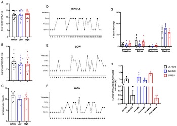
Twenty-one days after treatment interruption, the body (g - Figure 2A) and ovarian weights (Figure 2B) of naproxen-treated C57BL/6 females were similar to those of the vehicle-treated group (p > 0.05). Body weight: p = 0.5362, vehicle = 16.61 ± 0.621, low = 16.52 ± 0.735, high = 17.43 ± 0.532. Ovarian weight: p = 0.5666; vehicle: 3.350 ± 0.302, low = 2.936 ± 0.256, high = 2.957 ± 0.0331. Therefore, the gonadosomatic index (%) was also similar among groups (Figure 2C). Gonadosomatic index: p = 0.4327, vehicle = 11.61 ± 0.917, low = 9.948 ± 7.233, high = 10.56 ± 0.969.
Twenty-one days after naproxen administration, biometric and oestrous cycle parameters were unaffected. (A) Body (g) and (B) ovarian weight (mg) were similar among groups (p > 0.05), as well as (C) gonadosomatic index (%). The oestrous cycles of representative (D) vehicle (n = 6), (E) low (n = 7), and (F) high (n = 7) dose of naproxen-treated animals showed that after the treatment interruption, they recovered their oestrous cyclicity. Additionally, (G) frequencies of days in each stage (%) were not affected by treatments (p > 0.05). (H) The percentage of animals that ovulated or did not ovulate in C57BL/6 mice was not altered by naproxen treatment. Values are expressed as mean ± standard error of the mean (SEM) and were analysed by chi-square (X2) for parametric data, p < 0.05. The other values are expressed as mean ± SEM and were analysed by ANOVA for parametric data and by Kruskal–Wallis for nonparametric data, followed by Tukey’s test, p < 0.05.

Regarding the oestrous cycle, Figures 2D–F clearly showed, in a representative female, that from days 1–8 (during treatment), their cycle is disturbed by low and high doses of naproxen, compared to the vehicle. However, after treatment interruption (from day nine to 29), the oestrous cycle was restarted. Additionally, considering the entire experimental period, the percentage (%) of days in each stage (Figure 2G) was not influenced by treatment (proestrus: p = 0.5848, vehicle = 10.34 ± 2.953, low = 8.374 ± 3.089, high = 12.81 ± 3.076; oestrus: p = 2.836, vehicle = 24.71 ± 9.689, low = 9.852 ± 3.719, high = 20.20 ± 5.801; metoestrus: p = 0.0533, vehicle = 0.574 ± 0.574, low = 1.478 ± 1.478, high = 4.926 ± 1.821; dioestrus: p = 0.1723, vehicle = 64.37 ± 9.658, low = 80.30 ± 6,926, high = 62.07 ± 5.104).
Follicular quantification of C57BL/6 females 21 days after naproxen treatment interruption
The percentage (%) of primordial follicles in C57BL/6 females after treatment interruption (Figure 3A) was similar among groups (p = 0.0511, vehicle = 38.94 ± 4.13, low = 39.15 ± 1.46, high = 29.06 ± 2.58). However, the percentage of primary follicles (Figure 3B) in high-dose-treated females was significantly increased compared to low-dose-treated ones (p = 0.0082, high = 13.6 ± 2.585, low = 3.866 ± 0.662). Treatment also influenced the percentage of antral follicles (Figure 3B) in the high group compared to the low group (p = 0.0089, high = 8.965 ± 1.650, low = 2.782 ± 0.973, vehicle = 2.565 ± 0.0821) and the vehicle (p = 0.0071, high = 8.965 ± 1.650, vehicle = 2.565 ± 0.0821, low = 2.782 ± 0.973). The increased percentage of antral follicles of low and high treated animals after treatment interruption was qualitatively shown in the middle sections of the ovaries (Figure 3C–E).
Follicular quantification and morphometry in C57BL/6 mice after the interruption of naproxen treatment. The percentages of primordial follicles (A) did not show significant differences between the groups (p > 0.05). Even after treatment interruption, (B) high-dose naproxen (50 mg/kg)- treated animals showed an increased percentage of primary (p = 0.0082) and antral follicles (p = 0.0089) compared with vehicle. The rate of antral follicles of low-dose-treated animals was also increased in the high group compared to the control group (p = 0.0071). These quantitative data can also be qualitatively observed in mid-sections of the ovary of the animals (C–E), where red arrows denote antral follicles. Bars = 20 μm. After treatment pausing, (F) follicle diameters of all classes showed no significant difference between the groups (p > 0.05). Values are expressed as mean ± SEM and were analysed by ANOVA for parametric data and by Kruskal–Wallis for nonparametric data, followed by Tukey’s test, p < 0.05.

Figure 3. Long description
Panel A: A vertical bar graph shows the percentage of primordial follicles across three groups: Vehicle, Low-dose naproxen, and High-dose naproxen. The y-axis represents the percentage of primordial follicles, and the x-axis lists the treatment groups. The graph indicates no significant differences between the groups. Panel B: Another vertical bar graph displays the percentage of growing follicles, categorized into primary, secondary, and antral follicles. The y-axis represents the percentage of growing follicles, and the x-axis lists the follicle types. Significant increases in primary and antral follicles are observed in the high-dose naproxen group compared to the vehicle group. Panel C: An image of an ovary mid-section from the vehicle group shows antral follicles. Panel D: An image of an ovary mid-section from the low-dose naproxen group shows antral follicles. Panel E: An image of an ovary mid-section from the high-dose naproxen group shows antral follicles, with red arrows denoting antral follicles. Panel F: A scatter plot shows the follicle diameters of all classes across the three groups. The y-axis represents the follicle diameter in micrometers, and the x-axis lists the follicle types. No significant differences in follicle diameters are observed between the groups.
All classes of follicles showed similar diameters among groups (Figure 3F). Primordial: p = 0.1302, vehicle = 14.83 ± 0.0443, low = 13.74 ± 0.340, high = 14 ± 0.386; Primary: p = 0.8746, vehicle = 35.07 ± 1.882, low = 33.71 ± 1.895, high = 35.2 ± 2.296; Secondary: p = 0.2764, vehicle = 76.81 ± 3.301, low = 83.33 ± 3.398, high = 82.66 ± 2.673; Antral: p = 0.5465, vehicle = 177.5 ± 12.03, low = 167.9 ± 12.75, high = 196.4 ± 21.89.
Ovulatory response of C57BL/6 females 21 days after naproxen treatment interruption
All outcomes on the ovulatory response to a standard protocol, obtained 21 days after naproxen treatment interruption, indicated that treated C57BL/6 females restored their ovulatory capacity (Figure 4). The number of recovered oocytes (Figure 4A) was similar among groups (p = 0.3414, vehicle = 16 ± 6.361, low = 8.143 ± 2.052, high = 9.714 ± 2.347). Treatment did not influence the percentage of viable oocytes (Figure 4B; p > 0.05) (vehicle = 100 ± 0, low = 100 ± 0, high = 100 ± 0). A summary of ovulation results by animal is presented in Supplemental Table 1. The analysis of C57BL/6 animals that ovulated and those that did not ovulate revealed no significant differences (Figure 2H).
Oocyte and corpora lutea numbers of C57BL/6 mice after naproxen treatment interruption. After treatment pausing, (A) the number of oocytes, there was no significant difference among groups (p > 0.05), as well as (B) their viability (%; p > 0.05). The number of (C) corpora haemorrhagica and corpora lutea (D) was also similar among groups (p > 0.05), as also observed in (E–G) macroscopic pictures of ovaries. Black arrows denote corpora haemorrhagica, and red arrows indicate corpora lutea. Bars = 200 μm. Values are expressed as mean ± SEM and were analysed by ANOVA for parametric data and by Kruskal–Wallis for nonparametric data, followed by Tukey’s test, p < 0.05.

In the macroscopic evaluation of the ovaries, the number of corpora haemorrhagica (Figure 4C) and lutea (Figure 4D) was also unaffected by treatment (p > 0.05). Number of corpora haemorrhagica: p = 0.6757, vehicle = 4.5 ± 1.522, low = 4.286 ± 0.778, high = 3.143 ± 1.164. Number of corpora lutea: p = 0.7579, vehicle = 9.667 ± 2.305, low = 10.86 ± 1.933, high = 8.571 ± 2.369. Additionally, representative pictures of the ovaries (Figure 4E–G) qualitatively demonstrate the quantitative data. Red arrows indicate corpora lutea, and black arrows denote corpora haemorrhagica.
Ovulatory response of BALB/c and Swiss females treated with naproxen
Naproxen treatment, in both doses, did not influence (p > 0.05) the number of oocytes (Figure 5A) or corpora haemorrhagica (Figure 5B), and lutea (Figure 5C) in BALB/c females. Number of oocytes: p = 0.4339, vehicle = 7 ± 3,412, low = 3,917 ± 1,630, high = 7.83 ± 2,066; number of corpora haemorrhagica: p = 0.0743, vehicle = 7,778 ± 2,019, low = 4,667 ± 1,333, high = 2,833 ± 1,029; number of corpora lutea: p = 0.8424, vehicle = 8.667 ± 2.555, low = 7.750 ± 0.897, high = 7.333 ± 1.333. The ovulation results for the animals used are shown in Supplemental Table 2.
Oocyte and corpora lutea numbers of BALB/c and Swiss after naproxen treatment interruption. After treatment pausing in BALB/c females, (A) the number of oocytes, (B) corpora haemorrhagica, and (C) corpora lutea were similar among groups (p > 0.05). Similarly, in Swiss females, (D) the number of oocytes, (E) corpora haemorrhagica, and (F) corpora lutea showed no significant differences among groups (p > 0.05). Values are expressed as mean ± SEM and were analysed by ANOVA for parametric data and by Kruskal–Wallis for nonparametric data, followed by Tukey’s test, p < 0.05.

Similarly, Swiss females treated with low or high doses of naproxen were not affected (p > 0.05) in the numbers of oocytes (Figure 5D), corpora haemorrhagica (Figure 5E), or lutea (Figure 5F). Number of oocytes: p = 0.0599, vehicle = 24.67 ± 4.607, low = 13.56 ± 2.334, high = 14.92 ± 2.784; number of corpora haemorrhagica: p = 0.4794, vehicle = 4.556 ± 0.944, low = 5.25 ± 1.333, high = 3.455 ± 0.928; number of corpora lutea: p = 0.3855, vehicle = 7.750 ± 0.921, low = 8.444 ± 0.728, high = 7.091 ± 0.512.
Discussion
The search for new non-hormonal contraceptives has been an emerging field in Reproductive Biology. In this context, previous studies by Nascimento and collaborators (Reference Nascimento, Oliveira, Pereira, L.A.A., Lopes, Paula, R., Jorge and Campos-Junior2024) have indicated that naproxen treatment reduced late follicular growth and the number of ovulations in C57BL/6 mice. However, it is still necessary to better understand the reversibility of these effects after treatment interruption, as well as to conduct similar investigations in other strains. Thus, the present study demonstrated that interrupting naproxen administration for 21 days in C57BL/6 animals resulted in ovulation parameters similar to those in untreated animals. In addition, BALB/c and Swiss mice did not exhibit the reduction in ovulatory rate, indicating a dependence on genetic background for this phenotypic manifestation.
Regarding body weight, the results showed no significant differences among animals treated with naproxen. This parameter was also analysed by Grimes et al. (Reference Grimes, Kenney, Dominguez, Brayer, Guo, Wandinger-Ness and Hudson2021), who used R-ketorolac, an enantiomer of the NSAID ketorolac and observed no significant differences in body weight in mice. Regarding the gonadosomatic index parameter, no differences were observed in mice treated with naproxen. A study by Ezechukwu et al. (Reference Ezechukwu, Mbegbu, Mgbenka, Nwani, Nnamonu and Orji2024), which also analysed the gonadosomatic index using the ethanolic extract of Solanum macrocarpon leaves, found no significant differences in this parameter.
With respect to the oestrous cycle, the percentage of days in each stage did not differ significantly between C57BL/6 mice treated with naproxen and untreated controls. A similar parameter analysed by Ndeke et al. (Reference Ndeke, Mutembei, Kaingu, Muthee, Birech and Ondieki2022) was the frequency of oestrous cycle stages over 20 days following paracetamol treatment. This study demonstrated significant differences only during the proestrus stage in mice; no differences were observed in the oestrus, metoestrus, or dioestrus stages. Therefore, the finding that naproxen treatment does not induce significant changes in biometric patterns is of great importance, demonstrating that naproxen acts only during folliculogenesis and thereby preserves animal health.
Although the study of NSAIDs is critical, studies focusing on naproxen are still relatively scarce (Boizet-Bonhoure et al., Reference Boizet-Bonhoure, Déjardin, Rossitto, Poulat and Philibert2022). The data presented in this study indicate an increase in the percentage of primary and antral follicles in mice treated with 50 mg/kg naproxen for 8 days, followed by a 21-day interruption. A study by Nascimento et al. (Reference Nascimento, Oliveira, Pereira, L.A.A., Lopes, Paula, R., Jorge and Campos-Junior2024) demonstrated that an 8-day continuous treatment reduced the percentage of antral follicles at the high dose (50 mg/kg). In this context, the results obtained in the present study regarding the reversal of the low ovulation rate process, along with the change in treatment, can be explained by the inhibition or non-inhibition of COX and, consequently, of prostaglandins.
Antral follicle numbers increased after interruption of the high-dose treatment, which may reflect compensatory ovarian mechanisms triggered by prior follicular suppression. Prolonged exposure to high doses may transiently inhibit follicle growth or promote atresia of growing follicles, resulting in the accumulation of quiescent or slowly progressing preantral follicles (Adhikari and Liu, Reference Adhikari and Liu2009). Upon treatment withdrawal, the release from inhibitory signalling may induce a rebound effect, characterized by synchronized follicle recruitment and accelerated transition into the antral stage, a phenomenon previously described following endocrine or pharmacological suppression of ovarian activity (Fischer-Holzhausen and Röblitz, Reference Fischer-Holzhausen and Röblitz2022). In addition, delayed activation of follicular growth pathways, potentially involving altered sensitivity to gonadotropins and local ovarian regulators such as KIT ligand, GDF9 and BMP15, may further contribute to the post-treatment increase in antral follicles (Knight and Glister, Reference Knight and Glister2006; Edson et al., Reference Edson, Nagaraja and Matzuk2009). Collectively, these findings support the interpretation that the observed increase reflects a delayed and compensatory follicular response.
The inflammatory response is a mechanism adapted to physical injuries or infections (Wautier and Wautier, Reference Wautier and Wautier2023). Follicle rupture is an injury-generating process that is inherently associated with the ovulation mechanism (Espey, Reference Espey1981). As reported by Zhang et al. (Reference Zhang, Goods, Pattarawat, Wang, Haining, Zhang, Shalek, Duncan, Woodruff and Xiao2023), PGE2 is a prostaglandin that acts as a primary mediator of the inflammatory process during ovulation and is synthesized by the prostaglandin synthases COX-1 and COX-2, which catalyse the metabolism of arachidonic acid to produce prostaglandins. There are inhibitors of prostaglandin synthases, specifically COX-1 and COX-2, known as NSAIDs, which are widely used (Ibrahim et al., Reference Ibrahim, Moustafa, Almalki, Allam, Althagafi, Md and Mohamed2021). Therefore, continuous treatment with naproxen, which reduces ovulation in C57Bl/6 mice, likely results from inhibition of cyclooxygenase (COX), thereby preventing the synthesis of pro-inflammatory prostaglandins, such as PGE2, which are essential for ovulation.
COX, particularly the inducible isoform COX-2 (PTGS2), is expressed in the ovary predominantly in granulosa cells of preovulatory and luteinizing follicles, where its expression is rapidly induced by the luteinizing hormone (LH) surge and is essential for prostaglandin production during the ovulatory process (Tokuyama et al., Reference Tokuyama, Nakamura, Muso, Honda, Ishiko and Ogita2001). COX-2 knockout (Ptgs2-/-) mice showed profound reproductive defects, including impaired cumulus–oocyte complex expansion, failure of follicular rupture and consequent infertility, despite normal follicular growth and steroidogenesis (Davis et al., Reference Davis, Lennard, Lee, Tiano, Morham, Wetsel and Langenbach1999). Most studies investigating ovarian COX expression and function have been conducted in mice on a C57BL/6 genetic background, the strain most commonly used for COX-2-deficient models and reproductive studies (Wang et al., Reference Wang, Ma, Tejada, Zhang, Morrow, Das and Dey2004). Although COX-2 expression is a conserved feature of ovulation across murine models, differences in ovulatory capacity and inflammatory responsiveness among strains such as C57BL/6, BALB/c and outbred SWISS mice suggest potential genetic-dependent modulation of COX-2 expression or activity in the ovary; however, direct comparative analyses of ovarian COX-2 expression among these strains remain scarce in the literature (Galibert and Azzouzi, Reference Galibert and Azzouzi2023).
Duffy et al. (Reference Duffy, Ko, Jo, Brannstrom and Curry2019) investigate the inflammatory processes associated with ovulation and show a parallel between these two processes. One mechanism proposed by Duffy et al. (Reference Duffy, Ko, Jo, Brannstrom and Curry2019) is the role of immune cells in follicle rupture and oocyte release. Macrophages, mast cells and neutrophils have essential roles in recruiting other cells in the inflammatory process. They are also responsible for extracellular matrix degradation, which weakens the follicular wall and subsequently releases the oocyte. Based on this information, the proportions of immune cell subpopulations in different mouse strains may influence the inflammation associated with ovulation, potentially affecting oocyte release.
The present study showed that the genetic background influenced sensitivity to naproxen administration during late folliculogenesis. Differences in naproxen responsiveness among the C57BL/6, BALB/c and Swiss mouse strains may be linked to variations in inflammatory capacity and COX-1/2 expression. Studies indicate that female C57BL/6 mice exhibit lower leukocyte and monocyte counts than other strains (Silva-Santana et al., Reference Silva-Santana, Bax, Fernandes, Bacellar, Hooper, Dias, Silva, de Souza, Ramos and Santos2020) and reduced inflammatory competence, as evidenced by weaker mast cell induction in murine asthma models (Gueders et al., Reference Gueders, Paulissen, Crahay, Quesada-Calvo, Hacha, Van Hove, Tournoy, Louis, Foidart and Noël2009). Additionally, COX-2 expression decreases with age in several tissues, likely including the ovary, affecting all strains (Jung et al., Reference Jung, Yoo, Nam, Kim, Kim, Kwon, Lee, Choi, Kim and Yoon2019) and COX-2 deficiency directly impairs ovulation, as shown in C57BL/6 females (Lim et al., Reference Lim, Paria, Das, Dinchuk, Langenbach, Trzaskos and Dey1997). Therefore, the C57BL/6 strain appears more sensitive to naproxen-induced ovulatory inhibition due to its intrinsically lower inflammatory response and potentially more consequential COX-2 reduction, whereas BALB/c and SWISS may possess compensatory mechanisms that mitigate these effects.
Other studies demonstrate differences among strains in other observed contexts; for example, Culbreth et al. (Reference Culbreth, Biryukov, Shoe, Dankmeyer, Hunter, Klimko, Rosario-Acevedo, Fetterer, Moreau and Welkos2019) showed that C57BL/6 mice can be highly sensitive to analgesics, whereas BALB/c mice can be insensitive. Furthermore, Marques et al. (Reference Marques, Campos, Castro, Cardoso and Andrade2011) evaluated the response to injury and inflammation in mice of the three strains used in the current study and demonstrated that the responses of each strain were not uniform, being intrinsic to the genetic character of each strain, showing a greater capacity to respond to injury and inflammation in SWISS, intermediate in BALB/c and weak in C57BL/6. It was also demonstrated by Smith (Reference Smith2019) that mouse strains differ in sensitivity in inflammatory pain assays, suggesting that the C57BL/6 strain is more sensitive to this stimulus than the BALB/c strain. Considering this scenario, our study has broader implications for reproductive biology and drug discovery and repositioning, showing the importance of evaluating different mouse backgrounds in preclinical studies. Additionally, this study opens avenues for investigating genomic differences among mouse strains that may contribute to drug responsiveness.
Superovulation is a widely used artificial technique for inducing the release of multiple oocytes (Lamas et al., Reference Lamas, Carvalheira, Gartner and Amorim2021). In Swiss and BALB/c females, no changes in oocyte number were observed following treatment. The same treatment was used by Nascimento et al. (Reference Nascimento, Oliveira, Pereira, L.A.A., Lopes, Paula, R., Jorge and Campos-Junior2024) in C57BL/6 females, and, as reported by Clow et al. (Reference Clow, Hurst and Fleming2002), mice have a high ovulatory rate due to the short oestrous cycle length. However, the present study demonstrated that the drug produced different responses across the strains used. The lower ovulatory rate observed with the treatment is likely attributable to the isogenicity of the C57BL/6 mice; however, the BALB/c strain, which is also isogenic, did not respond similarly.
In this sense, Pankhurst et al. (Reference Pankhurst, Kelley, Sanders, Woodcock, Oorschot and Batchelor2018) demonstrated that C57BL/6 female mice ovulate 6 to 12 oocytes, indicating that, although isogenic, the ovulatory responses of individual animals are independent and lineage-specific. On the other hand, animals of the Swiss lineage are heterogeneous (Silva-Santana et al., Reference Silva-Santana, Bax, Fernandes, Bacellar, Hooper, Dias, Silva, de Souza, Ramos and Santos2020), which may explain the divergence in responses to naproxen treatment in the studied lineages, due to the level of sensitivity of the ovarian cycle and folliculogenesis of each lineage to the drug used, with the C57BL/6 lineage being the most affected, due to other characteristics of this lineage mentioned earlier in the text, such as the lower number of white blood cells in the organism and a possible overall decrease in the expression level of COX-1/2 resulting from the ageing of the animal, which affects all lineages. Further studies on this topic and the lineages are needed to confirm this hypothesis.
As mentioned by Kulesza et al. (Reference Kulesza, Paczek and Burdzinska2023), some NSAIDs present different degrees of reversibility, such as ibuprofen and aspirin, which represent the extremes between reversible and irreversible drugs, respectively. Since, in the present study, the number of recovered oocytes after naproxen treatment interruption was similar between groups, it is possible to infer that, at least for this parameter, there was reversibility. The results presented open new perspectives regarding potential drugs, such as naproxen, that could be redirected to more in-depth research in order to target their direct applications against ovulation, thus potentially creating a short-acting pericoital contraceptive method. Corroborating the article by Saja (Reference Saja2021), which states that NSAIDs can induce reversible infertility.
Taken together, these results demonstrate reversibility of the ovulation process in mice suspended from naproxen administration for 21 days, with ovulation rates normalized. The present study also showed that naproxen administration did not affect the number of ovulated oocytes in Swiss and BALB/c females. Therefore, this work indicates that naproxen administration can reversibly modulate murine follicular growth and ovulation, depending on the genetic background. This study offers new perspectives on the search for non-hormonal contraceptives through drug repositioning and underscores the importance of considering different mouse backgrounds in preclinical studies.
Supplementary material
The supplementary material for this article can be found at https://doi.org/10.1017/S0967199426100525
Data availability statement
The datasets generated during and/or analysed during the current study are available from the corresponding author (paulohenrique@ufsj.edu.br) upon reasonable request.
Acknowledgements
This work was supported by the National Council for Scientific and Technological Development, Brazil [406600, 2021]; the Minas Gerais State Research Foundation, Brazil [00638, 2022 and 29675, 2022]; and the Coordination for the Improvement of Higher Education Personnel, Brazil. JPLGC, VNG, PAB, APP, CSF, LAACP, SPO, KSSAD received scholarships from FAPEMIG, CNPq, or CAPES.
Author contributions
João Pedro Lobão Gomes Castro: Formal analysis-Equal, Investigation-Equal, Methodology-Equal; Vanessa Neves Guimarães: Formal analysis-Equal, Investigation-Equal; Patrick de Abreu Barbosa: Investigation-Equal, Methodology-Equal; Amanda Pereira Paz: Investigation-Equal, Methodology-Equal; Camila Stefane Ferreira: Methodology-Equal, Validation-Equal, Writing - original draft-Equal, Writing - review & editing-Equal; Luiza Aparecida Ansaloni Chagas Pereira: Investigation-Equal, Methodology-Equal, Supervision-Equal, Writing - original draft-Equal, Writing – review & editing-Equal; Stella Pollyanne Oliveira: Formal analysis-Equal, Investigation-Equal, Writing – original draft-Equal, Writing – review & editing-Equal; Karine Sthefany Serpa Amaral Dias: Investigation-Equal, Methodology-Equal, Writing – original draft-Equal, Writing – review & editing-Equal; Paulo Henrique Almeida Campos-Junior: Conceptualization-Equal, Funding acquisition-Equal, Investigation-Equal, Methodology-Equal, Project administration-Equal, Resources-Equal, Supervision-Equal, Visualization-Equal, Writing – original draft-Equal, Writing – review & editing-Equal.
PHACJ designed and supervised the experiments and reviewed the manuscript. CSF and LAACP also reviewed the manuscript and addressed questions regarding the study methods. VNG, PAB, APP, SPO, and KSSAD performed all experiments and analysis.
Competing interests
The authors declare that there are no known conflicts of interest associated with this publication and that no significant financial support for this work could have influenced its outcome.
Ethical standards
The Ethics Committee for the Use of Animals of the Federal University of São João Del Rei (protocol n° 009/2020) approved all procedures.