




No CrossRef data available.
Article contents
Electrocardiographic changes in patients with cardiac rhabdomyomas associated with tuberous sclerosis
Published online by Cambridge University Press: 24 May 2005
Abstract
Background: Cardiac rhabdomyomas associated with tuberous sclerosis induce various abnormalities in the electrocardiogram. Electrocardiographic evidence of ventricular hypertrophy may appear if the tumour is electrically active. To our knowledge, electrocardiographic evidence of ventricular hypertrophy has been reported only in association with congestive heart failure. Follow-up studies of changes in electrocardiographic findings are also lacking. Methods: We studied 21 consecutive patients with cardiac rhabdomyoma associated with tuberous sclerosis, 10 males and 11 females, aged from the date of birth to 9 years at diagnosis. The mean period of follow-up was 53 months. None of the patients developed congestive heart failure. We evaluated the electrocardiographic changes during the follow-up, and their association with echocardiographic findings. Results: Of the 21 patients, 12 showed one or more abnormalities on the electrocardiogram at presentation, with five demonstrating right or left ventricular hypertrophy. In all of these five cases, the tumours were mainly located in the respective ventricular cavity. In one patient with a giant tumour expanding exteriorly, there was marked left ventricular hypertrophy on the electrocardiogram. Followup studies showed spontaneous regression of the tumours in 12 of 19 patients, with abnormalities still present in only 7 patients. A gradual disappearance of left ventricular hypertrophy as seen on the electrocardiogram was noted in the patient with marked left ventricular hypertrophy at presentation in parallel with regression of the tumour. Conclusions: The presence of cardiac rhabdomyomas in patients with tuberous sclerosis might explain the ventricular hypertrophy seen on the electrocardiogram through its electrically active tissue without ventricular pressure overload or ventricular enlargement, although pre-excitation might affect the amplitude of the QRS complex. Even in cases with large tumours, nonetheless, the electric potential might not alter the surface electrocardiogram if the direction of growth of the tumour is towards the ventricular cavity. In many cases, electrocardiographic abnormalities tend to disappear, concomitant with regression of the tumours.
Information
- Type
- Original Article
- Information
- Copyright
- © 2003 Cambridge University Press
Rhabdomyomas, the most common primary cardiac tumours seen in infants and children, are often associated with tuberous sclerosis.1–4 The reported frequency of this association ranges from one-third in an autopsy study to almost half in some clinical series.5–7 A recent study showed that more than nine-tenths of the patients with rhabdomyomas also had tuberous sclerosis.8 The clinical manifestations of cardiac tumours vary from total absence of symptoms, when they are discovered incidentally, to intrauterine or postnatal sudden death.5, 9 The prognosis is favorable, as more than half of the tumours regress spontaneously, either partially or completely, with increasing age.10, 11
Rhabdomyomas can induce various cardiac arrhythmias,12, 13 including premature atrial contractions, supraventricular tachycardia, multifocal premature ventricular contractions,14 second or third degree atrioventricular block,15 sinus nodal dysfunction,16 and the pre-excitation syndrome.17, 18 In addition, the tumour is often associated with other electrocardiographic abnormalities, such as ventricular hypertrophy and disturbances of repolarization.12 The true mechanism of ventricular hypertrophy as seen on the electrocardiograms, however, remains to be elucidated. Furthermore, there are no follow-up studies that examined electrocardiographic changes, and no studies on the relationship between electrocardiographic and echocardiographic findings.
In the present study, we investigated retrospectively the electrocardiographic findings at presentation and follow-up in patients with cardiac rhabdomyomas associated with tuberous sclerosis. We also describe in detail the clinical course of one patient, with marked left ventricular hypertrophy on the electrocardiogram, which was not due to congestive heart failure but possibly to the giant tumour itself.
Subjects and methods
Our subjects were 21 consecutive patients, 10 males and 11 females, diagnosed with cardiac rhabdomyomas associated with tuberous sclerosis at the University Hospital of Tsukuba or Kanagawa Children's Medical Center between January 1986 and December 2001. Tuberous sclerosis was diagnosed based on the criterions described by Gomez.19 All our patients were confirmed as having characteristic subependymal hamartomas on computed tomograms or magnetic resonance imaging of the brain. Age at diagnosis of the cardiac rhabdomyomas ranged from birth to 9 years. Rhabdomyomas were diagnosed echocardiographically based on characteristic findings, including multiple tumours of high echo-dense and homogeneous pattern. In 16 patients, there were multiple tumours seen on the echocardiogram, and 13 of these had at least one giant tumour with the maximum diameter greater than 10 mm. None of the patients developed congestive heart failure. Surgical resection, or biopsy of the tumour for histopathological examination, was not performed. Electrocardiographic findings at presentation, and their association with echocardiographic findings, were evaluated retrospectively in each case. A follow-up electrocardiogram was available in 19 patients. The period of follow-up extended from 3 months to 13 years, with a mean of 53 months. Changes in the size of the tumours on echocardiograms, and the relation between electrocardiographic and echocardiographic findings, were both investigated. We also studied four patients with tuberous sclerosis, but without cardiac rhabdomyomas, three females and one male, aged from 5 months to 2 years, to confirm whether the tumours in the brain might distort the electrocardiographic waveforms.
Results
Electrocardiographic findings at presentation showed one or more abnormalities in 12 out of 21 patients (Table 1). They included right bundle branch block in 5 cases, right ventricular hypertrophy in 3 cases, left ventricular hypertrophy in 2 cases, premature atrial contraction in 3 cases, left axis deviation in 2 cases, and the pre-excitation syndrome in 2 cases. In three of the 5 cases with ventricular hypertrophy, the tumours were located predominantly in one or other of the ventricular cavities. These presented as ventricular hypertrophy on the electrocardiogram (Table 2). In one case with prominent left ventricular hypertrophy, a giant tumour was located in the apical region of the left ventricle. In 13 patients with giant tumours, only 3 showed ventricular hypertrophy on the electrocardiogram.
Table 1. Clinical findings of patients with cardiac rhabdomyoma.

Table 2. Patients who showed ventricular hypertrophy on electrocardiogram at diagnosis.

All four patients without cardiac rhabdomyomas had normal electrocardiograms. We will give a detailed description of the serial changes in the electrocardiographic and echocardiographic findings in our tenth patient, who had marked left ventricular hypertrophy:
Case 10: A 13-day-old boy was diagnosed as having cardiac rhabdomyomas on routine echocardiogram at a neighboring clinic. The newborn looked healthy, and no heart murmur was recognized on auscultation. On admission, the electrocardiogram showed single premature atrial contractions, left axis deviation, high-amplitude R wave in the left precordial leads with wide QRS duration and abnormal repolarization (Fig. 1a). The duration of PQ interval was 100 msec, but delta waves were not apparent. The echocardiogram revealed a giant highly echo-dense, tumour at the apex of the left ventricle, with a maximal diameter of 19 mm, extending toward the pericardium, as well as multiple small tumours inside the cavity of the left ventricle and the ventricular septum (Fig. 1b). No obstruction to either ventricular outflow or inflow was observed, and left ventricular volume and ejection fractions were normal. During the next 6 months, the giant tumour showed gradual and spontaneous regression on the echocardiogram (Fig. 2a). This was associated with a decrease in the amplitude of the R wave in the left precordial leads on electrocardiogram, although mild abnormalities of repolarization were still noted (Fig. 2b). The PQ interval did not change during the period.

Figure 1. (a) Electrocardiogram of our tenth patient at initial presentation, showing left axis deviation, prominently high voltage R waves in the left precordial leads, with wide QRS complexes and repolarization abnormalities. The PQ interval is 100 msec. Premature atrial contractions also appeared on the electrocardiogram, though they are not shown in this figure. (b) Echocardiogram of the same patient taken at first presentation. Note the giant echo-dense tumour at the apex of the left ventricle, with the maximal diameter of 19 mm, extending exteriorly towards the pericardium. Note also the presence of other small tumours in the left ventricular cavity and ventricular septum.

Figure 2. (a) Echocardiogram of our tenth patient at 6-month follow-up examination, showing regression of the tumour. (b) Electrocardiogram of the same patient on follow-up examination as a 6-month-old. Although mild repolarization abnormalities are still present, the voltage of the R waves in the left precordial leads is decreased.
Follow-up studies showed spontaneous regression of the tumours in 12 of our 19 patients (Table 1). In the remaining 7 cases, the size of the tumours did not change. None of the patients had enlargement of their tumours during follow-up. The abnormal electrocardiographic findings also disappeared in 7 of 12 cases. In 4 of the 5 patients with ventricular hypertrophy at the first presentation, normalization of the electrocardiogram was noted at their follow-up examination. On the other hand, two cases with an abnormal electrocardiogram at presentation showed normalization of the electrocardiogram without changes in the size of the tumour on the echocardiogram (Cases 11 and 14).
Discussion
Our patients with cardiac rhabdomyomas associated with tuberous sclerosis showed various electrocardiographic abnormalities, including ventricular hypertrophy. On follow-up examinations, spontaneous regression of the tumours was noted in many patients, together with normalization of the electrocardiogram.
With respect to ventricular hypertrophy on electrocardiogram, a recent report by Mühler et al.12 described ventricular hypertrophy in two of 21 patients with tuberous sclerosis. These authors, however, did not mention whether their patients had congestive heart failure caused by obstruction to ventricular outflow or inflow. Other studies of rhabdomyomas have described ventricular hypertrophy on the electrocardiogram,20–26 and many of these were associated with congestive heart failure. The main cause of ventricular hypertrophy in these studies was thought to be obstruction to ventricular outflow or inflow, resulting in pressure overload of the ventricle.27
In the tenth of our patients, prominent voltage of the R wave, and ST-T abnormalities, were demonstrated in the left precordial leads without any obstruction to outflow or inflow. The location of the giant tumour was at the apical region, so that block of a bundle branch was unlikely. These electrocardiographic findings regressed in parallel with diminution in size of the tumour. This suggests that the electrocardiogram of patients with cardiac rhabdomyomas can show ventricular hypertrophy without obstruction of the ventricular inflow or outflow tracts. Alternatively, it is possible that pre-excitation yielded the high-voltage R wave on the electrocardiogram, although a delta wave was not recognized.
The specific cell that gives rise to the cardiac rhabdomyoma is still controversial. Fenoglio et al.5 reported that the cardiac rhabdomyoma is a type of hamartoma originating from embryonic myoblasts, a theory widely accepted at present. If cardiac rhabdomyomas originate from myoblasts, then the rhabdomyomatous tissue itself could generate myocardial electric potential. On the other hand, Elliott et al.28 reported that the cardiac rhabdomyoma takes origin from the Purkinje cell. In fact, cases of rhabdomyoma are known to be associated with the pre-excitation syndrome,17, 18 as in our study. Rhabdomyomatous tissue traversing the atrioventricular junction might act as an accessory pathway.
Patients with giant cardiac rhabdomyomas do not necessarily show the electrocardiographic changes of ventricular hypertrophy. In our case with marked left ventricular hypertrophy, a giant tumour was located in the apex of the left ventricle, and expanded in the direction of the pericardium. In other cases with ventricular hypertrophy, tumours were mainly located in the ventricle. These patients showed electrocardiographic changes of ventricular hypertrophy, but the tumours did not expand to the pericardium. Rhabdomyomas can be embedded in the myocardium or can protrude into the cardiac cavity.5 Rhabdomyomas embedded in the myocardium do not cause any disturbance to the flow of blood, but may cause the electrocardiographic changes of ventricular hypertrophy. Not only the size of tumours, but also their location, could influence whether they show any features of ventricular hypertrophy on the electrocardiogram.
Because cardiac rhabdomyomas are known to regress spontaneously, surgical resection is not recommended unless the patients are symptomatic.10, 11 Our study supports this recommendation, since many electrocardiographic abnormalities disappeared with spontaneous regression of the tumours with increasing age.
Study limitations
Due to the retrospective nature of the study, the exact course of regression could not be determined. Furthermore, none of our patients required surgical resection. Biopsy material and histopathological examination of the tumour, therefore, was not performed. The diagnosis of rhabdomyoma is established by echocardiography, especially in those associated with tuberous sclerosis. Strictly speaking, nonetheless, we cannot exclude the possible presence of pathological differences in rhabdomyomatous tissue among our various patients.
Conclusions
Various arrhythmias and abnormalities on the electrocardiogram were observed in children with cardiac rhabdomyomas and tuberous sclerosis. Many of these abnormalities tended to disappear concomitant with the spontaneous regression of the tumours. Some patients showed ventricular hypertrophy on the electrocardiogram without obstruction to ventricular inflow or outflow. Cardiac rhabdomyomas themselves might explain ventricular hypertrophy on the electrocardiogram through the presence of electrically active tissue without ventricular pressure overload or ventricular enlargement. Even in patients with large tumours, however, the electric potential might not alter the surface electrocardiogram if the direction of growth of the tumour is towards the ventricular cavity.
References
Longino LA, Meeker IA Jr. Primary cardiac tumors in infancy. J Pediatr 1953; 43: 724–731.Google Scholar
Freedom RM, Lee KJ, MacDonald C, Taylor G. Selected aspects of cardiac tumors in infancy and childhood. Pediatr Cardiol 2000; 21: 299–316.Google Scholar
Becker AE. Primary heart tumors in the pediatric age group: a review of salient pathologic features relevant for clinicians. Pediatr Cardiol 2000; 21: 317–323.Google Scholar
Fenoglio JJ Jr, McAllister HA Jr, Ferrans VJ. Cardiac rhabdomyoma: a clinicopathologic and electron microscopic study. Am J Cardiol 1976; 38: 241–251.Google Scholar
Bass JL, Breningstall GN, Swaiman KF. Echocardiographic incidence of cardiac rhabdomyoma in tuberous sclerosis. Am J Cardiol 1985; 55: 1379–1382.Google Scholar
Smith HC, Watson GH, Patel RG, Super M. Cardiac rhabdomyomata in tuberous sclerosis: their course and diagnostic value. Arch Dis Child 1989; 64: 196–200.Google Scholar
Bosi G, Lintermans JP, Pellegrino PA, Svaluto-Moreolo G, Vliers A. The natural history of cardiac rhabdomyoma with and without tuberous sclerosis. Acta Paediatr 1996; 85: 928–931.Google Scholar
Shepherd CW, Gomez MR, Lie JT, Crowson CS. Causes of death in patients with tuberous sclerosis. Mayo Clin Proc 1991; 66: 792–796.Google Scholar
Smythe JF, Dyck JD, Smallhorn JF, Freedom RM. Natural history of cardiac rhabdomyoma in infancy and childhood. Am J Cardiol 1990; 66: 1247–1249.Google Scholar
Farooki ZQ, Ross RD, Paridon SM, Humes RA, Karpawich PP, Pinsky WW. Spontaneous regression of cardiac rhabdomyoma. Am J Cardiol 1991; 67: 897–899.Google Scholar
Mühler EG, Turniski-Harder V, Engelhardt W, von Bernuth G. Cardiac involvement in tuberous sclerosis. Br Heart J 1994; 72: 584–590.Google Scholar
Cosnett JE, Gibb BH. Tuberous sclerosis and cardiac arrhythmia in three Zulu patients. Br Med J 1969; 2: 672–673.Google Scholar
Taylor TR. Tuberous sclerosis presenting as cardiac arrhythmia. Br Heart J 1968; 30: 132–134.Google Scholar
Lessick J, Schwartz Y, Lorber A. Neonatal advanced heart block due to cardiac tumor. Pediatr Cardiol 1998; 19: 263–265.Google Scholar
Cowley CG, Tani LY, Judd VE, Shaddy RE. Sinus node dysfunction in tuberous sclerosis. Pediatr Cardiol 1996; 17: 51–52.Google Scholar
O'Callaghan FJK, Clarke AC, Joffe H, et al. Tuberous sclerosis complex and Wolff-Parkinson-White syndrome. Arch Dis Child 1998; 78: 159–162.Google Scholar
Mehta AV. Rhabdomyoma and ventricular preexcitation syndrome. A report of two cases and review of literature. AJDC 1993; 147: 669–671.Google Scholar
Gomez MR. Criteria for diagnosis. In: Gomez MR (ed.). Tuberous Sclerosis, 2nd edn. Raven Press, New York, 1988, pp 89–109.
Massumi RA, Adkins PC, Reichelderfer TR, Fraga JR, Sampson R. Congenital rhabdomyoma of the heart. Presentation of two cases. J Thorac Cardiovasc Surg 1968; 55: 711–718.Google Scholar
Kuehl KS, Perry LW, Chandra R, Scott LP. Left ventricular rhabdomyoma: a rare cause of subaortic stenosis in the newborn infant. Pediatrics 1970; 46: 464–468.Google Scholar
Van der Hauwaert LG. Cardiac tumours in infancy and childhood. Br Heart J 1971; 33: 125–132.Google Scholar
Simcha A, Wells BG, Tynan MJ, Waterston DJ. Primary cardiac tumours in childhood. Arch Dis Child 1971; 46: 508–514.Google Scholar
Shaher RM, Mintzer J, Farina M, Alley R, Bishop M. Clinical presentation of rhabdomyoma of the heart in infancy and childhood. Am J Cardiol 1972; 30: 95–103.Google Scholar
Shaher RM, Farina M, Alley R, Hansen P, Bishop M. Congenital subaortic stenosis in infancy caused by rhabdomyoma of the left ventricle. J Thorac Cardiovasc Surg 1972; 63: 157–163.Google Scholar
Tsakraklides V, Burke B, Mastri A, Runge W, Roe E, Anderson R. Rhabdomyomas of heart. Am J Dis Child 1974; 128: 639–646.Google Scholar
Elliott GB, McGeachy WG. The monster Purkinje-cell nature of so-called “congenital rhabdomyoma of heart”: a forme fruste of tuberous sclerosis. Am Heart J 1962; 63: 636–643.Google Scholar
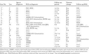
Table 1.
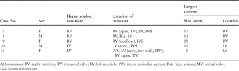
Table 2.

(a) Electrocardiogram of our tenth patient at initial presentation, showing left axis deviation, prominently high voltage R waves in the left precordial leads, with wide QRS complexes and repolarization abnormalities. The PQ interval is 100 msec. Premature atrial contractions also appeared on the electrocardiogram, though they are not shown in this figure. (b) Echocardiogram of the same patient taken at first presentation. Note the giant echo-dense tumour at the apex of the left ventricle, with the maximal diameter of 19 mm, extending exteriorly towards the pericardium. Note also the presence of other small tumours in the left ventricular cavity and ventricular septum.

(a) Echocardiogram of our tenth patient at 6-month follow-up examination, showing regression of the tumour. (b) Electrocardiogram of the same patient on follow-up examination as a 6-month-old. Although mild repolarization abnormalities are still present, the voltage of the R waves in the left precordial leads is decreased.
 You have
Access
- 14
- Cited by