Introduction
When spacecraft are sent to foreign celestial bodies, space agencies must ensure their cleanliness in order to prevent a forward contamination of extraterrestrial habitats and to ascertain the scientific integrity of life detection missions (COSPAR planetary protection policy; Anonymous 2002). Spores are frequent contaminants on spacecraft and may pose a risk for planetary protection considerations (Puleo et al. Reference Puleo, Fields, Bergstrom, Oxborrow, Stabekis and Koukol1977; Venkateswaran et al. Reference Venkateswaran, Satomi, Chung, Kern, Koukol, Basic and White2001; La Duc et al. Reference La Duc, Dekas, Osman, Moissl, Newcombe and Venkateswaran2007). Therefore these dormant, transient states of microbes are in the focus of this particular research area and their capabilities to survive and withstand decontamination procedures and space conditions are currently studied by many scientific groups worldwide. However, the evaluation of spore survival is currently based on cultivation assays only and is therefore time-consuming and biased by the fact that spore germination is a complex process influenced by several (environmental or methodical) factors.
Propidium monoazide (PMA) was introduced by Nocker et al. as a replacement for ethidium monoazide for selective detection of living cells in environmental samples (Nocker et al. Reference Nocker, Cheung and Camper2006). When applied to a microbial sample, PMA was shown to label naked DNA as well as DNA of dead cells after selective entry through their compromised membranes. Subsequent DNA extraction and PCR amplification resulted in amplicons of DNA of cells that were alive in the sample, while DNA of dead cells could not be amplified due to bound PMA. This technique allows the comparison of the living and dead communities within an environmental sample and has led to many subsequent studies (Nocker et al. Reference Nocker, Sossa and Camper2007a, Reference Nocker, Sossa-Fernandez, Burr and Camperb, Reference Nocker, Mazza, Masson, Camper and Brousseau2009, Reference Nocker, Richter-Heitmann, Montijn, Schuren and Kort2010). While the aforementioned surveys considered only vegetative cells in the analysis, another recent study has reported the application of PMA to (endo-) spores of Bacillus subtilis (Rawsthorne et al. Reference Rawsthorne, Dock and Jaykus2009). Its data suggested that PMA can enter heat-inactivated spores and bind their DNA, resulting in a decreased detection of spores via quantitative PCR.
Although molecular analytics of (environmental) samples are becoming more powerful, microscopy can still provide rapid information on aspects that are inaccessible to molecular tools, such as cell morphology, cell number or live/dead distinction using fluorescent dyes such as BacLight or PMA (Boulos et al. Reference Boulos, Prevost, Barbeau, Coallier and Desjardins1999; Nocker et al. Reference Nocker, Cheung and Camper2006). The properties of PMA, being fluorescently active and a strong DNA-masking reagent, are extraordinary features allowing a much more complete view of the viable and dead microbial part in environmental samples in combining molecular and microscopy-based viability staining methods.
The potential of DAPI 4',6-diamidino-2-phenylindole to enter the core of compromised spores has already been reported (Setlow et al. Reference Setlow, Loshon, Genest, Cowan, Setlow and Setlow2002), and also a successful usage of PMA to mask DNA from dead spores for quantitative PCR has been shown (Rawsthorne et al. Reference Rawsthorne, Dock and Jaykus2009). In this study, we took the next step and investigated the possible identification of inactivated bacterial endospores based on fluorescence microscopy and PMA as a fluorescent marker. Such a microscopy-based procedure could find wide applicability in testing decontamination protocols – for astrobiology, food, medical and general microbiology.
Material and methods
Bacterial strains
Overall, spores of five different Bacillus strains and one Geobacillus strain were examined. The strains Bacillus megaterium 2c1, Bacillus thuringensis E24 and Bacillus anthracis Sterne have already been used in previous publications (Probst et al. Reference Probst, Facius, Wirth and Moissl-Eichinger2010, Reference Probst, Facius, Wirth, Wolf and Moissl-Eichinger2011). Bacillus safensis DSM 19292T, Bacillus atrophaeus DSM 675T, and Geobacillus stearothermophilus DSM 22T were purchased from DSMZ (Deutsche Sammlung von Mikroorganismen und Zellkulturen).
Spore purification and cultivation
Spores were prepared as described elsewhere (Nicholson & Setlow Reference Nicholson and Setlow1990). In brief, strains were grown on TSA (Trypticase Soy Agar, BD, Heidelberg, Germany) or R2A plates (BD) for appropriate time until >90% spores were observed via phase contrast microscopy (B. anthracis Sterne 14 d, B. atrophaeus 5 d, B. megaterium 2c1 4 d, B. safensis 5 d, B. thuringiensis E24 4 d, G. stearothermophilus 21 d). Colonies were removed and spores were isolated via lysozyme and DNase treatment. Afterwards, enzymes were inactivated by a heat-shock step (15 min, 80°C), and spores were washed and stored in sterile, pure water.
In order to evaluate the percentage of colony-forming spores, appropriate dilutions were plated onto TSA and R2A, respectively. Colonies were counted after 24, 36 and 72 h of incubation and compared with the total number of spores in the solution (Thoma cell counting chamber). Ten replicates were performed for each spore preparation.
Spore inactivation
For heat inactivation of spores, appropriate aliquots of the stocks were autoclaved for 40 min (121°C, 200 kPa). Spore concentration and germinability were determined as described above. For γ-irradiation inactivation of G. stearothermophilus, spores were exposed to 25 kGy (BGS Beta-Gamma-Service GmbH & Co. KG, Wiel, Germany).
In order to purify spores more extensively from mother cell remnants, spores were subjected to ultracentrifugation as described elsewhere (336 239×g for 16 h; Fast Reference Fast1972; Probst et al. Reference Probst, Facius, Wirth and Moissl-Eichinger2010).
PMA treatment of spores
The method of Rawsthorne et al. (Reference Rawsthorne, Dock and Jaykus2009) with slight modification was used to carry out PMA treatment of spores. Prior to PMA staining, spore solutions were sonicated (3 min, 120 W, 35 kHz) and supplemented with sterile dithiothreitol (DTT; final concentration 10 mM). The preparation was incubated for 10 min at 65°C. Afterwards, PMA [in 20% (v/v) DMSO] was added to a final concentration of 50 μM. The suspension was incubated for 50 min at room temperature (RT) in the dark and occasionally shaken before being placed on ice. The solution was then exposed for 3 min to the light from a 500 W halogen lamp placed at a distance of 20 cm from the open tube to cross-link PMA and DNA. Washed twice with pure, sterile water, the preparations were stable for several days at 4°C protected from light.
Microscopy of PMA-treated spores
Epifluorescence microscopy was performed with an Axioskope 2 plus microscope using an excitation filter with 546 nm (±12 nm) and a long pass filter (>590 nm) for detection.
Confocal sections were scanned by an LSM510-Meta confocal microscope (Zeiss, Oberkochen, Germany). Fluorescence signals of PMA were detected (excitation: 514 nm; emission: 505 nm long pass) and merged with the phase contrast image. To determine the percentage of stained and unstained spores in a preparation, a minimum of 500 spores were categorized as stained or unstained based on their fluorescence signal in the spore core (if not stated otherwise). When necessary, preparations were placed on agarose slides [1% (w/v)] to ensure a proper scanning.
Due to safety reasons, B. anthracis Sterne spores were used for PMA staining and epifluorescence microscopy only and no quantitative experiments were performed.
Counter-staining of spores
Phase contrast microscopy was found to be not applicable for the detection of spores on surfaces. Succinimidyl esters of AlexaFluor 488 (Alf488) have been demonstrated to be fluorescence dyes that can be rapidly applied for the visualization of many vegetative cells (Wirth et al. Reference Wirth, Bellack, Bertl, Bilek, Heimerl, Herzog, Leiner, Probst, Rachel and Sarbu2011). Here, spore preparations in water were counter-stained by supplementing with 5 μg Alf488 and 50 mM NaHCO3 (final pH 8.0–8.5) as described previously (Turner et al. Reference Turner, Ryu and Berg2000; Wirth et al. Reference Wirth, Bellack, Bertl, Bilek, Heimerl, Herzog, Leiner, Probst, Rachel and Sarbu2011). To reduce the background fluorescence, spores were washed three times with sterile, pure water.
Image processing
Images (overlays) were processed using Adobe Photoshop CS4 (version 11). All other images were exported without modification from the LSM Image Browser (Zeiss, Germany). For some images, detector gain of the LSM 510 Meta Software was increased in order to show staining with weak fluorescence intensity, which may have led to an overshoot of those with higher intensity in the very same image; this was stated in the figure legend if necessary.
Detection of spores on surfaces
After successfully staining with Alf488 and PMA, spores were placed on an epoxy adhesive, Scotch-Weld™-2116 B/A (see below), and confocal scanning was performed. For this purpose, PMA spores were detected as described above; for the detection of the Alf488signal, the excitation was set to 488 nm and the emitted fluorescence was detected with a long pass filter (505 nm). Z-stacks were scanned with a 50% overlap of each 0.4 μm section.
Preparation of Scotch-Weld™-2116 B/A
A two-part structural adhesive (modified epoxy) was used as a surface on which to detect spores. The so-called Scotch-Weld™-2216 B/A (3M, Beauchamp, France) was prepared by mixing component A (accelerator/hardener) and component B (base) thoroughly for 5 min at RT in a ratio of 7:5 (w/w). Full curing was performed at RT for 7 days. The polymerized product was used as a model surface for spore detection.
Statistical analysis
For the comparison of regular and inactivated-spore preparations, statistical t-tests were performed on the number of colony forming units (CFU) of spores and PMA-stained spores. For pair-wise comparison a paired t-test and for non-pair-wise comparison a homoskedastic t-test were applied.
Results and discussion
When applied to a microbial solution, PMA intercalates to double-stranded DNA [similar to ethidium bromide (Waring Reference Waring1965; Nocker et al. Reference Nocker, Cheung and Camper2006)], which is free in the solution or can be accessed through compromised membranes of dead cells (Nocker et al. Reference Nocker, Cheung and Camper2006). After photoactivation of PMA, nitrogen is released from the azide, resulting in a highly reactive electrophile, a nitrene. This reacts with any C–H bond nearby, forming a covalent bond with its reactant. However, in general, a reaction with other organic molecules (C–H bonds) is also possible.
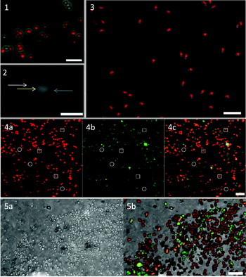
In this study, first we investigated the fluorescence activity of PMA-treated spores using epifluorescence microscopy. Fluorescence signals of different intensities were detected for almost all spores in a preparation and also for B. anthracis Sterne (Fig. 1). In order to exactly localize the signals, confocal laser scanning microscopy (CLSM) techniques were applied. Here, stained spores could be grouped into two categories: those with a stained outer layer only (‘doughnut-shape’) and others that had an additional and sometimes even brighter fluorescence signal in their core (particularly Figs. 2 and 3 and all other figures). The doughnut-shaped appearance was attributed to an unspecific binding of PMA to the outer layers of the spore, as already reported for DAPI, acridine orange and other dyes (Setlow et al. Reference Setlow, Loshon, Genest, Cowan, Setlow and Setlow2002; Magge et al. Reference Magge, Setlow, Cowan and Setlow2009). A possible autofluorescence of spores has been reported (LaFlamme et al. Reference LaFlamme, Verreault, Lavigne, Trudel, Ho and Duchaine2005), which could have explained this appearance also, but was not detected in this study (data not shown).
(Colour online) Epifluorescence microscopy of PMA-stained B. anthracis Sterne spores (red) merged with the phase contrast image (overlay). Scale bar: 10 μm.
Fig. 2. (Colour online) Confocal laser scanning micrograph of PMA-stained B. atrophaeus spore, 0.3 μm thick section; fluorescence in white. The white arrow points to stained sheath, the yellow arrow to the core (which is clearly stained) and the blue arrow to the unstained region, most probably the cortex. Scale bar: 1 μm.
Fig. 3. (Colour online) Confocal laser scanning micrograph of PMA-stained B. atrophaeus spores (ultracentrifuged). Overlay of the phase contrast image and PMA signal (red). PMA signal is concentrated in the spore core. Scale bar: 5 μm.
Fig. 4. (Colour online) Confocal laser scanning micrographs of B. safensis spores (a mixture of heat-inactivated and non-inactivated spores) on Scotch-Weld™-2116 B/A. Overlay of 13 optical sections (each section à 0.4 μm). White circles point at selected PMA-negative spores, white squares point at selected PMA-positive (dead) spores. (a) PMA signal (red); (b) Alf488 signal (green); (c) merged. Scale bar: 5 μm.
Fig. 5. (Colour online) Confocal laser scanning micrographs of B. safensis spores (a mixture of heat-inactivated and non-inactivated spores) on Scotch-Weld™-2116 B/A. Overlay of 27 optical sections (each section à 0.4 μm). (a) Phase contrast image; (b) overlay of phase contrast, PMA (red) and Alf488 (green) signal; scale bar: 5 μm.

When comparing an image of a PMA-stained spore to an ultrathin section of a Bacillus spore with common spore architecture (Driks Reference Driks1999), the conclusion was drawn that PMA had to penetrate the exosporium (if present), the (inner and outer) coat, the cortex and the (inner and outer) membrane to intercalate and bind DNA, resulting in this high fluorescence intensity in the spore core (Figs. 2 and 3); however, a damage to the inner membrane may be the crucial factor for PMA to enter the core, similar as already reported for DAPI (Setlow et al. Reference Setlow, Loshon, Genest, Cowan, Setlow and Setlow2002). The region between the core and the outer layer (likely the cortex) showed no fluorescence signal at all. Since all the spores analysed showed a weak, unspecific binding of PMA on the outer layers, a stained spore core detected via CLSM is referred to as a positively stained spore herein; this differentiation could be demonstrated for all bacterial strains studied in the course of this survey. Consequently, PMA was shown to be able to enter a spore with a compromised wall and bind to its DNA in the core, which was previously only assumed but not proven (Rawsthorne et al. Reference Rawsthorne, Dock and Jaykus2009). This fact is independent of the actual spore status – either alive and cultivable, superdormant or viable-but-non-culturable: all these spore viability statuses are characterized by an intact membrane as reported for superdormant spores which are capable of germination (Ghosh & Setlow Reference Ghosh and Setlow2009), and are therefore not stainable with PMA.
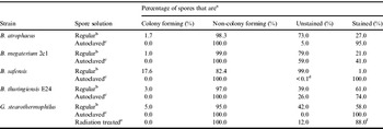
Comparing the number of non-colony-forming spores and those that were positively stained, no correlation could be found in regular preparations (Table 1). For instance, B. safensis spores showed a colony formation of 17.6%, although 99% were not stained.
Correlation of colony formation and PMA staining of the spore core. For each preparation 500 spores were enumerated and categorized as ‘stained’ or ‘unstained’ using CLSM (if not stated otherwise)

a Colony formation was determined via spread plating of appropriated dilutions and colony counting; ‘stained’ refers to PMA fluorescence signal detected in the core of the spore.
b Spore solution in pure water, purified according to Nicholson & Setlow (Reference Nicholson and Setlow1990).
c Spore solution in pure water, purified according to Nicholson & Setlow (Reference Nicholson and Setlow1990) and autoclaved for 40 min at 121°C.
d Six out of 500 spores unstained.
e Spore solution in pure water, purified according to Nicholson & Setlow (Reference Nicholson and Setlow1990) and γ-irradiated.
f Due to a large amount of cell debris, only 51 spores were enumerated.
The reasons for this observation can be manifold. For instance, one reason may be the low nutrient medium used for spore preparation as this can lead to the formation of superdormant spores. These spores require multiple triggers for germination and are therefore hardly detectable in standard spore cultivation assays (Ghosh & Setlow Reference Ghosh and Setlow2009). The same medium was used for the evaluation of CFU after purification, but a higher amount of nutrients could have led to higher yields. Besides nutrients, other triggers that stimulate and induce germination are ammonia or heat and could, therefore, also contribute to a better correlation of colony formation and PMA staining (Foerster & Foster Reference Foerster and Foster1966; Preston & Douthit Reference Preston and Douthit1984). Additionally, other factors such as hydrostatic pressure, sucrose, pH and L-alanine, and incubation temperature were reported to influence the germinability of Bacillus endospores (Smoot & Pierson Reference Smoot and Pierson1982; Blocher & Busta Reference Blocher and Busta1985; Raso et al. Reference Raso, Gongora-Nieto, Barbosa-Canovas and Swanson1998). For this study, the spores were heat-treated in the frame of the spore purification process, but the effect could have worn off until the determination of CFU; additionally, this heat step may not have been sufficient to activate all germinable spores of the thermophilic G. stearothermophilus strain. Experiments geared towards circumventing these limitations for our model organism B. safensis (using triggers such as heat shock or L-alanine and combinations thereof) did not result in an increase of CFU (data not shown) as other studies have also reported low amounts of germinable spores in regular preparations (Cook & Brown Reference Cook and Brown1964). Certainly, more studies are needed to optimize the germinability of the spores used in this work and to obtain a better correlation of PMA staining and CFU. However, it is very unlikely that in the case of B. safensis spores all unstained spores (99% in regular preparations) are also potentially germinable; other non-cortex-related damages to spores are plausible explanations for inhibition of germination and subsequent colony formation. Nevertheless, results gained from heat-inactivated spores showed a clear trend: for all the microbial strains analysed, the number of positively stained spores increased after inactivation and so did the percentage of non-colony-forming spores. However, the success of staining varied among the strains analysed. While B. megaterium 2c1 and B. thuringiensis showed only a slight increase of positively stained spores after heat inactivation, in B. safensis and G. stearothermophilus, almost all spores became positively stained. The differences in staining success can be attributed to different spore architectures, which have been shown to vary even within the genus Bacillus (Kondo & Foster Reference Kondo and Foster1967). To be able to bind to DNA, PMA has to enter the core of a spore and, therefore, pass through its different outer layers that may have not become completely permeabilized using the DTT treatment. In this work, DTT treatment was chosen in order to be able to follow a previous quantitative PCR-based approach and to allow a selective entry of PMA into the spore core (Rawsthorne et al. Reference Rawsthorne, Dock and Jaykus2009). However, different concentrations or incubation times with DTT or other chemicals (e.g. NaOH, SDS and combinations thereof) may be more suitable for spores with different architectures (Setlow et al. Reference Setlow, Loshon, Genest, Cowan, Setlow and Setlow2002). While B. safensis does not show any exosporium, B. anthracis (Liu et al. Reference Liu, Bergman, Thomason, Shallom, Hazen, Crossno, Rasko, Ravel, Read and Peterson2004) and B. thuringiensis (Bechtel & Bulla Reference Bechtel and Bulla1976) do, which may partially avoid the permeation of PMA into the spore core. In particular, the PMA staining success for B. megaterium was quite low (41%); its spores were, however, larger than others [0.8–1.2 μm×1.3–1.8 μm, determined using scanning electron microscopy by the method of Probst et al. (Reference Probst, Facius, Wirth, Wolf and Moissl-Eichinger2011)] and were reported to have various types of outer spore layers and up to two exosporia (Beaman et al. Reference Beaman, Pankratz and Gerhardt1972). Nevertheless, a P-value of 0.038 in the paired t-test across all strains indicates a significant change of positive PMA staining when comparing regular with heat-inactivated spore preparations. Furthermore, our results correlated with those published by Rawsthorn et al., although they used only B. subtilis for their tests.
Interestingly, G. stearothermophilus spores treated with a lethal dose of γ-radiation revealed also an increased PMA reaction (88%) compared with untreated spores (58%). A homoskedastic t-test including heat- and irradiation-inactivated spores compared with non-treated spores revealed then a P-value of 0.008, indicating a highly significant difference.
Most of the spores that are killed by wet heat undergo a critical damage resulting in sudden release of dipicolinic acid (DPA) (Coleman et al. Reference Coleman, Zhang, Li and Setlow2010). Nevertheless, since a small fraction of DPA-replete spores is still present after heat inactivation, the preliminary step in the killing process seems to be the denaturation of one or more (unknown) key proteins, which prevents successful outgrowth (Coleman et al. Reference Coleman, Zhang, Li and Setlow2010). In general, the killing process via wet heat does not affect the DNA structure, which is a considerable difference compared with radiation effects. But assuming a damage of the inner membrane being responsible for PMA to enter and stain the core, γ-irradiation must have caused a similar damage as heat treatment (damage of essential cell parts, including the membrane), although this necessitates further analysis.
In another experiment, B. atrophaeus spores were subjected to ultracentrifugation, with the result of a tremendous loss in viability (>99.9% non-colony-forming spores). Also, the number of PMA stained spores increased drastically (Fig. 3), demonstrating again a correlation of staining and inactivation of spores. However, the 336 236×g applied to the spores must have caused a mechanical damage to the spore wall (including the inner membrane), allowing PMA to enter and bind in the core.
In summary, the conclusion can be drawn that a positively stained spore is non-colony-forming and must be considered as dead. On the other hand, an unstained spore is not necessarily germinable. This differentiation must be considered when applying this method in future projects. Nevertheless, B. safensis was shown to be a good candidate for model experiments with respect to its behaviour before and after heat inactivation.
One special scientific field that necessitates differentiating between living and dead microbes is planetary protection, which involves the evaluation of cleanliness of spacecraft before sending them to a foreign celestial body. Spores are forms of microbial life that could possibly survive a space flight and are a significant contamination risk. Spores can be located on either the spacecraft surface, where they can be sampled quite easily using swabs or other devices (Probst et al. Reference Probst, Facius, Wirth and Moissl-Eichinger2010, Reference Probst, Facius, Wirth, Wolf and Moissl-Eichinger2011), but they could also be encapsulated and become even protected in spacecraft polymers (such as paint or adhesives). In the case of a hard landing on extraterrestrial targets, spores could be released and could compromise a mission's integrity.
In the literature, only two investigations have appeared so far that dealt with the detection of spores encapsulated in and released from a polymer (Mohapatra & La Duc Reference Mohapatra and La Duc2011), or the identification of microbes encapsulated in spacecraft material via cultivation (Vasin & Trofimov Reference Vasin and Trofimov1995). As already discussed above, evaluation of the number of germinable spores in a sample is complex and cannot be done simply by cultivation. Hence, we evaluated our novel staining protocol on a spacecraft polymer used frequently on spacecraft (Scotch-Weld™-2116 B/A) to demonstrate its possible application for encapsulated spore models. As spores on such polymer surfaces cannot be clearly visualized by phase contrast light microscopy, a counter-staining was necessary. Spores were stained with Alf488 before PMA staining and applied onto the material. By using CLSM techniques we were able to differentiate B. safensis intact and inactivated spores (Fig. 4) and also identify their localization on the polymer surface by including phase contrast images (Fig. 5). This is a crucial basis for further encapsulation model experiments: staining with Alf488 allows easy detection of spores on the surface and even in subsurface layers of polymers using CLSM. Additional PMA staining can then be used to investigate the intactness of surface-exposed and partially embedded spores. As we have shown here, B. safensis is suitable to serve as a model organism due to the clear staining reaction of its spores.
The novel microscopy method introduced here shows promise of becoming widely applicable in microbiology. Plate counting of CFU is the standard and well-accepted method since it is suitable also for samples with low biomass. Nevertheless, cultivation-based strategies are generally time-consuming (up to a few days incubation time) and biased by the above-mentioned aspects with respect to spore germination. In contrast, PMA staining and subsequent microscopic counting is certainly not suitable for low biomass, but can be performed reliably and rapidly (within a few hours). Consequently, PMA as the first fluorescence marker identifying non-viable spores and vegetative cells has the potential to become the method of choice to examine (spore) integrity in e.g. environmental samples of high biomass or cultures. Even more important, this PMA assay could allow a rapid evaluation of the efficiency of decontamination strategies (sterilization methods) and of spore resistance experiments (with respect to space flight), which are crucial for assessing inter-planetary contamination risk in terms of planetary protection. Besides applications in scientific microbiology, the PMA staining technique could find applicability in quality assessment procedures in pharmaceutical and general processing industry. Moreover, the possible combination of a microscopical inspection with a subsequent molecular analysis (Rawsthorne et al. Reference Rawsthorne, Dock and Jaykus2009) of the very same sample emphasizes another advantage of this innovative procedure.
Acknowledgements
The work carried out in this publication was supported by ESA contract no. AO/1-6079/09/NL/CP: ‘Evaluation of encapsulated bioburden on flight hardware’. We thank Dr Gerhard Kminek, Lobke Zuijderduijn, Professor Dr Reinhard Wirth, Mary Singer and Leonie Schaefer for critical discussions, support and review. Alexander Probst was supported by the German National Academic Foundation (Studienstiftung des deutschen Volkes). The authors are grateful to the critical reviewers who helped in improving the manuscript.