I. INTRODUCTION
Purification and recovery of recombinant proteins, after cell disruption, is achieved by techniques ranging from gel chromatography, ion exchange chromatography, and affinity chromatography to electrophoresis, precipitation, and membrane separation.Reference Langer and Ranck1, Reference Low, O’Leary and Pujar2 A versatile affinity chromatography approach for recombinant protein recovery is the immobilized metal ion affinity chromatography (IMAC), which provides a high recovery and selectivity to hexahistidine-tagged (His6-tagged) recombinant proteins. Another approach is the use of magnetic affinity adsorbents (MAAs) for their ability to retain the selectivity and recovery characteristics of IMAC with the additional advantage of being easily recovered using a magnetic field. These particles, however, are very expensive, making the cost-effectiveness a major concern in their large-scale commercial applications.
Several essential attributes are desired from inorganic magnetic core materials: (i) good response to the applied external magnetic field; (ii) small size (diameter) that provides large surface area for protein binding; (iii) reasonable cost; and (iv) ease of manufacturing.Reference Franzreb, Siemann-Herzberg, Hobley and Thomas3, Reference Horák, Babič, Macková and Beneš4 The ability of magnetic particles to aggregate rapidly in the presence of a magnetic field makes the utilization of nanoparticles ideal for separation. Furthermore, nickel nanoparticles (NNPs) are characterized by their large surface area and fast binding/adsorption kinetics in comparison to microparticles,Reference Franzreb, Siemann-Herzberg, Hobley and Thomas3 making them well suited for protein purification.
It has been well established that NNPs exhibit excellent magnetic behaviorReference Ai, Yao, Huang, Wang and Zhang5–Reference Zhang, Ding, Chow, Ran and Yi7 and nickel has been one of the most commonly used metal ions in IMAC.Reference Porath and Olin8, Reference Porath9 The polyol process in which metal salt precursors are reduced in a polyalcohol solvent has been used to produce a variety of elemental metal nanoparticles, which have applications in many fields such as in magnetic resonance imaging (MRI), catalysis or solar cells.Reference Carroll, Reveles, Shultz, Khanna and Carpenter10 In previous studies, it has been shown that a layer of nickel (II) oxide (NiO) was formed on the surface of the NNPs synthesized by the modified polyol method,Reference Couto, Klein, Schreiner, Mosca, de Oliveira and Zarbin6, Reference Baranova, Cally, Allagui, Ntais and Wüthrich11 probably by oxidization of Ni when the synthesized NNPs were exposed to air. In another study, Lee et al.Reference Lee, Lee, Park, Kim, Yi, Kim, Kim, Lee, Paik and Hyeon12 investigated the use of a complex type of nanoparticles that have a Ni core and a NiO shell for selective binding and magnetic separation of His-tagged green fluorescent protein (GFP). Furthermore, a magnetically recyclable protein separation system using three-layer magnetic nanocomposite spheres was investigated.Reference Kim, Piao, Lee, Park, Lee, Lee, Paik and Hyeon13 The magnetic nanocomposite spheres had a uniform hematite (α-Fe2O3) core, which was coated with a dense silica shell and then a porous silica shell. The porous silica shell was loaded with large amounts of NiO, which served as the binding sites for selectively purifying His-tagged GFP. The selective binding of His-tagged proteins to nickel oxide surfaces was also explored for bioactive protein nanoarrays.Reference Kim, Piao, Lee, Park, Lee, Lee, Paik and Hyeon13
In this research, we investigated the potential of NNPs synthesized from a modified polyol methodReference Ai, Yao, Huang, Wang and Zhang5, Reference Bai, Fan, Cao, Yuan, Zuo and Tang14 to serve as magnetic adsorbents for the purification a His6-tagged recombinant protein, ToxA5.1, a single-domain antibody (sdAb) neutralizing Clostridium difficile enterotoxin A (TcdA).Reference Hussack, Arbabi-Ghahroudi, Van Faassen, Songer, Ng, MacKenzie and Tanha15
II. MATERIALS AND METHODS
A. Synthesis of NNPs
NNPs were synthesized using a modified polyol methodReference Ai, Yao, Huang, Wang and Zhang5, Reference Bai, Fan, Cao, Yuan, Zuo and Tang14 by mixing 0.4 g nickel (II) hydroxide [Ni(OH)2] (Acros Organics, Fisher Scientific, Pittsburgh, PA), 160 mL ethylene glycol (EG; Fisher Scientific) and 0.1268 g of polyvinylpyrrolidone (PVP; Fisher Scientific) to get a 1:0.5 w/w ratio of Ni:PVP, at room temperature in a 1 L three-neck boiling flask for 30 min after which the solution was heated to 190 °C. Once the Ni(OH)2 in EG mixture reached 190 °C, a PdCl2 solution was quickly added to the flask. The PdCl2 (Sigma-Aldrich, St. Louis, MO) solution was prepared by mixing 0.0076 g palladium chloride (PdCl2) in 40 mL ethylene glycol (EG) in a beaker at room temperature for 45 min. Upon addition of the PdCl2 solution, the initial mixture of Ni(OH)2 in EG changed from a light green color to black within 15 min indicating the formation of metallic Ni. After 2 h of refluxing, the suspension was left to cool at room temperature and then the NNPs were separated from the spent EG solvent using a magnet. The recovered NNPs were thoroughly washed three times with 99% ethanol and separated using a magnet. The washed particles were then suspended in 10 mL of 99% ethanol and stored at room temperature.
B. Characterization of Ni nanoparticles
1. X-ray diffraction
The structure of Ni nanoparticles was determined by x-ray diffraction (XRD), using an Ultima IV powder XRD (Rigaku, The Woodlands, TX) with a copper source from 20 to 80° 2θ with a step of 0.02° 2θ.
2. Scanning electron microscopy
Scanning electron microscopy (SEM) was performed on the synthesized NNPs using a JSM-7500F FESEM (JEOL, Peabody, MA) instrument. To prepare the sample, 500 μL of particles suspended in ethanol was sonicated for 5 min in a Fisher Scientific FS140 ultrasonic cleaner (135 W and 42 kHz ± 6%) and a drop of the suspension was applied onto a carbon-coated copper grid. Average size and size distribution of the particles were calculated using ImageJ softwareReference Abràmoff, Magalhães and Ram16 based on the measurement of 260 particles distributed over two different areas of the sample (two different images).
3. Magnetic characterization
The magnetic measurements were obtained using a Quantum Design SQUID magnetometer MPMS-XL7 (Quantum Design, San Diego, CA) operating at 300 K for DC-applied fields ranging from −7 to 7 T. zero-field cooling (ZFC)/ field cooling (FC) analyses were performed on powdered samples of 17 mg of nanoparticles, wrapped in a polyethylene membrane.
4. Nickel quantification
Nickel (Ni) from NNPs was quantified using an iCE3300 Atomic absorption apparatus (Thermo Scientific, Waltham, MA) following appropriate sample dilutions. Quantification of the nickel was obtained by the average of five measurements of 4 s each at a wave length of 232 nm. Samples were prepared by pipetting 500 μL of NNPs suspended in ethanol into a beaker and subjecting them to a magnetic field to remove the ethanol. The NNPs were dissolved by adding sequentially 1 mL of concentrated hydrochloric acid (Fisher Scientific), 1 mL of concentrated nitric acid (Fisher Scientific) and 2 mL of deionized water. Upon dissolution, the solution was transferred to a 50 mL volumetric flask and the total solution volume was adjusted to 50 mL using deionized water.
C. Protein purification and regeneration of Ni nanoparticles
Escherichia coli TG1 cells bearing the plasmid for ToxA5.1 gene expression,Reference Hussack, Arbabi-Ghahroudi, Van Faassen, Songer, Ng, MacKenzie and Tanha15 were grown from a starting patch in sterile modified lysogeny broth (LB) media consisting of 20 g LB broth (Fisher Scientific), 6 g enzymatic Tryptone (Fluka, St. Louis, MO) and 5 g yeast extract (Oxoid, Lenexa, KS) for 1000 mL of distilled water overnight in a shaker at 37 °C and 200 rpm. Fresh sterile modified LB broth was inoculated with enough of the overnight culture to get an OD600 nm of 0.1 and grown at 37 °C until OD600 nm reached 0.6 at which point recombinant protein expression was induced using 2 mM isopropyl β-D-1-thiogalactopyranoside (IPTG; Promega, Madison, WI) at 37 °C for 18 h. Bacterial lysis was performed using Promega Fastbreak lysis system following the protocol provided by the manufacturer. Bacterial lysate was centrifuged at a relative centrifugal force (RCF) of 1150 for 10 min. The clarified lysate was used for the purification experiment.
For the purification experiments, 500 μL of NNPs suspension was added to a 2-mL microcentrifuge tube. The tube was inserted in a Promega magnetic holder for 1 min. The ethanol was removed and the NNPs were suspended in 300 μL of wash-bind buffer, which contained 100 mM 4-(2-hydroxyethyl)-1-piperazineethanesulfonic acid (HEPES) buffer and 10 mM imidazole (pH 7.5), by quick vortexing followed by sonication for 10 s to ensure dispersion prior to the binding step. The well-dispersed NNPs suspension is referred to as NNP slurry from now on. One milliliter of clarified bacterial lysate was added to the microcentrifuge tube containing the NNP slurry. To ensure proper binding, the tube was placed on a platform rocker (Boekel Scientific, Feasterville, PA) to mix for 2 min. The tube was then inserted in the magnetic holder for 1 min at which point the NNPs were separated from the bulk liquid by aggregation on the tube wall. The supernatant was then removed and kept for the analysis of the unbound fraction (UBF), which represents the proteins that did not bind to the NNPs. The tube was then removed from the magnetic holder and the NNPs were suspended in 300 μL of wash-bind buffer and mixed for 10 s. The tube was then inserted into the magnetic holder again to separate the NNPs from the supernatant, which was discarded. The washing step was repeated two more times. After the third wash, the NNPs were suspended in 100 μL of the elution buffer (100 mM HEPES and 500 mM imidazole, pH 7.5) to elute the adsorbed proteins. The suspension was sonicated for 10 s and put on a Boekel scientific rocking platform for 2 min. The tube was inserted into the magnetic stand for 1 min and the supernatant containing the recombinant proteins was removed and kept at 4 °C until analyzed. The NNPs were then washed three times using 1000 μL of wash-bind buffer each time. This completed the first purification with pristine NNPs. The same procedures were repeated three more times for a total of four binding cycles with the same NNPs and every time, the starting material was the clarified lysate obtained from the 18 h induced culture. An additional elution was performed on the NNPs after completing the fourth cycle by using elution buffer containing 1.0, 1.5, or 2.0 M imidazole.
For protein gel analysis, samples of appropriate dilutions were loaded on 4–15% discontinuous sodium dodecyl sulfate-polyacrylamide gel electrophoresis (SDS-PAGE) gels.Reference Laemmli17 Gels were run at 190 V for 45 min. Proteins were stained using Fermentas PageBlue Protein Staining Solution (Fermentas, Glen Burnie, MD) fast procedure as per manufacturer’s recommendations. Quantification of scanned gels was achieved via densitometry using ImageJ software.Reference Abràmoff, Magalhães and Ram16
To confirm that the purified protein was indeed the target protein and to assess the activity of purified ToxA5.1 via NNPs, an enzyme-linked immunosorbent assay (ELISA) was performed in comparison with a control. The control was the same ToxA5.1 but purified using commercial Novagen His·Bind column (EMD Millipore, Billerica, MA). Briefly, the column was equilibrated with 10 mL of 1x binding buffer (0.5 M NaCl Fisher Scientific, 20 mM Tris-HCl Fisher Scientific, 5 mM imidazole Alfa Aesar, Ward Hill, MA, pH 7.9) then 5 mL clarified cell lysate were loaded on the column. The column was then washed with 10 mL of binding buffer and 10 mL of wash buffer (0.5 M NaCl, 60 mM imidazole, 20 mM Tris-HCl, pH 7.9). ToxA5.1 was eluted using 5 mL of elute buffer (1 M imidazole, 0.5 M NaCl, 20 mM Tris-HCl, pH 7.9). ELISA was carried out as follows. First, a 96-well plate was coated overnight with C. difficile toxin A [2.5 μg/mL diluted in phosphate buffered saline (PBS), 100 μL/well, 4 °C]. The plate was then blocked with 1% casein (diluted in PBS, 200 μL/well) for 2 h at 37 °C. The primary antibodies (ToxA5.1, 100 μL/well) were serially diluted in PBS from a starting concentration of 0.5 μg/mL (31 nM), incubated at room temperature for 1 h, and then followed by 5 washes with PBS-Tween 20 (0.05%). Secondary antibody (rabbit anti-His6-HRP IgG, Cedarlane Laboratories, Burlington, ON) diluted 1:2500 in PBS was added at 100 μL/well, incubated for 1 h at room temperature, and then washed 5x with PBS-T. HRP substrate (KPL substrate kit Mandel Scientific, Guelph, ON) was added (100 μL/well) for 3 min. The reaction was stopped by the addition of 100 μL/well of 1 M phosphoric acid and plates were read at 450 nm wave length.
D. Calculations of binding capacities and ToxA5.1 recovery
The quantity of ToxA5.1 adsorbed per unit amount of NNPs (qn in mg/g) at the nth separation cycle was calculated using Eq. (1):
 $$<![CDATA[$$q_n {\rm{ }} = {\rm{ }}{{(V_{{\rm{broth}}} \times C_{{\rm{broth}}} ) - (V_{{\rm{UBF}}} \times C_{{\rm{UBF}}} ){\rm{ }}} \over {M_{\rm{p}} }} + q_{{\rm{residual}},{\rm{ }}\,n - 1} \quad,$$
where V broth is the volume of initial broth (L), C broth the concentration of ToxA5.1 in the initial broth (mg/L), V UBF the volume of the UBF (L), C UBF the concentration of ToxA5.1 in the UBF (mg/L), M p the mass of NNPs expressed as g of Ni.
The quantity of ToxA5.1 remaining on the NNPs after the elution (q residual in mg/g) of the nth cycle is calculated using Eq. (2):
 $$<![CDATA[$$q_{{\rm{residual}},\,{\rm{ }}n} = q_n - {{V_{{\rm{eluate}}} \times C_{{\rm{eluate}}} } \over {M_{\rm{p}} }}\quad,$$
where V eluate is the volume of the eluate fraction (L) and C eluate the concentration of ToxA5.1 in the eluate fraction (mg/L).
The protein percentage recovery is calculated using Eq. (3):
 $$<![CDATA[$${\rm{Recovery}} = {{V_{{\rm{eluate}}} \times C_{{\rm{eluate}}} } \over {V_{{\rm{broth}}} \times C_{{\rm{broth}}} }} \times 100\quad.$$
III. RESULTS AND DISCUSSION
A. NNP characteristics
1. XRD
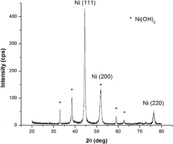
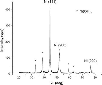
The structure of the resulting NNPs was confirmed by XRD as shown in Fig. 1. The diffraction lines of (1 1 1), (2 0 0), and (2 2 0) planes from the XRD profile confirm that NNPs with the face-centered cubic (fcc) structureReference Cullity18 were obtained. The XRD pattern also shows the presence of Ni(OH)2 phase with characteristic diffraction peaks at 33°, 38.5°, 52°, 59°, and 62.5° 2θ corresponding to (1 0 0), (1 0 1), (1 0 2), (1 1 0), and (1 1 1) crystalline planesReference Cullity18 indicating incompleteness of the reduction reaction in EG. The addition of PVP helped in slowing down the reduction of Ni(OH)2 to Ni by interacting with the nickel precursorReference Chou and Huang19 and controlling the size of the nanoparticles formed in the reaction which could explain the presence of unreacted Ni(OH)2. Using the full-width at half maximum (FWHM) of Ni (111) reflection and Scherrer formula,Reference West20 the crystallite size of Ni was calculated to be 24.3 nm.
XRD pattern of the NNPs (1% atomic Ni:Pd, 1:0.5 w/w Ni:PVP).
2. SEM
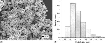
Figure 2 shows the SEM micrographs of the resulting NNPs and the corresponding histograms, respectively. It is seen that NNPs form large agglomerates consisting of small spherical-shaped particles. It should be further noted that the ability of NNPs to form agglomerates is beneficial for the intended application, i.e., purification of proteins from clarified or unclarified fermentation broth. As a matter of fact, it is much more difficult to separate the dispersed nanoparticles from the broth than to separate their larger counterparts.Reference Franzreb, Siemann-Herzberg, Hobley and Thomas3 The reagglomeration of some nanoparticles when mixing is stopped can greatly facilitate the separation of particles from the suspension and, as a result, be beneficial to large-scale application of the particles without a significant loss of binding surface area. From the SEM micrographs, the average NNP diameter was calculated to be 68 ± 28 nm; it was also found that 95.6% of the particles were in the range of 20–120 nm and 60.6% in the range of 40–80 nm. When comparing the measured diameter to the crystallite diameter calculated from the XRD data it can be said that the NNPs are constituted of several crystallites of 24.3 nm average size.
(a) SEM of NNPs with a magnification of 40,000 and (b) NNPs size distribution measured on 260 particles over two distinct areas of the sample. Average size of NNPs is 68 ± 28 nm.

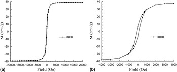
The magnetic behavior of the synthesized NNPs was measured using superconducting quantum interference device (SQUID) magnetometer. The NNPs were subjected to field-dependent magnetization at 300 K. Figure 3(a) shows that a magnetization saturation of 39.3 emu/g at 20,000 Oe was reached rapidly. Figure 3(b) shows the weak ferromagnetic behavior of the NNPs as indicated by the hysteresis loop between −1000 and 1000 Oe. This behavior is desired for a magnetic adsorbentReference Kim, Piao, Lee, Park, Lee, Lee, Paik and Hyeon13 as high magnetization values enable fast and easy removal from solution and the low coercivity enables rapid dispersion of the NNPs upon removal of the magnetic field. Magnetic properties of the NNPs were also confirmed by visually observing that it takes less than 5 s after applying a magnet to the suspension of NNPs contained in a 2 mL microcentrifuge tube to obtain a clear solution.
(a) Field-dependent measurements (SQUID) for the NNPs at 300 K from the field range of −20,000 to 20,000 Oe and (b) from −4,000 to 4,000 Oe to show the hysteresis loop.

B. Selective binding of the His-tagged ToxA5.1 by the NNPs
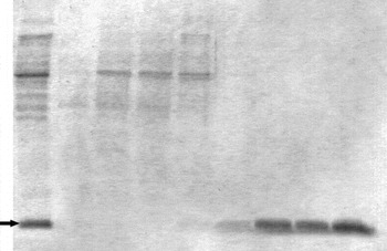
In this study, the synthesized NNPs were tested as MAAs for the purification of hexahistidine-tagged single-domain antibody (His6-tagged sdAb) ToxA5.1Reference Hussack, Arbabi-Ghahroudi, Van Faassen, Songer, Ng, MacKenzie and Tanha15 from the whole lysate of recombinant E. coli TG1 cells overexpressing the protein after clarification of the cell lystate by centrifugation. Protein purification experiments were carried out using the pristine particles (the 1st cycle) and recycled particles, i.e., the NNPs after elution and regeneration. For each purification cycle, the NNPs went through the following steps sequentially: (i) binding by mixing the NNPs with clarified cell lysate (CCL); (ii) three washes using wash-bind buffer; and (iii) elution using buffer containing 0.5 M imidazole. No special regeneration step was included in the experiments other than washing three times the used NNPs with 1000 μL of the wash-bind buffer. Samples of the CCL, UBF, and the eluate were all subjected to SDS-PAGE gel analysis. As shown on the electrophoresis gel in Fig. 4, lane 1 is the CCL sample and lanes 2–5 are the UBF samples from the 1st, 2nd, 3rd, and 4th purification cycles, respectively. Lanes 6–9 are samples of eluates from the first to the fourth purification cycles. It is clear that the band representing ToxA5.1 (∼16 kDa molecular weight) was absent from lanes 2 to 4 (the UBF samples of the 1st to 3rd purification cycle) and was very faint in lane 5 (the UBF sample of the fourth purification cycle), indicating that undetectable (lanes 2–4) or trace amount (lane 5) of ToxA5.1 were remaining in the UBF. Comparing the protein bands in lane 1 (CCL) to lanes 2 to 5 (UBF), it is clear that a lower number of protein appeared in the UBF samples than that in the CCL samples, indicating that some of the native proteins existing in the CCL were adsorbed on to the NNPs and therefore absent from the UBF. This observation could be explained by the binding of some native proteins with natural metal-binding motifs, e.g., proteins with histidine clusters or motifs rich in glutamic acid (Glu), aspartic acid (Asp), tyrosine (Tyr), cysteine (Cys), arginine (Arg), lysine (Lys), or methionine (Met)Reference Gaberc-Porekar and Menart21 on their surfaces or proteins that bind to heterologously expressed His-tagged proteinsReference Block, Maertens, Spriestersbach, Brinker, Kubicek, Fabis, Labahn and Schäfer22 which in our study is ToxA5.1. Nonspecific binding of native proteins to the NNPs by mechanisms other than affinity interaction between the His-tag and the nickel ions could also occur. Possible nonspecific binding mechanisms include weak physical forces such as van der Waals forces and the hydrophobic interaction between proteins and the PVP layer surrounding the NNPs.
SDS-PAGE analysis of successive purification cycles of ToxA5.1 sdAb. Lane 1: CCL arrow showing ToxA5.1 sdAb, Lanes 2–5:UBF of first, second, third, and fourth purification cycles using pristine or regenerated NNPs, Lanes 6–9: eluates from the first to the fourth purification cycles.

Figure 4 also shows that only ToxA5.1 was detected in lanes 6 to 9, indicating that the eluates contained undetectable level of contaminating proteins. This seems to highlight that the experimental washing procedure was sufficient to remove the loosely bound nonspecific proteins and therefore prevented them from contaminating the eluate. These results demonstrate that the protein recovery from the clarified whole cell lysate using NNPs was highly selective under the tested conditions.
It is worth mentioning that both the binding of ToxA5.1 onto the NNPs in the binding step and the elution of proteins from the NNPs occurred rapidly and equilibrium was established within 5 min in both cases (data not shown). The rapid binding and elution of NNPs can be explained by the fact that (i) the NNPs are very small with an average diameter of 68 nm and (ii) the binding was limited to the surface of the particles. On the contrary, in conventional adsorbents such as ion changers, most binding sites are located inside the pores of the particles, requiring slow intraporous diffusion for binding and elution.
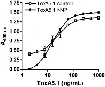
For confirming that the NNP-purified proteins were the targeted His6-tagged ToxA5.1 and that the antibody still had the ability to bind to the target toxin, ELISA was performed. As shown in Fig. 5 the ELISA binding data indicate that the purified proteins were indeed active ToxA5.1 as they bound to the C. difficile toxin A coated on the plate. Furthermore, the binding curve of the NNP-purified ToxA5.1 was very similar to the control, which was obtained with ToxA5.1 purified using commercial Novagen His·Bind column (EMD Millipore, Billerica, MA) following the manufacturer’s protocol. These results indicate that purification using NNPs did not degrade nor affect the folding or functionality of ToxA5.1. If the purification scheme had affected the integrity of ToxA5.1, the NNP-purified curve would have been shifted to the right, as a higher concentration of ToxA5.1 would have been needed to obtain the same absorbance reading as the control sample.
ELISA demonstrating NNP-purified ToxA5.1 retains its ability to bind C. difficile toxin A. Included in the ELISA was a control preparation of ToxA5.1 purified using a commercially available affinity resin.

The high selectivity displayed by the NNPs to His6-tagged antibodies and the clear separation of the His6-tagged antibody using the imidazole elutions indicate that specific metal chelation by the His-tag, which is analogous to IMAC, was probably at play on the surface or interstitial layers of the NNPs. It is worth mentioning that in a previous study,Reference Baranova, Cally, Allagui, Ntais and Wüthrich11 it was confirmed via surface characterization using Fourier transform infrared spectroscopy and x-ray photoelectron spectroscopy that the NNPs synthesized by the modified polyol method possess an external layer of NiO. It is hypothesized that the binding of the recombinant His6-tagged protein ToxA5.1 to the NNPs can be attributed to the interaction of an electron-donating group at the surface of the protein with metal, which is in this case the histidine tag and nickel, via coordination bonding such as what occurs in conventional IMAC.Reference Gaberc-Porekar and Menart21, Reference Chen, Liu, Lin and Ho23
The observation that the NNPs, which had a NiO surface layer, could selectively purify His-tagged ToxA5.1 is compatible with the results of Lee et al.,Reference Lee, Lee, Park, Kim, Yi, Kim, Kim, Lee, Paik and Hyeon12 who used a different and more complex approach to synthesize Ni/NiO core/shell nanoparticles. The capacity of NiO surfaces to selectively bind His-tagged proteins was also demonstrated by Kim et al.,Reference Kim, Piao, Lee, Park, Lee, Lee, Paik and Hyeon13 who used magnetic nanocomposite spheres decorated with NiO for selective purification of His-tagged GFP and by the work of Nam et al.,Reference Nam, Han, Lee, Liu, Ratner and Mirkin24 who developed bioreactive protein nanoarrays exploiting the selective binding of His-tagged proteins to nickel oxide surfaces formed by dip-pen nanolithography.
C. Reusability of the NNPs
As shown in lanes 2 to 5 of Fig. 4, the residual ToxA5.1 in UBF became noticeable only in the fourth purification cycle, indicating that the used NNPs recovered from previous cycles were able to capture almost all the ToxA5.1 in the following cycles and the loss of proteins to the UBF became observable only after three cycles. Given the fact that no optimized regeneration steps were involved in the adsorption/elution cycles, these results suggest that the NNPs are reusable. The development of an optimized regeneration step using specially designed regeneration buffer is underway, which is expected to further improve the reusability of the particles.
It is to be noted that the eluate from the first cycle using pristine NNPs as shown in lane 6 of Fig. 4 contained less target protein than the eluates from the subsequent purification cycles (i.e., the 2nd to 4th cycles), which are shown in lanes 7 to 9 of Fig. 4. The protein concentrations in eluates were 6 mg ToxA5.1/L for the first purification cycle and 18, 22, and 21 mg ToxA5.1/L for the 2nd, 3rd, and 4th purification cycles, corresponding to recoveries of 14, 41, 50, and 48%, respectively. As only undetectable levels of ToxA5.1 were left in the UBF after the binding step (lanes 2–5 in Fig. 4) and the washing supernatants (data not shown), it is reasonable to assume that most of the missing proteins remained on the NNPs, which were not eluted using the elution buffer containing 0.5 M imidazole. The observation implies that the development of a more efficient elution procedure (e.g., elution in a column rather than in a stirred container), or the combination of the two, is necessary for better elution efficiency. It should also be noted that both the adsorption and the elution were carried out in well-mixed test tubes. Therefore, results of the binding and elution steps reflect the distribution of protein on the adsorbents and the aqueous solutions when equilibrium was established in the corresponding conditions. Using packed or fluidized columns for elution, which is a common practice in ion exchange and affinity absorption, is also expected to increase the protein recovery and NNP reusability.
IV. CONCLUSION
In conclusion, we have demonstrated that NNPs synthesized using a straightforward modified polyol method could serve as a selective and effective magnetic adsorbent for the purification of His6-tagged recombinant proteins from CCLs. The results show that the NNPs had an average diameter of 68 nm, exhibited ferromagnetic behavior, were highly selective to His6-tagged proteins and were reusable without a significant loss in binding capacity. ELISA binding data also demonstrated that the purification scheme utilizing the NNPs did not cause any degradation, unfolding or loss of activity to the target protein ToxA5.1. Studies are underway to improve the efficiency and reusability of processes using such particles.
ACKNOWLEDGMENTS
The authors wish to thank Dr. Yun Liu for the SEM pictures, Dr. Tara Kell for the XRD scan, and Dr. Muralee Murugesu’s laboratory for the SQUID measurements. Financial support from the Natural Science and Engineering Council of Canada (NSERC) and Canadian Foundation of Innovation (CFI) is gratefully acknowledged.