INTRODUCTION
Giardiasis is an illness caused by the flagellate protozoan Giardia intestinalis. This parasite is the most frequent enteric parasite both humans and domestic and wild animals (Feng and Xiao, Reference Feng and Xiao2011) and the infectious organism most associated with waterborne outbreaks of diarrhoea at day care centres, and with illnesses in international travellers (Okhuysen, Reference Okhuysen2001). Giardia intestinalis can cause both acute and chronic diarrhoea (Newman et al. Reference Newman, Moore, Lima, Nataro, Guerrant and Sears2001). Giardia species are endemic in most areas with poor sanitation and giardiasis is a public health problem, especially in developing countries (Kappus et al. Reference Kappus, Lundgren, Juranek, Roberts and Spencer1994; Hollm-Delgado et al. Reference Hollm-Delgado, Gilman, Bern, Cabrera, Sterling, Black and Checkley2008). In 2004 giardiasis was included in the WHO neglected diseases initiative (WHO, 2004; Savioli et al. Reference Savioli, Smith and Thompson2006). The mode of transmission of this parasite is in the cyst form via contaminated food and water (Dawson, Reference Dawson2005; Daly et al. Reference Daly, Roy, Blaney, Manning, Hill, Xiao and Stull2010), and ingestion of as few as 10 Giardia cysts may be enough to cause infection (Yoder et al. Reference Yoder, Gargano, Wallace and Beach2012). Giardiasis can be asymptomatic or symptomatic (Prado et al. Reference Prado, Cairncross, Strina, Barreto, Oliveira-Assis and Rego2005; Nikolić et al. Reference Nikolić, Klun, Bobić, Ivović, Vujanić, Živković and Djurković-Djaković2011). Patients with symptomatic giardiasis have acute or chronic diarrhoea, anorexia, abdominal pain, intestinal malabsorption and weight loss. There may be other non intestinal manifestations as well, such as eye pain, arthralgia, allergic reactions and urticaria (Müller and von Allmen, Reference Müller and von Allmen2005; Buret, Reference Buret2007; Mørch et al. Reference Mørch, Hanevik, Rortveit, Wensaas, Eide, Hausken and Langeland2009). In Mexico a total of 10 650 new cases of giardiasis were reported in 2012 (Secretaría de Salud, 2013). Metronidazole (MTZ) is the drug most commonly used to treat giardiasis (Busatti et al. Reference Busatti, Santos and Gomes2009; Solaymani-Mohammadi et al. Reference Solaymani-Mohammadi, Genkinger, Loffredo and Singer2010). Although effective, MTZ has undesirable side effects such as nausea, stomachache, anorexia and a metallic taste in the mouth (Misra et al. Reference Misra, Kumar, Agarwal and Jagota1995). There are additional reports of adverse effects on the nervous system, including encephalopathy, peripheral neuropathy, headache, confusion, vertigo, depression, optic neuritis and blindness (Bradley et al. Reference Bradley, Karlsson and Rassol1977; Heaney et al. Reference Heaney, Campeau and Lindell2003; McGrath et al. Reference McGrath, Kent-Smith and Franzco2007). MTZ is mutagenic in laboratory animals (Bendesky et al. Reference Bendesky, Menéndez and Ostrosky-Wegman2002), treatment failure occurs in up to 20% of cases, in laboratory studies Giardia can readily acquire laboratory-induced resistance to MTZ and cross-resistance to other 5-nitroimidazole drugs (Upcroft and Upcroft, Reference Upcroft and Upcroft2001; Lopez-Velez et al. Reference Lopez-Velez, Batlle, Jiménez, Navarro, Norman and Perez-Molina2010; Tejman-Yarden et al. Reference Tejman-Yarden, Millman, Lauwaet, Davids, Gillin, Dunn, Upcroft, Miyamoto and Eckmann2011). More recently, the FDA approved a new thiazolideantiparasitic agent, nitazoxanide, for the treatment of diarrhoea caused by Cryptosporidium species and Giardia intestinalis. Nitazoxanide has eradication rates of over 70% for G. intestinalis in clinical studies (Romero et al. Reference Romero, Guerrero, Muñóz and Geyne1997; Abaza et al. Reference Abaza, El-Zayadi, Kabil and Rizk1998; Escobedo et al. Reference Escobedo, Alvarez, González, Almirall, Cañete, Cimerman, Ruiz and Pérez2008). There are alternative therapies for the treatment of giardiasis, but in many cases these are less effective than 5-nitroimidazoles (Gardner and Hill, Reference Gardner and Hill2001). Medicinal plants could be a source of new antiprotozoal drugs. Several plants used in Mexican traditional medicine, such as Lippia spp., Justicia spicigera, Geranium mexicanum, Dorstenia contrajerva, Senna villosa, Ruta chalepensis, Psidium guajava, Punica granatum, Mangifera indica, Plantago major, Castela tortuosa and Haematoxylum campechianum, show activity against Giardia (Ponce-Macotela et al. Reference Ponce-Macotela, Navarro, Martínez and Álvarez1994, Reference Ponce-Macotela, Rufino-González, de la Mora-de la Mora, González-Maciel, Reynoso-Robles and Martínez-Gordillo2001, Reference Ponce-Macotela, Rufino-González, González-Maciel, Reynoso-Robles and Martínez-Gordillo2006; Calzada et al. Reference Calzada, Cervantes-Martínez and Yépez-Mulia2005, Reference Calzada, Yépez-Mulia and Aguilar2006). Heterotheca inuloides (common name: Mexican arnica) is a plant that belongs to the Asteraceae family and is widely distributed in Mexico (Calderón and Rzedowski, Reference Calderón and Rzedowski2005; Martínez et al. Reference Martínez, Alvarado, Mendoza and Basurto2006; Semple, Reference Semple2008). An infusion of dried flowers has been used to treat contusions (Lozoya et al. Reference Lozoya, Aguilar and Camacho1987; Hersch-Martínez, Reference Hersch-Martínez1997), colic, muscular pain, skin inflammation and wounds (Andrade-Cetto, Reference Andrade-Cetto2009). For internal administration, the flowers are boiled in water and consumed as tea. For external administration, the plant is applied to unbroken skin as a cream or ointment (González-Stuart, Reference González-Stuart2010). Several types of compounds have been isolated from this plant, including cadinane-type sesquiterpenes, phenolic compounds and phytosterols. Extracts and some metabolites have demonstrated antimicrobial, antifungal (Kubo et al. Reference Kubo, Muroi, Kubo, Chaudhuri, Sánchez and Ogura1994; Castillo-Juárez et al. Reference Castillo-Juárez, González, Jaime-Aguilar, Martínez, Linares, Bye and Romero2008; Rosas-Piñón et al. Reference Rosas-Piñón, Mejía, Díaz-Ruiz, Aguilar, Sánchez-Nieto and Rivero-Cruz2012), anti-inflammatory (Segura et al. Reference Segura, Freixa, Ringbom, Vila, Perera, Adzet, Bohlin and Cañigueral2000; Delgado et al. Reference Delgado, Olivares, Chávez, Ramírez-Apan, Linares, Bye and Espinosa-García2001) and cytotoxic activity (Villarreal et al. Reference Villarreal, Alonso and Melesio1992; Kubo et al. Reference Kubo, Chaudhuri, Kubo, Sanchez, Ogura, Saito, Ishikawa and Haraguchi1996). The aim of this study was to evaluate the giardicidal activity of selected H. inuloides metabolites and some semisynthetic derivatives using an in vitro experimental model of giardiasis. We also used electron microscopy to investigate their effects on the morphology and ultrastructural organization of G. intestinalis.
MATERIALS AND METHODS
Giardia isolate (WB)
The reference strain WB (American Type Culture Collection 30957) was provided by Dr G. Ortega (Centro de Investigación y Estudios Avanzados, Mexico). This strain was originally isolated from 27-year-old white American patient with chronic diarrhoea infected in Afghanistan (Smith et al. Reference Smith, Gillin, Spira and Nash1982). First genome of Giardia WB was published in 2007 (Morrison et al. Reference Morrison, McArthur, Gillin, Aley, Adam, Olsen, Best, Cande, Chen, Cipriano, Davids, Dawson, Elmendorf, Hehl, Holder, Huse, Kim, Lasek-Nesselquist, Manning, Nigam, Nixon, Palm, Passamaneck, Prabhu, Reich, Reiner, Samuelson, Svard and Sogin2007) and recently, chromosome-wide maps were established by optical mapping (Perry et al. Reference Perry, Morrison and Adam2011). Trophozoites were grown in axenic culture at 37°C in TYIS-33 medium supplemented with 10% fetal bovine serum and bovine bile (Keister, Reference Keister1983). Log phase (48–72 h) cultures were harvested by cooling the culture vials at 4°C for 15 min and centrifuging them (5 min at 3500 rpm). The trophozoites were counted in a haemocytometer (Neubauer cell-counter chamber) and used in experiments.
Plant material
Dried flowers of H. inuloides Cass was provided by Mixim Laboratories (Mexico). Plant material was collected in 2010, in the town of Mesas Altas de San Juan Xoconusco, municipality of Donato Guerra (State of Mexico). The plant material was authenticated by MS AbigailAguilar-Contreras and a voucher was deposited in the Medicinal Plant Herbarium of the InstitutoMexicano del Seguro Social (IMSS, Mexico City), where the voucher specimen is conserved under reference code number IMSSM-16064.
Extraction and fractionation of constituents
Dried and powdered plant material (10 kg) was extracted with acetone at room temperature (3 times/24 h), to afford, after evaporation of the solvent, 450 g of residue. The residue was purified by vacuum liquid chromatography (VLC) (Coll and Bowden, Reference Coll and Bowden1986; Delgado et al. Reference Delgado, Olivares, Chávez, Ramírez-Apan, Linares, Bye and Espinosa-García2001) using hexane and mixtures of hexanes–EtOAc. The fractions (1 L each) were monitored by thin layer chromatography (TLC) and pooled into 9 (A–I) fractions according their similarity. Fraction A (86 g, eluted with hexanes–EtOAc, 9·5:0·5) was rechromatographed by VLC, and some fractions were further chromatographed on Si gel to afford 2·682 g of 7-hydroxy-3,4-dihydrocadalene (1) mp 100–103°C [lit. 103·5°C] (Bohlmann and Zdero, Reference Bohlmann and Zdero1976); Rf 0·5 (n-hexanes–EtOAc, 8:2)], and 520 mg of 7-hydroxycadalene (2) mp 117–118°C [lit. 118–119°C], (Gallagher and Sutherland, Reference Gallagher and Sutherland1965); Rf 0·43 (hexanes–EtOAc, 8:2). Fraction D (9 g) was purified by successive VLC on silica gel (hexanes–EtOAc gradient) to afford 3,7-Dihydroxy-3(4H)-isocadalen-4-one (3) (205 mg) mp 200–202°C [lit. 198–200°C] (Delgado et al. Reference Delgado, Olivares, Chávez, Ramírez-Apan, Linares, Bye and Espinosa-García2001); Rf 0·32 (n-hexanes–EtOAc, 7:3) and 24·5 mg of eriodictyol-7,4′-dimethyl ether (8), mp 168–170°C; Rf 0·45 (n-hexanes–EtOAc, 6:4). The melting point of this compound agrees with data from literature [165–168°C] (Vasconcelos et al. Reference Vasconcelos, Silva and Cavaleiro1998). Fraction E was chromatographed using VLC with mixtures of hexanes–EtOAc to afford 15 mg of quercetin-3,7,3′,4′-tetramethyl ether (7) mp 150–152°C [lit. 148–150°C] (de la Torre et al. Reference de la Torre, Rodrigues, Tomé, Silva and Cavaleiro2004); Rf 0·29 (n-hexanes–EtOAc, 6:4). Fraction F (17·5 g, eluted with n-hexanes–EtOAc, 8:2) was chromatographed using VLC with mixtures of hexanes–EtOAc to afford 48·5 mg of (1R,4R)-1-hydroxy-4H-1,2,3,4-tetrahydrocadalen-15-oic acid (4), mp 198–200°C [lit. 203–205°C] (Delgado et al. Reference Delgado, Olivares, Chávez, Ramírez-Apan, Linares, Bye and Espinosa-García2001); Rf 0·29 (n-hexanes–EtOAc, 6:4), and 17 mg of quercetin-3,7,3′-trimethyl ether (6), mp 170–172°C [lit. 168–170°C] (Ali et al. Reference Ali, Chowdhury, Rahman, Borkowski, Nahar and Sarker2008); Rf 0·30 (n-hexanes–EtOAc, 6:4). Fraction G (127·5 mg, eluted with hexanes–EtOAc, 6·5:3·5) was rechromatographed using VLC with mixtures of hexanes–EtOAc to afford 3·110 g of quercetin (5), mp greater than 300°C [lit. 309–311°C] (Dutta et al. Reference Dutta, Mazumdar, Mishra, Dastidar and Park2007); Rf 0·68 (Isopropyl alcohol–water, 3:2). The identification of the isolated compounds was determined by comparison of the spectroscopic and spectrometric data with those published previously and confirmed by direct comparison with authentic samples from our laboratory (Delgado et al. Reference Delgado, Olivares, Chávez, Ramírez-Apan, Linares, Bye and Espinosa-García2001).
Esterification
Compounds 7-acetoxy-3,4-dihydrocadalene (9), 7-acetoxycadalene (11) and quercetin pentaacetate (13) (Picq et al. Reference Picq, Prigent, Némoz, André and Pacheco1982) were obtained by treating the natural products 1, 2 and 5, respectively, with acetic anhydride and pyridine at room temperature. Compounds 7-benzoxy-3,4-dihydrocadalene (10) and 7-benzoxycadalene (12) were obtained by treating 1 and 2 with benzoyl chloride and pyridine. Product formation was monitored by TLC using n-hexane–EtOAc (8·5:1·5) as elution solvent system. The reactions were stopped by adding ice. After 1 h of stirring the reaction mixture was extracted 3 times with EtOAc. The combined organic phases were treated first with 10% HCl, then with a saturated solution of NaHCO3, and finally with saturated NaCl. The organic layer was dried with anhydrous sodium sulphate and concentrated under vacuum. The resulting residue was chromatographed on a silica gel column using a n-hexane–EtOAc gradient elution system to afford derivatives 9, 10, 11, 12 and 13 in 86, 82, 83, 80 and 81% yield, respectively. 9–12 are novel compounds that have not been reported previously in the literature.
7-acetoxy-3,4-dihydrocadalene (9): oily substance; [α]25 D-44·4 (c 0·1, MeOH); IR (KBr) ν max 3552, 2962, 2931, 2862, 1720, 1600, 1580, 1491, 1463, 1381, 1290, 1233, 1222, 1216, 1128, 1074, 1037, 956, 918 cm−1; 1H and 13C NMR see Table 1, EIMS m/z 258 [M]+ (9), 215 (10), 214 (5), 199 (5), 186 (4), 174 (16), 173 (100), 172 (9), 158 (14), 155 (5), 128 (5), 115 (3). HRFABMS m/z 258·1621 [M]+ (calcd for C17H22O2, 258·1620). 7-benzoxy-3,4-dihydrocadalene (10): oily substance; [α]25 D-28 (c 0·1, MeOH); IR (KBr) ν max 3030, 2960, 2928, 2871, 1732, 1602, 1567, 1495, 1452, 1382, 1261, 1175, 1130, 1084, 1066, 1026, 900 cm−1; 1H and 13C NMR see Table 1, EIMS m/z 320 [M]+ (5), 277 (7), 175 (3), 106 (6), 77 (13). HRFABMS m/z 320·1770 [M]+ (calcd for C22H24O2, 320·1776). 7-acetoxycadalene (11): colourless needles (EtOAc/hexane), mp 68–70 °C; IR (KBr) ν max 3073, 3037, 3021, 2966, 2929, 2869, 1750, 1464, 1441, 1236, 1149, 1126, 1041, 1017, 914 cm−1; 1H and 13C NMR see in Table 1, EIMS m/z 256 [M]+ (20), 215 (15), 214 (55), 184 (13), 169 (6), 155 (7), 153 (6), 128 (5). HRFABMS m/z 256·1466 [M]+ (calcd for C17H20O2, 258·1463). 7-benzoxycadalene (12): colourless needles (EtOAc/hexane), mp 138–140°C; IR (KBr) ν max 2962, 2924, 2862, 1734, 1599, 1508, 1447, 1381, 1317, 1266, 1241, 1149, 1062, 1024, 714 cm−1; 1H and 13C NMR see Table 1, EIMS m/z 318 [M]+ (35), 303 (3), 170 (9), 128 (3), 155 (9), 141 (100), 128 (3), 77 (25). HRFABMS m/z 318·1628 [M]+ (calcd for C22H22O2, 318·1620).
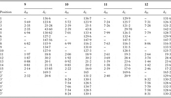
1H (400 MHz) and 13C NMR (100 MHz) spectral data for compounds 9, 10, 11 and 12 (CDCl3, δ)

Hydrogenation
Compound 1, which is (4R)-7-hydroxy-3,4-dihydrocadalene (36 mg, 0·163 mmol), was dissolved in 6 mL of ethyl acetate in a 50 mL flask. The flask was sealed with a septum and the air was replaced with argon. The catalyst (10% Pd/C, 4 mg) was added, and the flask was placed in the hydrogenation equipment. The system was purged with H2 to replace the argon. The mixture was stirred at 25°C for 2 h and the reaction progress was monitored by TLC using n-hexane–EtOAc (8·5:1·5). The reaction mixture was purified using a silica gel chromatography column, using a n-hexane–EtOAc gradient to afford compound 14 in 93% yield, whose enantiomer was previously reported (Pereira et al. Reference Pereira, Chaves, Pinto, Leitão and Bizzo2011).
Bioassays
The in vitro assays were performed as follows. All compounds were dissolved in DMSO. Serial dilutions (100, 50, 25, 12·5, 6·25 and 3·125 μg mL−1) were prepared in TYIS-33 medium in 96-well plates, and 1×10⁵ G. intestinalis trophozoites were added for a final volume of 300 μL (0·4% v/v DMSO) of final volume. Wells without compounds (0·4% DMSO alone) served as negative controls. In addition, the susceptibility of trophozoites to MTZ (40, 20, 10, 5, 2·5 and 1·25, 0·6125, 0·312, 0·156 μg mL−1) also was quantified. Trophozoites were incubated for 24 h at 37°C. After the incubation period, the culture medium was removed by inverting the plate and shaking it gently and the trophozoites were fixed with methanol and stained with 0·1% methylene blue for 10 min. The plate was washed and incubated for 20 min with HCl 0·1M, the dye removed and the absorbance was determined at 655 nm (Hounkong et al. Reference Hounkong, Sawangjaroen and Phongpaichit2011). Growth inhibition was calculated as a percentage by comparison with negative controls (Rufino-González et al. Reference Rufino-González, Ponce-Macotela, González-Maciel, Reynoso-Robles, Jiménez-Estrada, Sánchez-Contreras and Martínez-Gordillo2012). Each experiment was performed six times.
Transmission electron microscopy
Giardia trophozoites treated with compound 1 (7-hydroxy-3,4-dihydrocadalene at 50 μg mL−1) and negative control trophozoites (no compounds) were fixed in 3% glutaraldehyde buffered with phosphate buffer saline (0·1 M, pH 7·2), post-fixed in osmium tetroxide (1%) and dehydrated in an ethanol series. The samples placed in propylene oxide and embedded in EPON resin, were incubated at 60°C for 48 h. Thin sections (60–90 nm) were cut with an ultramicrotome (Reichert-JungLeica, GmbH, Heidelberg, Germany), mounted on Formvar-coated copper grids and counter stained with uranyl nitrate and lead citrate. For each sample, structural changes were examined in 50 fields using a Jeol 1200EXII transmission electron microscope (Jeol Ltd, Tokyo, Japan).
Statistics
Percentage of dead trophozoites were plotted against concentration. The curves obtained were analyzed by Origin 8 software (OriginLab Co., Northampton, Massachusetts). The 50% (IC50) and 90% (IC90) were determined by graphic extrapolation of the concentration–response curves using a nonlinear regression analysis. Standard error of the mean were calculated from data of six experiments.
RESULTS
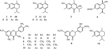
Compounds 1–8 were isolated from H. inuloides flowers. The identities of the natural compounds were confirmed by comparing their spectroscopic data with those published in the literature and by direct comparison with authentic samples previously isolated in our laboratory (Delgado et al. Reference Delgado, Olivares, Chávez, Ramírez-Apan, Linares, Bye and Espinosa-García2001). Four novel compounds were synthesized by conventional esterification procedures: 7-acetoxy-3,4-dihydrocadalene (9), 7-benzoxy-3,4-dihydrocadalene (10), 7-acetoxycadalene (11) and 7-benzoxycadalene (12). The known compounds quercetin pentaacetate (13) and 7-hydroxycalamenene (14) (Fig. 1) were obtained from 5 and 1, respectively, by conventional chemical methods. Their structures were determined by 1H and 13C NMR, HRMS, IR and UV spectroscopic analyses. The 1H and 13C NMR data for 9–12 were consistent with the expected changes due to esterification and are shown in Table 1. In particular, the 1H NMR signals of the new acetyl groups (in 9 and 11) were evident by the appearance of the signals at 2·32 and 2·40 ppm, respectively, corresponding to the methyl protons, and at 20·8 and 20·9 ppm for methyl carbons in 13C NMR experiments. For compounds 10 and 12 the signals of the carbonyl group appeared at 165·0 and 165·2 ppm, respectively, in the 13C experiments, and the appearance of the corresponding aromatic signals were in agreement with the structures.
Natural products isolated from H. inuloides and semisynthetic derivatives that were assayed for activity against Giardia intestinalis.

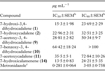
A colorimetric assay was used to evaluate the in vitro anti-giardial activity of compounds isolated from H. inuloides flowers as well as the activity of synthesized compounds. Cadinane-type compounds, but not flavonoid-type compounds, showed activity against Giardia trophozoites. Compounds 7-hydroxy-3,4-dihydrocadalene (1) and 7-hydroxycalamenene (14) displayed the best antigiardial activity at IC50 and IC90 (Table 2). Compounds 7-hydroxycadalene (2), 7-acetoxy-3,4-dihydrocadalene (9), 7-acetoxycadalene (11), and 7-benzoxycadalene (12) only achieved similar activity levels at concentrations two to four times the of compounds 1 and 14. Compounds 3,7-dihydroxy-3(4H)-isocadalen-4-one (3), and (1R,4R)-1-hydroxy-1,2,3,4-tetrahydrocadalen-15-oic acid (4) showed no significant antigiardial activity (Table 2).
Antigiardial activity of H. inuloides metabolites and derivatives a

a Results are expressed as mean (n=6).
b Standard error of the mean.
c Positive control.
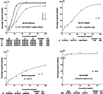
Nonlinear regression of concentration–response curves showing different behaviours were described by four equations (Fig. 2). Concentration–response curves for compounds 1, 2, 9 and 14 showed a sigmoid pattern described by the Boltzmann equation (Fig. 2a), and particularly, 1 and 14 produced around 90% death in trophozoites at 25 μg mL−1. Compounds 10 and 11 showed Slogistic and asymptotic curves, respectively (Fig. 2b and c). The concentration–response curve obtained by the inhibitory effect of MTZ upon Giardia tophozoites displayed a rapid increase in mortality of trophozoites at low concentrations (Fig. 2d). The behaviour of MTZ was consistent with previous reports in the literature (Busatti and Gomes, Reference Busatti and Gomes2007).
Effect of H. inuloides metabolites and drug reference (MTZ) concentration upon the growth of trophozoites of G. intestinalis. The equations describe the best fit curves obtained by nonlinear regression.

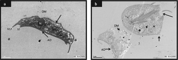
After 24 h in the in vitro activity assay, untreated trophozoites showed morphology that was normal for Giardia, as shown in Fig. 3a; undamaged cytoplasmic membranes, adhesive discs, a nucleus and flagellar axonemes. Compounds 1 (7-hydroxy-3,4-dihydrocadalene) and 14 (7-hydroxycalamenene) had anti-Giardia activity in vitro; consistent with this there were alterations in the cell membrane that caused irreversible damage to the cellular structure. Trophozoites exposed to 50 μg mL−1 (230 μM) of 7-hydroxy-3,4-dihydrocadalene (1) showed increased volume, vacuoles in the cytoplasm, dense material accumulated around the edges of the nuclei, adhesive disc fragmentation and lateral membrane disruption (Fig. 3b).
Transmission electron microscopy of G. intestinalis trophozoite in (a) the negative control group (no treatment). A coronal view of a trophozoite shows the integrity of the adhesive disc (AD), dorsal membrane (DM), nucleus (N) and flagellar axonemes (FA). The ventrolateral edge (VLE) and lateral crest (LC) are shown for the ventral disc; (b) exposed to 50 μg mL−1 of compound 1. The trophozoite showed structural changes, an increase in volume, dorsal membrane destruction (DM) and adhesive disc fragmentation (AD). The ventral region of the trophozoite has a large vacuole near the nucleus (*). Rupture of the cytoplasmic membrane (short arrow) and loss of cytoplasmic material in the caudal region.

DISCUSSION
Giardiasis is currently treated mostly with nitroimidazoles, primarily MTZ and other drugs such as albendazole, nitazoxanide, paromomycin and nitrofurans (Gardner and Hill, Reference Gardner and Hill2001; Müller and von Allmen, Reference Müller and von Allmen2005). However, many of these drugs have side effects and non-negligible failure rates (Gardner and Hill, Reference Gardner and Hill2001; Müller and von Allmen, Reference Müller and von Allmen2005). The failure rates reflect the need for more alternatives for treating giardiasis. Several experimental studies have investigated the effects of medicinal plants used to treat the gastrointestinal disorders against Giardia trophozoites. These studies have evaluated more than 100 natural products from diverse plant species. Such products fall into the following main chemical groups: flavonoids, triterpenes, alkaloids and sesquiterpenes. Other compounds have been tested, but to a lesser extent (Amaral et al. Reference Amaral, Ribeiro, Barbosa-Filho, Reis, Nascimento and Macedo2006). Several Mexican plants have been evaluated as remedies for Giardia (Calzada et al. Reference Calzada, Meckes, Cedillo-Rivera, Tapia-Contreras and Mata1998, Reference Calzada, Yépez-Mulia and Aguilar2006; Arrieta et al. Reference Arrieta, Reyes, Calzada, Cedillo-Rivera and Navarrete2001; Peraza-Sánchez et al. Reference Peraza-Sánchez, Poot-Kantún, Torres-Tapia, May-Pat, Simá-Polanco and Cedillo-Rivera2005; Barbosa et al. Reference Barbosa, Calzada and Campos2007).
Several methods can be used to evaluate the bio-activity of isolated compounds including: cell morphology assays, trophozoite adhesion, radiolabelling and direct counting. These are laborious and time consuming, and also depend on the observer's experience. We evaluated the giardicidal activity of H. inuloides metabolites and some derivatives in an experimental in vitro assay involving a methylene blue-based colorimetric method. This method is convenient and reliable and has been used previously to assess the antigiardial activity of drugs (Busatti et al. Reference Busatti, Santos and Gomes2009; Hounkong et al. Reference Hounkong, Sawangjaroen and Phongpaichit2011). The results showed that cadinane-type compounds had activity against Giardia. In particular, compounds 7-hydroxy-3,4-dihydrocadalene (1) and 7-hydroxycalamenene (14) significantly reduced the survival of the G. intestinalis trophozoites in vitro. Although several compounds isolated from H. inuloides have anti-inflammatory activity (Delgado et al. Reference Delgado, Olivares, Chávez, Ramírez-Apan, Linares, Bye and Espinosa-García2001) and it has been found that the 7-hydroxycalamenene shows activity against Leishmania chagasi promastigotes with a minimum inhibitory concentration of 15·6 μg mL−1 (Rodrigues et al. Reference Rodrigues, Azevedo, Chaves, Bizzo, Corte-Real, Alviano, Alviano, Rosa and Vermelho2013), this is the first report that 1 and 14 can act as antigiardial agents.
Modification of the molecular structure of the compounds changed their in vitro activity. Specifically the aromaticity of ring B decreased the activity of compound 2 and its derivatives with respect to compound 1. Esterification of the hydroxyl residue at position 7 in compounds 1 and 2 decreased their antigiardial activity. Hydrogenation product 14 showed antigiardial activity as strong as that of compound 1. Acetylated derivatives showed slightly better activity than benzoylated ones, possibly due to steric hindrance in the latter.
Previous studies reported that some flavonoids inhibit the growth of trophozoites of Entamoeba histolytica and Giardia lamblia (Calzada et al. Reference Calzada, Meckes, Cedillo-Rivera, Tapia-Contreras and Mata1998). Another study observed that kaempferol had a strong activity; to a lesser extent, so did quercetin flavonoids isolated from Cuphea pinetorum, plant used by the Tzotzil and Tzetzal communities in Mexico as antidiarrhoeals and to treat dysentery, and they show activity against Giardia (Calzada Reference Calzada2005). The antiprotozoal activity of flavonol glycosides isolated from Helianthemum glomeratum showed that tiliroside was the most potent antiamoebic and antigiardial compound (Calzada and Alanís, Reference Calzada and Alanís2007). However, the antiprotozoal activity of flavonoids was moderate in an evaluation of flavonoids isolated from Mimosa tenuiflora (Bautista et al. Reference Bautista, Calzada, Ortega and Yépez-Mulia2011). In the present study the tested flavonoids did not show significant antigiardial activity.
This is the first study to evaluate the activity of the natural products and derivatives of H. inuloides against Giardia trophozoites. The transmission electron microscopy results revealed that trophozoites of G. intestinalis treated with H. inuloides metabolites showed marked changes in cellular architecture accompanied by ultrastructural disorganization (Fig. 3b). G iardia intestinalis is a protozoan with a two-stage life cycle, i.e. the trophozoite and cyst stages, the trophozoite is the cause of giardiasis in patients and asymptomatic carriers. This protozoan divides asexually; making it important in the in vitro assays that the population is completely killed, eliminating the possibility that the organism population will rebound after a short time.
The present study evaluated the anti-giardial activity of H. inuloides flower constituents and of some semisynthetic derivatives. It demonstrated the in vitro antigiardial efficacies of 7-hydroxy-3,4-dihydrocadalene and 7-hydroxycalamenene. However, the mechanism of action involved in the lysis and death of Giardia cells is not yet known, therefore, complementary studies are needed to clarify the mechanism of action of the compounds.
ACKNOWLEDGEMENTS
We thank Chem. J. Ebrard (LaboratoriosMixim) for providing the plant material and Dr G. Ortega (Centro de Investigación y Estudios Avanzados, Instituto Politécnico Nacional) for providing the Giardia intestinalis isolate (WB). We also thank M. I. Chávez, B. Quiroz, R. Patiño, R. Gaviño, L. Velasco, J. Pérez, A. Peña, E. Huerta, Instituto de Química, UNAM for technical assistance.
FINANCIAL SUPPORT
J.L.R.-Ch. thanks CONACyT (México) and Programa de Doctorado en Ciencias Biomédicas (UNAM) for scholarship for doctoral studies. We acknowledge UNAM-DGAPA-PAPIIT (Project IG200514) and CONACyT (Project 102158) for support.