Significant outcomes
-
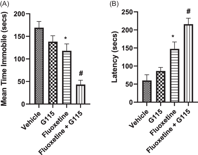
Chronic (14-day) treatment with G115® significantly potentiated fluoxetine-induced reductions in immobility time and increased latency to immobility in the FST.
-
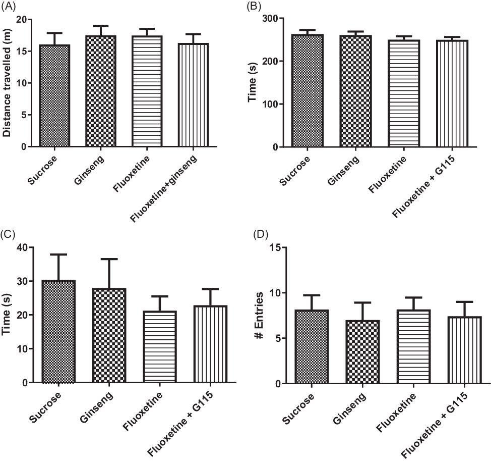
No significant effects on general locomotor activation or anxiety behaviours were observed.
-
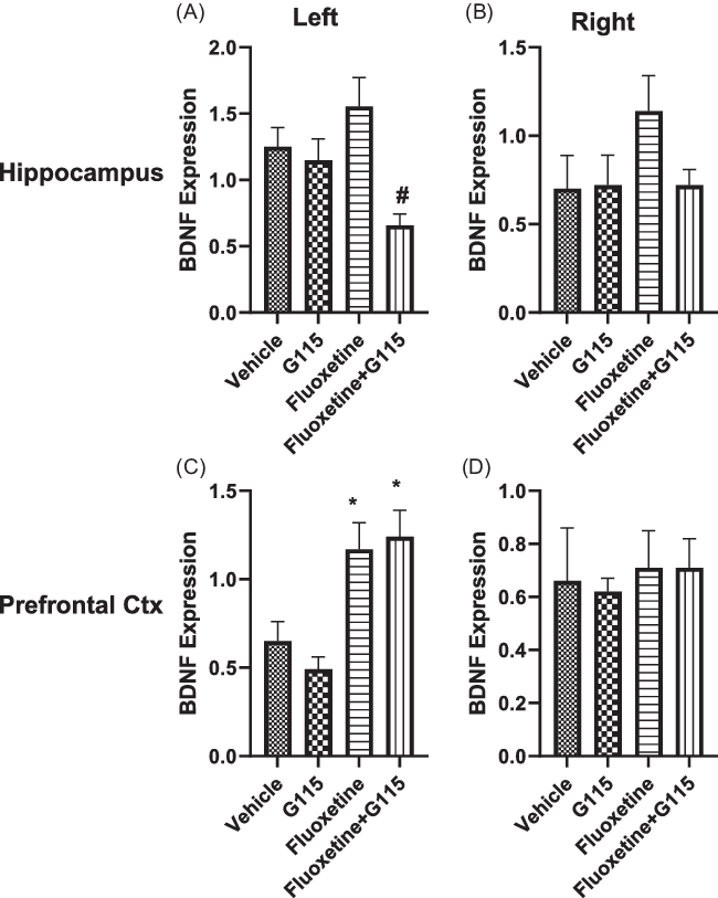
Significant changes in hippocampal and prefrontal cortex BDNF but not TrkB were found.
Limitations
-
Only male Sprague-Dawley (SD) rats were used. Effects of sex and strain are unknown.
-
Effects of varying doses or duration of treatment with G115® and fluoxetine are unknown.
-
The drug combinations were not tested in an established rat model of depression.
Introduction
Depression is one of the most common mental health problems in today’s society, affecting over 320 million people worldwide (WHO, 2017), which represents a global prevalence of 4.7% (Ferrari et al., Reference Ferrari, Somerville, Baxter, Norman, Patten, Vos and Whiteford2013) although this varies with age (Costello et al., Reference Costello, Erkanli and Angold2006) and gender (Schuch et al., Reference Schuch, Roest, Nolen, Penninx and de Jonge2013). Although there are many antidepressant therapies currently being prescribed to alleviate symptoms associated with depression, as many as 30–50% of patients suffering from depression fail to show a substantial clinical response to these conventional treatments including the widely prescribed selective serotonin reuptake inhibitors (SSRIs) (Mrazek et al., Reference Mrazek, Hornberger, Altar and Degtiar2014; Kohler et al., Reference Kohler, Cierpinsky, Kronenberg and Adli2015). In fact, a 2016 meta-analysis of 34 clinical trials in children and adolescents concluded that only the SSRI fluoxetine was significantly more effective than placebo in these age groups (Cipriani et al., Reference Cipriani, Zhou, Del Giovane, Hetrick, Qin, Whittington, Coghill, Zhang, Hazell, Leucht, Cuijpers, Pu, Cohen, Ravindran, Liu, Michael, Yang, Liu and Xie2016). This lack of treatment efficacy demonstrates a significant gap in our understanding of depression and the neurochemical systems involved, as evidenced by the multiple current theories of depression aetiology.
Panax ginseng has been widely used in traditional Asian medicine to alleviate a variety of ailments, including many symptoms commonly associated with depression (for review, see Jin et al., Reference Jin, Cui, Zhao, Fan and Li2019). This traditional use of ginseng typically uses all parts of the ginseng plant (roots, stems and leaves) in certain formulations; however, most modern studies involving ginseng tend to utilise mainly the ginseng root. Ginseng root is believed to be the source of the plant’s antidepressant properties, and contains a number of biologically active ingredients, including over 60 ginsenosides and a number of polysaccharides, fatty acids, oligopeptides and polyacetylenic acids (Qi et al., Reference Qi, Ignatova, Luo, Liang, Jun, Wang and Sutherland2010). With so many possible combinations of biologically active ingredients potentially contributing to the reported antidepressant effects, most studies have chosen to focus on compounds or formulations that have shown successful antidepressant outcomes in the past (see Lee et al., Reference Lee, Bahk, Lee and Pae2020), including the purified ginsenosides Rg1 (Jiang et al., Reference Jiang, Xiong, Yang, Wang, Wang, Hu, Wang and Chen2012), Rg3 (Cui et al., Reference Cui, Jiang and Xiang2011) and Rb1 (Yamada et al., Reference Yamada, Araki and Yoshimura2011). Rg1 and Rb1, along with several others (i.e. Rb2, Rc, Rd and Re), can be found in the P. ginseng C.A. Meyer extract G115® (Cui, Reference Cui1995), which is marketed in a number of jurisdictions and widely consumed. Given the antidepressant effects associated with several of its constituents, it can be hypothesised that G115® would also show a reduction in depressive-like symptoms, such as behavioural despair.
Behavioural despair (also referred to as learned helplessness) is a measure by which depression is commonly scored in rodent models using the forced swim test (FST) (Porsolt et al., Reference Porsolt, Anton, Blavet and Jalfre1978). This model, which examines the amount of time that the rodent spends immobile when exposed to an inescapable stressor (a column of water), is responsive to most conventional antidepressant therapies (Slattery & Cryan, Reference Slattery and Cryan2012; Abbas et al., Reference Abbas, Naqvi and Dar2012). While an increased proportion of time spent swimming or struggling in the FST, and a corresponding decrease in immobility time, can be indicative of an antidepressant treatment effect, this result could be confounded by differences in locomotor abilities and/or anxiety state. To account for possible confounding effects from these areas of potential difference, tests of locomotor activity and anxiety should accompany the FST (Bourin et al., Reference Bourin, Fiocco and Clenet2001).
In addition to effects on depressive behaviour, upregulation of brain-derived neurotrophic factor (BDNF) has been reported following administration of Rg1 (Lu et al., Reference Lu, Shen, Zhang, Xu, Fan, Cheng and Dong2010) and several lines of evidence support an important role for BDNF in the response to antidepressants (Nestler et al., Reference Nestler, Barrot, DiLeone, Eisch, Gold and Monteggia2002; Shirayama et al., Reference Shirayama, Chen, Nakagawa, Russell and Duman2002; Adachi et al., Reference Adachi, Barrot, Autry, Theobold and Monteggia2008). Moreover, daily injections of Rg1 over 14 days have been shown to reverse the reductions in BDNF and neurogenesis that accompany the chronic mild stress (CMS) model of depression in rats (Jiang et al., Reference Jiang, Xiong, Yang, Wang, Wang, Hu, Wang and Chen2012).
Our objective in the current study was to determine whether or not the administration of G115® through voluntary drinking, either alone or in combination with the commonly used and efficacious SSRI fluoxetine (Cipriani et al., Reference Cipriani, Zhou, Del Giovane, Hetrick, Qin, Whittington, Coghill, Zhang, Hazell, Leucht, Cuijpers, Pu, Cohen, Ravindran, Liu, Michael, Yang, Liu and Xie2016), would yield an effect on the behaviour of rats as measured in the FST. Further, we aimed to determine whether or not this effect would also manifest itself through changes in cortical and/or hippocampal BDNF signalling.
Materials and methods
Detailed methods information according to ARRIVE guidelines is included in Supplemental Table 1.
Drug formulations
Commercially available ginseng extract G115® (Ginsana®, Switzerland), a standardised extract of P. ginseng C.A. Meyer was used throughout. The content of each soft gelatin capsule was removed and dissolved in a solution of 10% sucrose water to a concentration of 12.5 mg/ml. The sucrose solution was added to increase the hedonic value of the G115® extract for the animals, which helped ensure that the solution would be consumed within a reasonable timeframe.
Fluoxetine HCl (Sigma-Aldrich, Oakville, Canada) was dissolved in 10% sucrose water either alone or in combination with G115® at a concentration of 0.25 mg/ml.
Animals and treatment
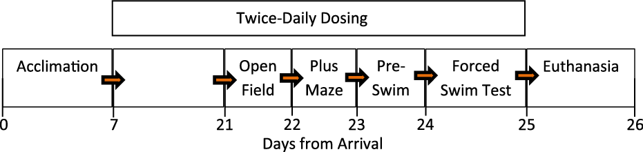
Male Sprague-Dawley (SD) rats (Charles River; 225–250 g; N = 54) were pair-housed in a temperature-controlled environment with a 12 h light/dark cycle (lights on at 06:00) with standard chow and water available ad libitum. Following their arrival, the animals were acclimated to voluntarily consume a 10% sucrose solution in a pre-determined time (2 min). Three rats failed to meet the criterion during the acclimation phase and were excluded from further treatment and testing. Subjects were randomly divided into four groups and received twice-daily (08:00 and 16:00) treatments of either a 20 mg/kg G115® (n = 14), 5 mg/kg fluoxetine (n = 12), 20 mg/kg G115® with 5 mg/kg fluoxetine (n = 12) or vehicle alone (10% sucrose solution; n = 13). Groups were administered these doses each day throughout both a 14-day feeding period and the subsequent 4-day behavioural testing protocol. The experimental timeline is depicted in Fig. 1.
Timeline of the experimental design.

All animals were cared for following procedures approved in advance by the University of Prince Edward Island Animal Care Committee and in accordance with the Canadian Council on Animal Care guidelines. All possible efforts were made to minimise animal suffering and the number of animals used.
Behavioural testing
All behavioural testing was conducted experimenter blind between the two daily feeding sessions during the light phase of the light/dark cycle. The testing rooms were kept at the same temperature as the room holding the home cages and were dimly lit with indirect light. All behavioural trials were video-recorded for later experimenter-blinded analysis. Open field (OF) and elevated plus maze (EPM) data were acquired using AnyMaze® software (Stoelting, Wood Dale, USA). Specifics of each test paradigm are described below.
Open field
To examine possible treatment effects on motoric abilities that could confound any potential differences in the FST, animals were tested in an OF arena to examine locomotor activity (Seibenhener & Wooten, Reference Seibenhener and Wooten2015). The animals were placed in a square black box (1 m × 1 m) and given 5 min to explore the arena. Total distance travelled in the maze and time spent in the perimeter of the maze were recorded.
Elevated plus maze
In addition to locomotor abilities, differences in anxiety states between groups could confound potential differences in the FST. The EPM is often used as a rodent model for human anxiety as it measures the internal conflict between voluntarily entering the naturally feared open and elevated arms of the maze versus retreating to the closed arms of the maze (Walf & Frye, Reference Walf and Frye2007). Therefore, animals were tested in an EPM to examine the effects of treatment on anxiety state. The animals were placed in the centre of the maze (arms 55 cm × 10 cm; elevation 1.5 m) facing an open arm and were given 5 min to explore the maze. Anxiety state was assessed by the time spent in the open arms of the maze and the total number of open arm entries.
Forced swim test
Following the OF and EPM, animals were subjected to a 2-day FST according to a well-established protocol (Porsolt et al., Reference Porsolt, Anton, Blavet and Jalfre1978; Slattery & Cryan, Reference Slattery and Cryan2012). On day 1, the animals were exposed to a 15-minute pre-swim session in the FST (cylinder 20 cm in diameter, 60 cm in height; water temperature 24˚C ± 1˚C). The following day, a 5-minute probe trial was administered and video recordings of this trial were scored by two independent investigators who were blind to treatment for time spent immobile, swimming or struggling by recording the predominant behaviour in each 5-second interval. A state of immobility was defined as when the animal showed no forelimb or hindlimb movement other than that necessary to avoid sinking, while swimming was scored when the animal moved horizontally in the water and struggling was marked by vertical bursts of movement.
Western blotting
Following the FST, a pseudorandomly selected (based on home cage) subset of rats (n = 8/group) were placed in a closed chamber containing isoflurane until deeply anaesthetised (approximately 1 min), decapitated and brains were rapidly removed and divided into right and left hemispheres. The prefrontal cortical region and the hippocampus were dissected from each and flash-frozen using liquid nitrogen. Tissue was stored in labelled tubes at −80°C until further processing.
Tissues were weighed and placed in ice-cold homogenisation buffer, containing 30 ul/ml of protease inhibitor (Roche ‘Complete’ 33×, Roche Diagnostics, IN, USA). Tissue was homogenised at 30 000 rpm for 30 s. The homogenate was centrifuged at 500 g for 20 min at 4 °C and the supernatant (membrane/cytosolic fraction) was removed and aliquoted. The concentration of protein was determined using the Pierce BCA Protein Assay Kit (Thermo Scientific, IL, USA).
For western blotting of BDNF and its cognate receptor tropomyosin receptor kinase B (TrkB), 20 ug of protein per sample was denatured using Laemmli buffer (Bio-Rad Laboratories Inc., CA, USA) and boiling for 3 min. The sample was then loaded into 4–15% gradient acrylamide gels (Bio-Rad Laboratories Inc., CA, USA) for protein separation by gel electrophoresis. Separated proteins were transferred by electrophoresis to polyvinylidene difluoride (PVDF) membranes (4–15% Mini-PROTEAN TGX stain-free protein gel) (Bio-Rad Laboratories Inc., CA, USA). Membranes were blocked with 5% milk in PBS, containing 0.1% Tween, for 1.5 h at room temperature. Each blot was then incubated with two primary/secondary antibody combinations:
-
1. Anti-TrkB (1:1000, Santa Cruz sc-377218): Anti-mouse IgG-HRP (1:5000, Sigma #A9917).
-
2. Anti-BDNF (1:500, Abcam ab108319): Anti-rabbit IgG-HRP (1:10000, Sigma #A0545).
All primary antibodies were diluted in blocking buffer and incubated overnight at 4 °C. All secondary antibodies were diluted in PBS-Tween and incubated for 1.5 h at room temperature. All bands were visualised using clarity-enhanced chemiluminescence (ECL) substrate (Bio-Rad Laboratories Inc., CA, USA) and visualised using the ChemiDoc® UV imaging system (Bio-Rad Laboratories Inc., CA, USA). Image analysis was performed using the Image Lab Software (Bio-Rad Laboratories Inc., CA, USA). A loading control common to all blots was generated by combining equal volumes of each fraction of the study samples that were then divided into equal 20 µg aliquots. All experimental samples were run in duplicate and data were expressed as the ratio of protein of interest to the loading control, normalised to total protein.
Statistical analysis
All data are presented as mean ± standard error of the mean (SEM). Statistical analysis was performed using SPSS (Version 23; SPSS Inc., Chicago, IL, USA). Data were confirmed as being homogenous for variance using Levene’s test and analysed using one-way Analysis of Variance with post hoc comparisons using Tukey’s Multiple Comparison Test, with p < 0.05 considered as statistically significant.
Results
There was no significant difference between groups with respect to mean weight during the 4 days of testing (data are not shown).
Forced swim test
Analysis of the FST data revealed significant main effects for time spent struggling (F 3,47 = 7.65; p = 0.0003), swimming (F 3,47 = 6.42; p = 0.001) and immobile (F 3,47 = 16.23; p < 0.0001) as well as a significant main effect for the latency to 10 consecutive seconds of immobility (F 3,47 = 20.09; p < 0.0001). Post hoc analysis of the immobility time data revealed that fluoxetine alone produced a significant reduction relative to sucrose vehicle (p < 0.05), but not compared to G115® alone. Further, the combination of G115® and fluoxetine resulted in reductions in immobility time that were significantly different from all other groups (p < 0.01) (Fig. 2A). When data were analysed for the latency to the first bout of 10 s of immobility, there was no significant difference between sucrose vehicle and G115® alone, but fluoxetine alone significantly increased latency relative to both vehicle (p < 0.01) and G115® (p < 0.05) and co-administration of fluoxetine and G115® produced significantly increased latency relative to vehicle (p < 0.01), G115® (p < 0.01) and fluoxetine (p < 0.05). These data are presented graphically in Fig. 2B.
Performance in the FST. A. Total time immobile. B. Latency to 10 consecutive seconds of immobility. Values are represented as mean +/− SEM time (s). N = 12–14* p < 0.05 relative to vehicle control; # p < 0.01 relative to all other groups.

Open field and elevated plus maze
Performance in the FST can be influenced by both changes in overall locomotor activation as well as by increases or decreases in anxiety. To determine if treatment altered either or both of these parameters, we measured behaviour in an OF arena and an EPM prior to testing in the FST (see Fig. 1). Analysis of the OF data indicated no significant main effects of treatment on total distance travelled (F 3,47 = 0.263; p = 0.85); an indicator of gross locomotor behaviour (Fig. 3A).
A. Total distance (m) travelled in the OF. B. Time (s) spent in thigmotaxis in the OF. C. Time (s) in the open arm of the EPM. D. Number of entries into the open arm of the EPM. All values are represented as mean +/− SEM. N = 12–14.

Thigmotaxis in an OF is also considered an indicator of anxiety. Analysis of time spent adjacent to the wall of the maze revealed no significant effects of treatment (F 3,47 = 0.701; p = 0.556) (Fig. 3B). In the EPM, both time in the open arm and entries into the open arm of the maze were found not to differ between treatment groups (F 3,47 = 0.394; p = 0.758 and F 3,47 = 0.127; p = 0.944, respectively) (Fig. 3C and D).
BDNF and TrkB expression
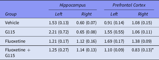
Changes in the expression of both BDNF and its cognate receptor TrkB have been reported previously following the exposure to individual ginsenosides as well as to antidepressant drugs. In the current study, we found a significant main effect for BDNF expression in the left hippocampus (F 3,21 = 6.03; p = 0.004). Post hoc comparisons revealed that the difference was between the fluoxetine and fluoxetine + G115® groups with neither group being significantly different from any other (Fig. 4A). Analysis of right hippocampal BDNF expression revealed no significant treatment effect (Fig. 4B). In contrast, BDNF expression in the left prefrontal cortex was increased in both the fluoxetine and fluoxetine + G115® groups relative to G115® alone (Fig. 4C); an effect not seen in the right hemisphere (Fig. 4D). Analysis of TrkB expression detected a significant group effect in the right prefrontal cortex (F 3,21 = 4.48; p = 0.014) due to the fluoxetine + G115® group being significantly lower than the fluoxetine group although this was not different from the vehicle or G115® alone groups. No other changes were observed (Table 1). Representative western blots of BDNF and TrkB are shown in Supplemental Fig. 1.
Expression of BDNF in the left (A) and right (B) hippocampus and the left (C) and right (D) prefrontal cortex as measured by western blot. Values are mean +/− SEM density relative to loading control. *p < 0.05 relative to G115; # p < 0.01 relative to fluoxetine. N = 8 per group.

Regional expression of TrkB as measured by western blot. Values are mean +/− SEM density relative to loading control

* p < 0.05 relative to fluoxetine. N = 8 per group.
Discussion
In previous studies, P. ginseng extract G115® has been found to attenuate ethanol-induced depression after oral gavage (Boonlert et al., Reference Boonlert, Benya-Aphikul, Welbat and Rodsiri2017). Administering the compound through voluntary drinking is inherently less stressful for the animal and more closely mimics how ginseng has been consumed in traditional Asian medicine. Therefore, in the present study, we first observed the effects of voluntary drinking of G115®, either alone or in combination with the SSRI fluoxetine, twice daily for 14 days on behavioural despair/learned helplessness by measuring both the time spent immobile (Fig. 2A) and the latency to immobility (Fig. 2B) in the FST. To determine the specificity of these effects, we also examined locomotor and anxiety behaviour in an OF and EPM paradigm, respectively. And lastly, we investigated both BDNF and TrkB expression in the hippocampus and prefrontal cortex.
Behavioural despair/learned helplessness begins when an organism is put in an aversive situation from which it cannot escape, and then can be observed during subsequent encounters with this situation, as the organism will put less effort into trying to escape (Seligman & Maier, Reference Seligman and Maier1967). Behavioural despair and, by proxy, depression may be studied in rodents by use of the FST (Porsolt et al., Reference Porsolt, Anton, Blavet and Jalfre1978; Castagne et al., Reference Castagne, Moser, Roux and Porsolt2011) and measurement of the time spent immobile as opposed to struggling or swimming. Because this behaviour is increased in rodent models of depression, including the Flinders line of depression-prone rats (Wegener et al., Reference Wegener, Matthe and Neumann2012) and reduced by most current antidepressant drugs and therapies (for review, see Petit-Demouliere et al., Reference Petit-Demouliere, Chenu and Bourin2005), immobility time is commonly used as a measure of depressive behaviour in rodents.
In the current study, investigator-blinded analysis of the behaviour of the rats during a 5-minute FST conducted 24 h after a 15-minute pre-swim revealed a significant reduction in immobility time (and a corresponding increase in struggling and swimming time; data are not shown) in the fluoxetine alone and fluoxetine + G115® groups (Fig. 2A). Further, animals in these two groups had a significantly longer latency to the first bout of 10 s of immobility (Fig. 2B). These results could be interpreted as a reduction in behavioural despair (an indicator of depression), but could have also been caused by drug-treated rats experiencing greater locomotor stimulation than their vehicle-treated counterparts. To investigate this, we analysed various indicators of overall locomotor behaviour in an OF test conducted prior to the FST (see Fig. 1). There were no differences between any of the four groups in total distance travelled (Fig. 3A) or average velocity (data are not shown), indicating that reduced immobility was not due to a generalised increase in locomotor behaviour. Similarly, it can be argued that rodents prone to heightened anxiety will struggle more in the FST resulting in a corresponding reduction in immobility time or latency to immobility. To examine this possibility, in addition to the OF test, we conducted an EPM test prior to the FST (see Fig. 1). Heightened anxiety manifests as both increased thigmotaxis in the OF and reductions in time and entries in the open arm of the EPM. Our data indicate no differences between any of these parameters, between any of the four groups (Fig. 3B–D). Consequently, we conclude that the effect of fluoxetine and fluoxetine + G115® treated rats in the FST was, in fact, a reduction in the degree of behavioural despair exhibited by these rats.
As expected, we observed a reduction in immobility time in the group treated chronically with fluoxetine (Fig. 2A). The use of the FST to screen antidepressant drugs, and particularly SSRIs, is well established (for review, see Cryan et al., Reference Cryan, Valentino and Lucki2005). More importantly, however, we found that co-administration of G115® significantly potentiated the antidepressant effect of fluoxetine as measured by total immobility time (Fig. 2A) and latency to immobility (Fig. 2B). G115® alone did not produce this effect (Fig. 2) suggesting a mechanistic overlap between the ginsenoside formulation and the SSRI. As an initial attempt to further elucidate this interaction, we measured protein concentrations of BDNF and its cognate receptor TrkB in the hippocampus and prefrontal cortex of treated rats.
Elevations of neurotrophins, particularly BDNF, as well as other components of the BDNF signalling cascade have been previously reported following both acute and chronic administration of purified ginsenosides. Rg1 has been shown to increase BDNF expression both in vitro and in vivo (Liang et al., Reference Liang, Ge, Yang, Yang, Ye, Yan, Du and Luo2010; Shi et al., Reference Shi, Huang, Chen, Pan, Zhang, Zhu and Chen2010) and Jiang et al. (Reference Jiang, Xiong, Yang, Wang, Wang, Hu, Wang and Chen2012) reported that chronic administration of Rg1 reverses both immobility time in the FST and tail suspension test as well as reductions in BDNF and TrkB in a mouse model of CMS. Similarly, the ginsenoside Rg3 has been reported to both reduce depression and increase BDNF in mouse models (Zhang et al. Reference Zhang, Zhou, Chen, Zhong and Zhong2017; You et al., Reference You, Yao, Shen, Gu, Xu, Wu, Chen and Li2017). Reductions in hippocampal BDNF and TrkB have also been reported in various rodent models of depression (Aso et al., Reference Aso, Ozaita, Valdizan, Ledent, Pazos, Maldonaldo and Valverde2008; Badowska-Szalewska et al., Reference Badowska-Szalewska, Spodnik, Klejbor and Morys2010). Accordingly, we chose to measure both BDNF and TrkB expression in prefrontal cortex and hippocampus following chronic consumption of G115 alone or in combination with fluoxetine. The data presented in Fig. 4 and Table 1 show that in the hippocampus fluoxetine did not significantly increase BDNF expression (although there was a non-significant tendency towards an increase in both hemispheres) (Fig. 4A and 4B). This is in contrast to some literature demonstrating that antidepressant therapies elevate BDNF (Mondal & Fatima, Reference Mondal and Fatima2019; Deyama & Duman, Reference Deyama and Duman2020), but it should be noted that these studies use various rat models of depression, that have reduced BDNF expression that is subsequently normalised by therapy, as opposed to the rats in the current study that presumably began treatment with normal BDNF expression. Hence, it is possible that there is a ‘ceiling’ effect for hippocampal BDNF. In combination with G115®, however, there is a highly significant decrease only in the left hemisphere relative to fluoxetine alone (Fig. 4A). This was not observed in the right hemisphere although the group means followed a similar trend. In the prefrontal cortex, chronic fluoxetine significantly increased BDNF in the left, but not the right hemisphere (Fig. 4C and 4D) but there was no effect of concurrent administration of G115®. Western blot analysis of TrkB expression in these tissues indicated no significant difference amongst treatment groups with one minor exception that is probably a type 1 error given the lack of consistency with the BDNF data (Table 1). Taken collectively, the BDNF and TrkB data are difficult to interpret, but it clearly demonstrates that the observed potentiation of fluoxetine by G115® is not due to further enhancement of BDNF expression, although enhanced BDNF signalling by, for example, increased phosphorylation of TrkB and/or CREB are possibilities requiring further investigation.
In conclusion, we have demonstrated a significant potentiation of the antidepressant-like action of fluoxetine by G115® in male SD rats. Marked changes in immobility in the FST, a measure of behavioural despair, were not confounded by drug actions on either locomotor activity or anxiety. The behavioural changes produced by G115® appear to reflect at least a partial mechanistic overlap with the actions of fluoxetine, but within the constraints of the current study, they do not appear to be due to changes in BDNF signalling in the hippocampus or prefrontal cortex. Further investigation in both normal rats and in rat models of depression is warranted with the ultimate aim of determining if G115® (or other ginsenoside formulations) might have a useful role as adjunctive therapy to SSRIs in the treatment of clinical depression.
Supplementary material
To view supplementary material for this article, please visit https://doi.org/10.1017/neu.2021.2.
Acknowledgements
The authors thank Melissa Perry and Sweta Daboo for technical assistance.
Funding for this research was provided by Fondation J. Louis Levesque in support of a Research Professorship awarded to RAT.
Author contributions
DJT and RAT designed the experiments and wrote the manuscript; DJT and DSM performed the experiments and collected and analysed the data; all authors edited the manuscript; RAT secured the funding.
Conflict of interest
The authors declare no conflict of interest.