




No CrossRef data available.
Neuropsychological sequelae of obstructive sleep apnea-hypopnea syndrome: A critical review
Published online by Cambridge University Press: 01 September 2004
Abstract
Obstructive sleep apnea-hypopnea syndrome (OSAHS) is a well-recognized clinical sleep disorder that results in chronically fragmented sleep and recurrent hypoxemia. The primary daytime sequelae of the disorder include patient reports of excessive daytime sleepiness, depression, and attention and concentration problems. It has been well established that OSAHS negatively impacts certain aspects of cognitive functioning. The primary goals of this article are to (1) clarify the pattern of cognitive deficits that are specific to OSAHS; (2) identify the specific cognitive domains that improve with treatment; and (3) elucidate the possible mechanisms of cognitive dysfunction in OSAHS. At the conclusion of the paper, we propose a potential neurofunctional theory to account for the etiology of cognitive deficits in OSAHS. Thirty-seven peer-reviewed articles were selected for this review. In general, findings were equivocal for most cognitive domains. Treatment, however, was noted to improve attention/vigilance in most studies and consistently did not improve constructional abilities or psychomotor functioning. The results are discussed in the context of a neurofunctional theory for the effects of OSAHS on the brain. (JINS, 2004, 10, 772–785.)
Information
- Type
- CRITICAL REVIEW
- Information
- Journal of the International Neuropsychological Society , Volume 10 , Issue 5 , September 2004 , pp. 772 - 785
- Copyright
- 2004 The International Neuropsychological Society
INTRODUCTION
Obstructive sleep apnea-hypopnea syndrome (OSAHS) is a well-recognized clinical sleep disorder that affects 2% of middle-aged women, 4% of middle-aged men (Young et al., 1993), and up to 42% of individuals over the age of 65 years (Ancoli-Israel et al., 1991). It may also be more prevalent among African-Americans (Kripke et al., 1997; Redline et al., 1997). The pathophysiology of OSAHS is characterized by repeated complete (apnea) or partial (hypopnea) cessations of breathing during sleep caused by narrowing at various sites along the upper airway. During these breathing events, blood oxygen saturation (SaO2) can drop to dangerously low levels, resulting in increased respiratory effort and arousals from sleep to resume breathing. Recurrent hypoxemia and fragmented sleep are therefore significant consequences of the disorder (Bassiri & Guilleminault, 2000). The primary daytime sequelae of the disorder include excessive daytime sleepiness, mood changes, and self-reported cognitive problems. OSAHS is also associated with an increased risk of co-morbid medical illnesses, such as hypertension and vascular disease, making it a potentially costly disorder (Nieto et al., 2000; Shahar et al., 2001).
Diagnostic Considerations
The diagnosis of OSAHS requires an overnight assessment of sleep (polysomnography) in a sleep laboratory. A full diagnostic study typically includes continuous monitoring of neurophysiological variables (electrooculography, submental electromyography, electroencephaolography, and anterior tibialis electromyography) in addition to measures related to respiration, including airflow, respiratory effort, pulse oximetry, and electrocardiography. The measures most commonly used to summarize respiratory event frequency are the Respiratory Disturbance Index (RDI) and Apnea/Hypopnea Index (AHI), which are both indices of the number of apneas and hypopneas per hour of sleep. These indices indicate the degree of sleep fragmentation. Measures of oxygen saturation during the night provide an indication of the severity of hypoxemia during the respiratory disturbances. Nadir SaO2 during sleep, mean SaO2 during sleep, and the percent of sleep time below a threshold saturation level (often 80% or 90%) are common measures of hypoxemia. Syndrome definitions for OSAHS were published by an American Academy of Sleep Medicine Task Force in 1999. Their recommendations included that a minimal threshold of five breathing-related events per hour of sleep be used to diagnose clinically-significant OSAHS, and that the severity of the disorder be classified as follows: mild: 5 to 15 events/hr; moderate: 15 to 30 events/hr; and severe: greater than 30 events/hr (American Academy of Sleep Medicine Task Force, 1999).
Medical, Psychological, and Functional Consequences of OSAHS
OSAHS has been associated with an increased risk for serious medical conditions, particularly vascular diseases, such as hypertension, heart disease and stroke (Guilleminault & Robinson, 1997; Hudgel et al., 1993; Partinen & Guilleminault, 1990; Schäfer et al., 1998). Psychiatric disorders, such as depression and anxiety (Aikens et al., 1999; Aikens & Mendelson, 1999; Beutler et al., 1981; Kales et al., 1985), have also been linked to OSAHS. Even mild levels of OSAHS are associated with self-reported lower general health status independent of age, sex, body mass index, tobacco use, alcohol use, or history of cardiovascular disease (Finn et al., 1998). The daytime functional consequences of OSAHS can be severe. For example, OSAHS patients are more likely to be involved in motor vehicle accidents than normal controls, with the risk of having an accident increasing as the severity of the disorder increases (Aldrich, 1989; George et al., 1987; George & Smiley, 1999). Moreover, the decrements in performance on a sustained attention task for individuals with OSAHS have been found to be comparable to those of moderately intoxicated normal controls (Powell et al., 1999).
Previous research also suggests that individuals with OSAHS utilize more health care services than normal controls. Kapur et al. (1999), for instance, found that the mean annual health care cost for 238 OSAHS patients was $2,720 compared with $1,384 for age- and sex-matched controls. The increased consumption of health care resources for OSAHS patients may be evident as many as 7 years prior to the diagnosis (Ronald et al., 1999). With adherent treatment, physician claims and hospital stays both decline significantly (Bahammam et al., 1999).
Clinical Management of OSAHS
Although behavioral (e.g., weight loss) and surgical options exist for the clinical management of OSAHS, continuous positive airway pressure (CPAP) is considered the treatment of choice (Sullivan et al., 1981). CPAP consists of a nasal mask attached to a pneumatic pump by a flexible air tube, and held in place by a harness that fits over the head. The pump supplies constant positive air pressure to the upper airway, preventing it from collapsing during sleep. Once the appropriate pressure is determined, CPAP generally eliminates most or all nocturnal breathing disturbances, thereby improving oxygen saturation and reducing sleep fragmentation (American Thoracic Society, 1994).
With proper use, CPAP dramatically reduces health care utilization (Bahammam et al., 1999) and the risk of mortality (He et al., 1988; Keenan et al., 1994). Despite these benefits, CPAP is not a cure per se, but provides only a “pneumatic splint” to the upper airway. One study found that as little as one night without CPAP returns breathing disturbances and daytime symptoms to baseline levels (Kribbs et al., 1993). Unfortunately, research has revealed that long-term adherence to CPAP is less than optimal for reasons that remain to be fully elucidated, but which include barriers associated with the equipment (e.g., mask discomfort, nasal congestion, nasal dryness) and psychological barriers to behavior change (Aloia et al., 2001a; Hoffstein et al., 1992; Waldhorn et al., 1990). Treatment adherence may have direct implications on the magnitude of improvements in daytime functioning that patients experience with treatment.
Neuropsychological Consequences of OSAHS
Decreased mental acuity can have significant negative consequences for occupational performance, automobile safety, educational pursuits, and in-home functioning. Most studies of cognition in OSAHS have focused on attention and vigilance since these are the primary complaints reported by OSAHS patients. In a recent review article, Décary et al. (2000) argued for the battery approach using standardized neuropsychological measures to determine the pattern of cognitive dysfunction in OSAHS. Studying multiple cognitive domains in a single sample allows for comparisons between these domains, and thus between assumed brain regions, which can lead investigators to develop a more comprehensive neurofunctional theory of OSAHS. The battery approach also has clinical application. The presence of OSAHS, particularly in elderly populations, may contribute to diagnostic uncertainty for the practitioner, given the overlap in cognitive impairment related to OSAHS with more common neurocognitive disorders. It is therefore important for neuropsychologists and other healthcare practitioners to be aware of the potential impact of OSAHS on cognitive functioning in order to improve diagnostic sensitivity and specificity, and to provide appropriate treatment recommendations. Important questions remain, however, concerning the cognitive domains that are specifically affected in OSAS, treatment-related improvements, and the etiology of cognitive deficits.
The primary goals of this article are (1) to clarify the pattern of cognitive deficits that are specific to OSAHS; (2) to identify the specific cognitive domains that improve with treatment; and (3) to elucidate the possible mechanisms of cognitive dysfunction in OSAHS. At the conclusion of the paper, we propose a potential neurofunctional theory for the effects of OSAHS on the brain.
METHODS
Data Collection
Data for this review consisted of empirical articles published in peer-reviewed journals over the last 17 years (1985–2002). An extensive computer-assisted literature search was conducted using the Medline and PsychInfo databases. Search terms for OSAHS were apnea and sleep-disordered breathing. Neuropsychological search terms included cognition, cognitive ability, mental status, neuropsychology, memory, attention, vigilance, executive, and psychomotor. Searches were conducted using all possible combinations of the sleep and neuropsychological terms. Additional published studies were identified from the reference sections of the initial group of identified studies. Excluded from consideration were non-English articles, book chapters, monographs, commentaries, review articles, case studies, dissertations, abstracts, letters to the editor, and any non data-analytic or non peer-reviewed reports. This initial search yielded 187 articles for consideration. Since we were interested in reviewing data on OSAHS and neuropsychological functioning specifically, we reviewed all of these articles and imposed the following additional exclusion criteria:
- Studies that specifically recruited non-OSAHS participants, but that subsequently made comparisons on outcome measures based on OSAHS status (five articles excluded).
- Studies without verification of OSAHS by overnight polysomnography (21 articles excluded). For instance, some studies included samples of self-reported snorers, but we aimed to establish clearly the relationship between indices of disease severity (i.e., sleep fragmentation and hypoxemia) and cognitive functioning.
- Studies including special medical populations with OSAHS, such as dementia, chronic obstructive pulmonary disease, and Down's syndrome (39 articles excluded).
- Studies including children and/or adolescents, defined as participants less than 18 years of age (18 articles excluded).
- Studies including patients with central sleep apnea only (four articles excluded). We excluded these studies because central sleep apnea is relatively uncommon and represents a completely different sleep disorder than OSAHS.
- Studies assessing only one cognitive domain or exclusive use of non-validated instruments (63 articles excluded). We were particularly interested in examining patterns of cognitive deficits in OSAHS, and consistent with the recommendations by Décary et al. (2000), have included only articles assessing multiple cognitive domains that utilized standardized cognitive tests with published normative data. The reader should be advised that there have been numerous peer-reviewed publications on attention and vigilance that were not included in this review because the tests were not standardized. These additional restrictions resulted in a final pool of 37 articles for inclusion in this review.
Study Categorization
Studies meeting the inclusion/exclusion criteria were then divided into three, non-exclusive categories: pre-treatment group comparison studies (n = 12), treatment efficacy studies (n = 18), and correlational studies (n = 11); a total of 6 studies met criteria for inclusion in two or more categories. To ensure reliability of categorization, three of the authors (M.S.A., J.T.A., & D.B.) independently categorized all 37 articles. Where there was disagreement, all authors were consulted to reach group consensus.
Methodological Concerns
The authors had several methodological concerns about the studies reviewed. These concerns, listed below, limited our confidence in the results of some studies.
Failure to control for practice effects with repeated measurement
Test experience is a common threat to the internal validity of any longitudinal study and is best addressed by employing tests with multiple forms or with little demonstrated practice effects. Even tests with high test–retest reliability can result in spurious “improvements” with multiple assessments. Many studies failed to utilize alternative measures, and some studies exposed participants to test items before actually collecting data.
Inconsistent use of diagnostic criteria for apnea
This issue is crucial when reviewing the effects of OSAHS severity on performance. Severity measures vary greatly with the inclusion of hypopneas, for example. Although it is common practice to use indices that include these partial obstructive events, some studies reported only complete airway obstructions (AI). Clearly, these measures are quite different and can make comparison between studies difficult.
Inconsistent methods for controlling demographic factors
Some studies controlled for demographic factors by using a comparison group while others adjusted scores based on age and education using published norms. It is more scientifically rigorous to recruit a representative control group for each study, however, normative comparisons are useful when determining the clinical meaningfulness of findings. Lack of control of demographic factors makes it difficult for cross-study comparisons.
Failure to address treatment compliance
Compliance with CPAP treatment is a common problem in OSAHS. Compliance at prescribed levels has been estimated as low as 28% (Pack, 1994). It remains unclear whether or not greater use would result in more obvious changes on neuropsychological test performance. In this review, 11/17 CPAP treatment studies measured compliance. The average compliance rate was about 4.5 hr of CPAP per night. However, only one study sample used more than 6 hr/night on average, and only four studies examined change in cognitive functioning by compliance. We believe that this type of analysis is more meaningful, as neglecting to examine results by compliance could result in erroneously assuming that treatment does not improve cognition. We have studied this issue in a small sample and found that greater use was associated with greater improvements on cognitive test scores (Aloia et al., 2001a).
Due to these limitations in the existing literature we chose to conduct a qualitative review by tabulating impaired performance by cognitive domain for each study. Cognitive domains were defined by the authors of this review, with the guidance of common texts of neuropsychological assessment (Lezak, 1983; Spreen & Strauss, 1998), and included global cognitive functioning, attention/vigilance, executive functioning, memory, construction, and psychomotor functioning (see Appendix for domain descriptions). In summarizing the pattern of cognitive deficits from each study, we evaluated the findings for each test of each domain. Performance was considered impaired in a particular domain if the authors found group differences on a task. When more than one test was used in a single domain (e.g., Trails A, Digit Span, and PASAT for attention/vigilance), we required evidence of impairment from a majority of the tests to conclude that the domain was affected. This approach was adopted in an effort to minimize Type I error.
RESULTS
Pattern of Neuropsychological Deficits in OSAHS
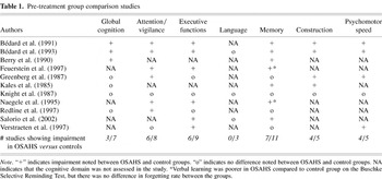
Pre-treatment group comparisons were used to identify the specific cognitive domains affected by OSAHS in comparison to normal controls (see Table 1).
Pre-treatment group comparison studies

The majority of studies found spared global cognitive functioning (four of seven studies) and language functions (all of three studies). In contrast, attention/vigilance, executive functioning, and memory were impaired in at least 60% of the studies reviewed. Construction and psychomotor functioning impairments were noted in 80% of the studies (four of five studies finding impairment in each). We allowed tests of both psychomotor speed and fine motor coordination to be grouped under the psychomotor functioning construct, although they may tap different brain systems. When we separated findings out by the two sub-constructs, it was apparent that simple psychomotor speed was intact in OSAHS, with neither of the two studies assessing finger tapping showing a difference between the OSAHS and the control groups. Fine motor coordination, however, was consistently impaired, with all of four studies assessing performance on the pegboard task showing impairment of the OSAHS group.
Cognitive Changes With Treatment
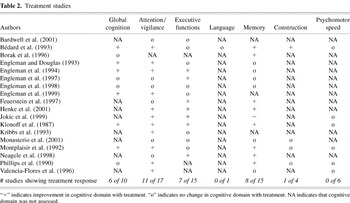
Treatment studies were examined to determine whether pre-treatment cognitive impairments were permanent in OSAHS or if they remitted with treatment (see Table 2). Examining treatment-related improvements and residual deficits can help to generate hypotheses about the long-term effects of apnea on the brain.
Treatment studies

All but one study evaluated the effects of the most commonly prescribed treatment, CPAP, on cognitive functioning. The one non-CPAP study found improvements in global cognitive functioning, attention/vigilance, executive functioning, and memory with surgical treatment for OSAHS (Klonoff et al., 1987). Only the domain of psychomotor functioning did not improve with treatment. Among the entire group of treatment studies, attention/vigilance improved with treatment in 11 of 17 studies assessing this cognitive domain. Changes in global functioning, executive functioning, and memory improved in about half of the studies reviewed: 6/10, 7/15, and 8/15 studies, respectively. In contrast, tests of psychomotor function (none of six studies) and construction (one of four studies) failed to improve with treatment in the majority of studies.
As is evident from Table 1, impairments in psychomotor functioning and construction were noted in 80% of the studies, making an examination of treatment effects on these constructs of particular interest. Identifying residual impairments is important because it provides insight into possible neuropathological mechanisms associated with the disorder. One could argue that the cognitive problems that do not remit with treatment are likely the effects of chronic intermittent hypoxemia, as any cognitive effects of sleep fragmentation should be less permanent. One way to address this possibility is to examine the differential relationships of sleep fragmentation and hypoxemia with cognitive performance. Below we review those studies that examined this issue specifically.
Identifying the Mechanisms of Cognitive Dysfunction in OSAHS
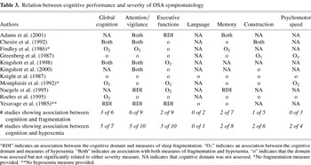
Eleven studies examined the association between cognitive status and clinical severity indicators of OSAHS (see Table 3). Fragmentation and hypoxemia measures may be associated with different patterns of cognitive change and response to treatment, and may reflect different neuropathological mechanisms. Of the 11 studies, eight examined the relationship between cognitive performance and measures of both sleep fragmentation (AHI, RDI, or AI) and hypoxemia (i.e., blood oxygen nadir SaO2 during sleep or time SaO2 ≤ 80%), 1 (Yesavage et al., 1985) reported only sleep fragmentation measures, and 2 (Findley et al., 1986; Montplaisir et al., 1992) reported only hypoxemia measures. Two of the studies examined (Findley et al., 1986; Roehrs et al., 1995) used a between groups design to examine the relationship between cognitive functioning and clinical indicators of OSAHS severity; the remainder used correlational designs.
Relation between cognitive performance and severity of OSA symptomatology

Of the 11 studies reviewed, 10 found a significant association between apnea severity variables (either fragmentation or hypoxemia) and at least one domain of cognitive performance. Only the Knight et al. (1987) study failed to find any association between apnea severity and cognitive functioning; however, global cognitive functioning and attention/vigilance were the only domains to demonstrate any consistent association with either severity measure across studies. For these two cognitive domains, the majority of studies found a negative relationship between severity of OSAHS and cognitive functioning, with more severe OSAHS clinical indicators associated with poorer cognitive performance. Hypoxemia was more associated with performance on tests of global cognitive functioning (five of seven studies), and fragmentation measures were associated with performance on tests of Attention/Vigilance in the majority of studies reviewed (six of nine studies). Psychomotor functioning was of particular interest given its apparent lack of improvement with treatment above. This domain was related to hypoxemia in only two of four studies, however, both of these studies assessed fine motor coordination (i.e., Grooved Pegboard) rather than simple psychomotor speed (i.e., Finger Tapping Test). There was no relationship between fine motor coordination and measures of fragmentation or between simple psychomotor speed and sleep fragmentation or hypoxemia.
Two studies specifically examined the effects of hypoxemia and sleep fragmentation on cognition using a group comparison rather than a correlational design. Findley et al. (1986) compared OSAHS patients with and without coexisting hypoxemia on a battery of neuropsychological tests measuring global cognitive functioning, attention/vigilance, executive functioning, and memory. They found that patients with hypoxemia performed more poorly on measures of attention/vigilance, memory, and global cognitive functioning. Roehrs et al. (1995) compared patients with sleep apnea to those with chronic obstructive pulmonary disease (COPD), hypothesizing that differences between the groups would be attributable to sleep fragmentation since both groups experienced some degree of hypoxemia. OSAHS patients were more impaired than COPD patients on tests of sustained attention, supporting the notion that attention/vigilance is most likely a result of sleep fragmentation than it is of hypoxemia in this population.
DISCUSSION
The goal of this paper was to review and summarize the relevant literature on the neuropsychological functioning of patients with OSAHS. We specifically wanted (1) to clarify the pattern of cognitive impairment in individuals with OSAHS; (2) to identify the specific cognitive domains that may improve with treatment; and (3) to elucidate the possible mechanisms of cognitive dysfunction in OSAHS. Below we address these goals by answering key questions raised in the study of the cognitive effects of sleep apnea. At the conclusion of the discussion, we pull upon existing cardiovascular literature to offer a potential explanation for the cerebrovascular effects of chronic, intermittent hypoxemia in OSAHS.
Are Patients With OSAHS Cognitively Impaired and Why?
Our approach to this critical question was to review literature that specifically examined cognitive function in OSAHS using standardized neuropsychological measures. We chose to include only well-validated neuropsychological tests assessing a minimum of three cognitive domains for this review in order to examine the pattern of cognitive deficits evident in this population. In general, we found that language is spared in OSAHS. Interestingly, four of seven group comparison studies found that global cognitive functioning was spared; however, global cognitive improvement following treatment was noted in 6/10 treatment studies. Only one study made both between and within group comparisons to compare first untreated apnea patients to controls and then pre–post treatment changes in the apnea group (Bédard et al., 1993). The study found no differences between untreated apnea patients and controls on the WAIS–R Full Scale IQ or Verbal IQ, but reported a significantly lower WAIS–R Performance IQ in the untreated apnea patients. This latter deficit improved significantly with treatment. Thus, apnea patients may exhibit relatively few deficits in the global cognitive domain when compared to normal controls, but treatment may produce within-subjects improvements in performance. It is difficult to determine why these improvements would exist, especially since the array of tests included in this domain is quite vast. With regard to language, too few pre-treatment and treatment studies examined this construct to draw firm conclusions about the effects of OSAHS on this domain, however, there is no theoretical reason to believe that language skills would be affected by either sleep fragmentation or repeated hypoxemia.
Attention/vigilance, executive functioning, and memory were impaired in the majority of studies reviewed (six of eight, six of nine, and 7/11, respectively). Attention/vigilance is the most commonly assessed cognitive construct in OSAHS, and multiple studies assessing sustained attention using tests developed in the sleep field (e.g., Psychomotor Vigilance Test, PVT; Dinges & Powell, 1985; Steer Clear; Findley et al., 1989) have found deficits in this domain (e.g., Findley et al., 1999; Kribbs et al., 1993). Thus, attention/vigilance appears to be the most consistently affected cognitive domain in these patients. The relationships between OSAHS and measures of executive functioning and memory have been less thoroughly studied. Both of these cognitive domains present a challenge to investigators in that they are complex domains. For example, executive functioning can be tested using measures of set shifting (e.g., Trails B), working memory (e.g., Wisconsin Card Sorting Test), or verbal fluency (e.g., Controlled Oral Word Association). All are accepted tests of “executive functioning” and have been categorized similarly here, but each tests a distinctly different aspect of the various functional systems that lie within the frontal lobes of the brain. Because they represent different systems, performance on these tests is likely to be less consistent in any single cohort than would multiple tests of a different domain, such as attention. Memory testing suffers from the same methodological concerns, although to a lesser degree. In this case, test performance can be impaired for a number of reasons that would implicate distinctly different brain regions. For example, memory performance deficits can be attributable to complications in initial learning, free recall, or forgetfulness, each of which has its own functional implications relating to different brain regions. In fact, in this review, two studies (Feuerstein et al., 1997; Naegele et al., 1995) found primary learning impairments in the tally of studies finding “memory” impairments, while the remainder of the studies found deficits in free recall. Despite these limitations, patients with OSAHS appear to perform more poorly on tests of executive functioning and memory than do matched controls. Clearly, these findings have implications for the potential involvement of certain brain regions in this disorder. It is, however, difficult to determine the degree to which attention and executive functioning, which are frontally mediated, contribute to impairments in memory.
Although assessed less frequently than most other domains, psychomotor functioning and construction were impaired in OSAHS patients relative to normal controls in the majority of studies reviewed. Interestingly, these domains have rarely been discussed as primary sources of impairment in this population. The mechanism for psychomotor dysfunction in OSAHS is not obvious. Certainly, excessive sleepiness could result in slow motor performance, which would result in deficits on psychomotor tasks. If this were the case, however, tests of fine motor coordination (e.g., Grooved Pegboard) should be impaired to the same degree as tests of simple motor speed (e.g., Finger Tapping). All four studies assessing fine motor coordination found OSAHS patients to be impaired relative to control subjects (Bédard et al., 1991, 1993; Greenberg et al., 1987; Verstraeten et al., 1997), while neither study examining motor speed identified impairments (Knight et al., 1987; Verstraeten et al., 1997). Deficits on tests of fine motor coordination suggest the possible involvement of specific brain regions (e.g., the basal ganglia) in OSAHS that have not previously been the focus of investigation (for review, see Ring & Serra-Mestres, 2002). The constructional findings are more complex and have not been addressed specifically in the literature. Executive and motor coordination problems could account for a large percentage of the variance in constructional performance in this population, however, studies are needed to address this question directly.
The two most commonly implicated etiological mechanisms of cognitive dysfunction in OSAHS are sleep fragmentation (indexed by the RDI) and hypoxemia. There were no strong and consistent relationships between either measure of severity and any cognitive domain. Although fragmentation measures were consistent across studies (i.e., RDI and AHI) this was not the case for measures of hypoxemia. Some studies used nadir oxygen (e.g., Kingshott et al., 2000), while others used the number of desaturations beyond a specified criterion (e.g., Cheshire et al., 1992). While these measures are likely correlated, they represent very different aspects of the hypoxemia in OSAHS and may limit the likelihood of finding relationships. All but one study, however, identified a relationship between one or both of these clinical indicators of severity and at least one cognitive domain. In general, attention/vigilance and global cognitive functioning were most strongly related to some measure of severity. The other domains (executive functioning, language, memory, psychomotor functioning, and construction) appeared unrelated to these clinical indicators. It should be noted, however, that the two studies of Psychomotor functioning that used tests of fine motor coordination both found a relation between performance and measures of hypoxemia (Greenberg et al., 1987; Montplaisir et al., 1992). The only entirely negative study, which examined older adults with apnea (Knight et al., 1987), may have suffered from a restricted range in their OSAHS sample, since 5/10 participants exhibited only mild apnea and only 1 participant met criteria for severe apnea (RDI = 37.6). Given that apnea severity increases with age, mild apnea in an elderly group might not be clinically significant. It is possible, therefore, that cognitive dysfunction depends on a greater severity of apnea, especially in a group of older adults. Studies recruiting moderate to severe OSAHS patients consistently reported differences in performance between apnea and control groups. One study (Bédard et al., 1991) compared moderate and severe apnea patients to each other as well as to normal controls. The authors found that individuals with OSAHS performed more poorly than did normal controls on tests of executive functioning, memory, and psychomotor functioning. Severe patients were, however, more impaired than moderate patients on tests of global cognitive functioning, executive functioning and psychomotor functioning, suggesting a clear relationship between severity and cognition. It is possible that the relationship between cognition and apnea severity is not linear, even within a specified age range. Cognitive deficits might require a threshold of apnea severity, but there is clear evidence that a relationship exists at least among moderate to severe patients.
Does Treatment for OSAHS Improve Cognition?
Identifying the pattern of cognitive impairment in patients with untreated OSAHS can lead investigators to hypothesize about the etiology of cognitive deficits in this population. In particular, it would be interesting to identify if those cognitive functions related most to hypoxemia were also least likely to remit with treatment. This would suggest lasting effects of hypoxemia on cognitive functions via specific brain regions. The above findings, however, do not present a clear and consistent pattern of deficits in OSAHS. The most consistent findings may be in the domains less frequently tested (e.g., psychomotor functioning and construction). In fact, the findings of relatively consistent deficits in fine motor coordination and construction raise some interesting questions about the possible brain regions involved in the disorder. Psychomotor functions are in part reliant on the integrity of subcortical grey matter structures (Ring & Serra-Mestres, 2002). These structures are preferentially affected in other hypoxic populations and may be particularly susceptible to the repeated, intermittent hypoxemia seen in OSAHS (Caine & Watson, 2000). Moreover, fluctuations in oxygen availability, rather than fragmented sleep, are more likely to be associated with tissue damage.
One would expect that the functions associated with sleep fragmentation would improve rapidly with effective treatment; on the other hand, those associated with hypoxemia would likely not change rapidly and, indeed, may not change at all. This is only partially supported by the above review. Attention/Vigilance was associated with both sleep fragmentation (six of nine studies) and hypoxemia (5/10 studies), but it tended to remit with treatment (11/17 studies). Psychomotor functioning, on the other hand, was most resistant to the effects of treatment, suggesting that it may represent the more permanent effects of OSAHS. We would expect this domain to be most associated with hypoxemia. Table 3 shows that psychomotor functioning is equivocally associated with hypoxemia (two of four studies). We found, however, that the two studies using the grooved pegboard test both found associations with hypoxemia. It is therefore possible that psychomotor functions, and specifically fine motor coordination, represent a domain of deficit in OSAHS that may be the result of chronic, intermittent hypoxemia and may be more resistant to the effects of treatment. The brain regions subserving fine motor coordination may be particularly vulnerable to the hypoxemia resulting from OSAHS. These findings are consistent with those in other hypoxic populations (Cummings et al., 1984; Earnest et al., 1980; Kotila & Kajaste, 1984) and suggest that the subcortical grey matter might be preferentially affected by oxygen loss in apnea (see Caine & Watson, 2000, for review). Other candidate regions, including the cerebellum, have never been studied directly in OSAHS.
The hippocampus is often implicated as vulnerable in other hypoxic populations (Cummings et al., 1984; Petito et al., 1987; Rempel-Clower et al., 1996; Volpe & Petito, 1985), and memory was impaired in 7/11 studies in this review. It was not, however, consistently related to measures of hypoxemia, nor was it resistant to the effects of treatment (remitting in 8/15 studies), making it difficult to imply a cause and effect relationship between memory and oxygen loss in OSAHS. It should also be emphasized that it is difficult to ascribe a neuroanatomical substrate to the memory deficits noted in the studies reviewed because most studies evaluated free recall only and did not evaluate retention and/or recognition memory. More comprehensive assessment of memory processes in the future will provide greater insight into the brain regions that may underlie the memory deficits noted in OSAHS.
In summary, cognitive findings in OSAHS are not consistent, but findings regarding fine motor coordination may provide insight into the potentially lasting effects of intermittent hypoxemia in this disorder. The effects of hypoxemia on the brain are of particular interest to neuropsychologists. Specifically, based in part on the neuropsychological literature in OSAHS and the literature in other hypoxic populations, we are interested in the involvement of subcortical brain regions. The downstream effects of hypoxemia in OSAHS have been best studied in terms of cardiovascular involvement. Using this framework and existing theories of the effects of hypoxemia on vascular integrity in OSAHS, we can begin to develop plausible explanations of the possible permanent effects of OSAHS on the brain.
A Potential Mechanism of the Effects of OSAHS on the Brain
Investigators studying the cardiovascular effects of OSAHS have suggested that the disorder results in changes to the vascular structure and function, and that these changes are commonly seen in other hypoxic populations (Parkinson et al., 2002). Such theories have direct implications for the neuropsychologist studying OSAHS, as the same mechanisms that are implicated in the heart have implications for the vessels of the brain. Hypoxia typically results in almost immediate vasodilation, which serves as a protective mechanism to better distribute oxygen to the necessary organs. Researchers have shown, however, that people with OSAHS lack this protective response (Kato et al., 2000). One potential reason that OSAHS patients do not respond to hypoxemia as expected is that they do not suffer single, sustained hypoxic events; instead, they suffer repeated events (by definition more than 5/hr) where recovery time is often limited. In very severe cases, people suffer oxygen desaturations more than 100 times/hr. This results in breathing disturbances with approximately four seconds recovery time per episode. It is impossible to predict the response, protective or not, to such repeated hypoxemic events, but it seems that the vasculature would sustain some damage with such repeated events. Apnea investigators have already suggested that vasculopathy may be a direct result of OSAHS and have posited potential mechanisms. Lanfranchi and Somers (2001) have suggested that an increase in sympathetic vasoconstriction together with a decrease in vascular protective mechanisms result in changes to the structure and function of the blood vessel. Their model is described in detail elsewhere, but is summarized below (Lanfranchi & Somers, 2001).
The Lanfranchi and Somers model suggests that the hypoxemia seen in OSAHS results in a number of autonomic, humoral, and neuroendocrine responses that can lead to vasculopathy. The first, they believe, is chemoreflex activation, which results in an increase in sympathetic vasoconstriction in the peripheral vessels. This, in turn, results in increases in both blood pressure and the release of circulating catecholamines, both of which have a negative effect on the vessel. In one study, CPAP markedly reduced sympathetic activity and increases in blood pressure (Faccenda et al., 2001). The second mechanism of vasculopathy involves the pressor effect. Hypoxemia typically results in an increase in endothelin, a potent vasoconstrictor. Endothelin has sustained hypertensive effects and is increased in individuals with OSAHS. Increased vasoconstriction has obvious immediate implications for blood pressure and, if chronic, may cause permanent damage to the blood vessel itself. The third mechanism by which the vessel is damaged is through endothelial dysfunction. The endothelial cells line the wall of the vessel and, typically, produce nitric oxide in response to hypoxemia. Nitric oxide acts as a vasodilator, protecting the vessel from the constricting effects outlined above. Impaired endothelial function is characteristic of hypertension and atherosclerosis. These investigators have shown that individuals with sleep apnea have less of a protective nitric oxide response than do matched controls, suggesting that vasodilation is less likely to occur. The final mechanism put forth by Lanfranchi and Somers involves a decrease in cardiovascular variability with an increase in blood pressure variability. These authors suggest that this combination of cardiovascular variability derangements sets the stage for cardiovascular dysfunction and even heart disease. Together, this cascade of responses in OSAHS is thought to have profound, and possibly lasting, effects on both the structure and function of the blood vessel. Small vessels may be more susceptible to hypertension in general as well as these mechanisms of vasculopathy.
Recent data suggest that there is an association between apnea severity and small vessel ischemic brain disease (Aloia et al., 2001b; Colrain et al., 2002). Kamba et al. (1997) were the first to investigate cerebral metabolism in individuals with sleep apnea. These investigators utilized Magnetic Resonance Spectroscopy and showed that cerebral metabolism was lower in the cerebral white matter in participants with moderate–severe sleep apnea than in those with mild sleep apnea. In a follow-up study, Kamba et al. (2001) demonstrated that the observed metabolic change was specific to the cerebral white matter and not to the cortex, and that this relationship was independent of age. Consistent with these findings, Colrain et al. (2002) demonstrated a relationship between severity of subcortical white matter hyperintensities and level of hypoxemia in 41 identical twin pairs. The presence of these hyperintensities within the subcortical grey and deep white matter suggests the involvement of endothelial damage in these regions, where vascular hypoperfusion is more common.
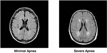
Although investigators have developed theoretical models of the vascular effects of OSAHS and the presence of subcortical hyperintensities in patients with OSAHS, these models do not directly address the cognitive effects of the disorder. We believe that damage to the small vessels of the brain can result in a number of functional cognitive problems, without absolute specificity of dysfunction in any particular domain. Many patients with OSAHS do not show obvious cerebrovascular disease, but increases in the risk for cerebrovascular disease have been established independent of other risk factors like age and obesity (Lanfranchi & Somers, 2001). Preliminary data from our laboratory supports a relationship between white matter hyperintensities on brain MRI and aspects of cognition in a small sample of patients with OSAHS. Four older adults (age > 65) with severe OSAHS (AHI > 30) and 4 with minimal OSAHS (AHI < 10) were recruited from a sleep apnea clinic and matched for age and body mass index. All participants had controlled hypertension. Subcortical white matter hyperintensities were measured using a semi-automated quantification procedure. Results indicated that the group with severe OSAHS had more subcortical white matter hyperintensities on brain MRI than those with minimal apnea (Aloia et al., 2001b; see Figure 1). Moreover, there was a trend for a negative association between subcortical hyperintensities and free recall of a word list (Table 4). There have been no specific studies of the basal ganglia (a likely candidate given its predilection for damage in hypoxic states) and/or the cerebellum, but our group has begun to investigate these structures as well.
Correlations of MRI data with sleep, cognition, and mood


Subcortical hyperintensities among individuals with severe versus minimal apnea.
Taken together, this review identifies definite areas of cognitive compromise in OSAHS. We also believe that logical etiologies exist for such dysfunction and can lead investigators to a better understanding of the mechanisms of the disease itself. We expect to study the validity of the above theory over the next several years. We also hope that other laboratories begin to explore the functional consequences of this disorder, bearing in mind the benefits of a comprehensive, standardized neuropsychological battery for identifying valid and reliable patterns of cognitive dysfunction and response to treatment.
ACKNOWLEDGMENTS
The authors would like to thank Jaime Skrekas and Leisha Smith who served as assistants in preparing the final manuscript. Grant support for work conducted on this paper was provided by the National Heart Lung and Blood Institute of NIH (R01 HL 67209) and by the Ocean State Research Institute. Raine Riggs is now at the Dartmouth-Hitchcock Medical Center (Hanover, New Hampshire) and Desiree Byrd is now at Columbia University (New York, New York).
APPENDIX
GLOBAL COGNITIVE (GC)
Global cognitive functioning was defined as reliable and valid tests that tapped functioning across numerous cognitive domains. These tests most often included scores of full scale IQ and/or global verbal or performance IQ measures. Other measures included the MMSE and estimates of premorbid intelligence, such as word-reading tasks.
ATTENTION (ATTN/VIG)
Perhaps one of the most important cognitive domains in apnea, these tests were comprised of valid tests of either attention or vigilance. Clearly these two constructs are not identical, but we felt it important to combine them because many sleep researchers do not differentiate between them. It is common for researchers in the field of sleep medicine to opt for true vigilance testing, though some use more abbreviated tests of complex attention. These tests most often included measures such as the Digit Span subtest of the Wechsler Adult Intelligence Scale, Continuous Performance Test, Paced Auditory Serial Addition Test, and letter cancellation tasks.
EXECUTIVE FUNCTION (EXFUNC)
For this domain, we chose tests with high construct validity, realizing this domain, like that of attention, can be divided into numerous subdomains. However, we believe that most “executive” tests are accepted in the field as tests of global Executive Functioning (e.g., Wisconsin Card Sorting Test, Halstead Categories Test, Trails B, Controlled Oral Word Fluency, Stroop Test, and Mazes).
LANGUAGE (LANG)
Language tests were relatively few in number but easily defined as a construct. The Boston Naming Test and Category Fluency were the most commonly used measures among the studies evaluated.
MEMORY (MEM)
As a category, memory was broadly defined as measures of learning, retention, and recognition memory performance were not evaluated separately. This was largely due to the fact that most studies assessed only free recall memory. Commonly used measures included the California Verbal Learning Test, Rey-Osterrieth Complex Figure, and Logical Memory and Visual Reproduction Subtests of the Wechsler Adult Intelligence Test.
CONSTRUCTION (CONSTR)
Again, few investigators used constructional tests in these studies. We have, however, found correlations between these ability and levels of hypoxemia in a previous study (CIT) and believe that this is an important and, as yet, relatively poorly investigated cognitive domain in the study of sleep apnea. Construction measures often included intersecting pentagons from the Mini Mental State Exam (MMSE), Block Design subtest from the Wechsler Adult Intelligence Scale, and the Rey-Osterrieth Complex Figure.
PSYCHOMOTOR FUNCTIONING (PM)
Motor functions were defined primarily by tests of fine motor coordination and psychomotor speed. Grooved Pegboard and the Finger Tapping Test were the most commonly used measures of psychomotor functioning.
References
REFERENCES
Adams, N.,
Strauss, M.,
Schluchter, M., &
Redline, S.
(2001).
Relation of measures of sleep-disordered breathing to
neuropsychological functioning.
American Journal of Respiratory and Critical Care Medicine,
163,
1626–1631.CrossRefGoogle Scholar
Aikens, J.E.,
Caruana-Montaldo, B.,
Vanable, P.A.,
Tadimeti, L., &
Mendelson, W.B.
(1999).
MMPI correlates of sleep and respiratory disturbance in obstructive
sleep apnea.
Sleep,
22,
362–369.CrossRefGoogle Scholar
Aikens, J.E. &
Mendelson, W.B.
(1999).
A matched comparison of MMPI responses in patients with primary
snoring or obstructive sleep apnea.
Sleep,
22,
355–359.CrossRefGoogle Scholar
Aldrich, M.S.
(1989).
Automobile accidents in patients with sleep disorders.
Sleep,
12,
487–494.CrossRefGoogle Scholar
Aloia, M.S.,
DiDio, P.,
Ilniczky, N.,
Perlis, M.L.,
Greenblatt, D.W., &
Giles, D.E.
(2001a).
Improving compliance with nasal CPAP and vigilance in older adults
with OSAHS.
Sleep and Breathing,
5,
13–21.Google Scholar
Aloia, M.S.,
Arnedt, J.T.,
Davis, J.,
Malloy, P.,
Salloway, S., &
Rogg, J.
(2001b).
MRI White matter hyperintensities in older adults with obstructive
sleep apnea.
Sleep,
24,
A55.Google Scholar
American Academy of Sleep Medicine Task Force.
(1999).
Sleep-related breathing disorders in adults: Recommendations for
syndrome definitions and measurement techniques in clinical
research.
Sleep,
22,
667–689.Google Scholar
American Thoracic Society.
(1994).
Indications and standards for use of nasal continuous positive
airway pressure (CPAP) in sleep apnea syndromes.
American Journal of Respiratory and Critical Care Medicine,
150,
1738–1745.Google Scholar
Ancoli-Israel, S.,
Kripke, D.F.,
Klauber, M.R.,
Mason, W.J.,
Fell, R., &
Kaplan, O.
(1991).
Sleep-disordered breathing in community dwelling elderly.
Sleep,
14,
486–495.CrossRefGoogle Scholar
Bahammam, A.,
Delaive, K.,
Ronald, J.,
Manfreda, J.,
Roos, L., &
Kryger, M.H.
(1999).
Health care utilization in males with obstructive sleep apnea
syndrome two years after diagnosis and treatment.
Sleep,
22,
740–747.CrossRefGoogle Scholar
Bardwell, W.A.,
Ancoli-Israel, S.,
Berry, C.C., &
Dimsdale, J.E.
(2001).
Neuropsychological effects of one-week continuous positive airway
pressure treatment in patients with obstructive sleep apnea: A
placebo-controlled study.
Psychosomatic Medicine,
63,
579–584.CrossRefGoogle Scholar
Bassiri, A.G. &
Guilleminault, C.
(2000).
Clinical features and evaluation of obstructive sleep
apnea-hypopnea syndrome. In
M.H. Kryger,
T. Roth, &
W.C. Dement (Eds.),
Principles and practice of sleep medicine–Third Edition (pp.
869–878).
Philadelphia:
W.B. Saunders Company.
Bédard, M.,
Montplaisir, J.,
Richer, F.,
Rouleau, I., &
Malo, J.
(1991).
Obstructive sleep apnea syndrome: Pathogenesis of
neuropsychological deficits.
Journal of Clinical and Experimental Neuropsychology,
13,
950–964.CrossRefGoogle Scholar
Bédard, M.-A.,
Montplaisir, J.,
Malo, J.,
Richer, F., &
Rouleau, I.
(1993).
Persistent neuropsychological deficits and vigilance impairment in
sleep apnea syndrome after treatment with continuous positive airway
pressure (CPAP).
Journal of Clinical and Experimental Neuropsychology,
15,
330–341.CrossRefGoogle Scholar
Berry, D.T.R.,
Phillips, B.A.,
Cook, Y.R.,
Schmitt, F.A.,
Honeycutt, N.A.,
Arita, A.A., &
Allen, R.S.
(1990).
Geriatric Sleep Apnea Syndrome: A preliminary description.
Journal of Gerontology,
45,
169–174.CrossRefGoogle Scholar
Beutler, L.E.,
Ware, J.C.,
Karacan, I., &
Thornby, J.I.
(1981).
Differentiating psychological characteristics of patients with
sleep apnea and narcolepsy.
Sleep,
4,
39–47.CrossRefGoogle Scholar
Borak, J.,
Cieslicki, J.K., &
Koziej, M.
(1996).
Effects of CPAP treatment on psychologic status in patients with
severe obstructive sleep apnea.
Journal of Sleep Research,
5,
123–127.CrossRefGoogle Scholar
Caine, D. &
Watson, J.D.G.
(2000).
Neuropsychological and neuropathological sequelae of cerebral
anoxia: A critical review.
Journal of the International Neuropsychological Society,
6,
86–99.Google Scholar
Cheshire, K.,
Engleman, H.,
Deary, I.,
Shapiro, C., &
Douglas, N.J.
(1992).
Factors impairing daytime performance in patients with sleep
apnea/hypopnea syndrome.
Archives of Internal Medicine,
152,
538–541.CrossRefGoogle Scholar
Colrain, I.M.,
Bliwise, D.L.,
DeCarli, C., &
Carmelli, D.
(2002).
The contribution of oxygen desaturation to the development of white
matter hyperintensities in elderly male twins.
Sleep,
25,
A3.Google Scholar
Cummings, J.L.,
Tomiyasu, U.,
Read, S., &
Benson, D.F.
(1984).
Amnesia with hippocampal lesions after caridopulmonary arrest.
Neurology,
34,
679–681.CrossRefGoogle Scholar
Décary, A.,
Rouleau, I., &
Montplaisir, J.
(2000).
Cognitive deficits associated with sleep apnea syndrome: A proposed
neuropsychological battery.
Sleep,
23,
369–381.Google Scholar
Dinges, D.F. &
Powell, J.E.
(1985).
Microcomputer analyses of performance on a portable, simple visual
RT task during sustained operations.
Behavior Research Methods, Instruments and Computers,
17,
652–655.CrossRefGoogle Scholar
Earnest, M.P.,
Yarnell, P.,
Merrill, A., &
Knapp, G.L.
(1980).
Long-term survival and neurologic status after resuscitation from
out-of-hospital cardiac arrest.
Neurology,
30,
1298–1302.CrossRefGoogle Scholar
Engleman, H. &
Douglas, N.J.
(1993).
Cognitive effects and daytime sleepiness.
Sleep,
16,
S79.Google Scholar
Engleman, H.M.,
Kingshott, R.N.,
Wraith, P.K.,
Mackay, T.W.,
Deary, I.J., &
Douglas, N.J.
(1999).
Randomized placebo-controlled crossover trial of continuous
positive airway pressure for mild sleep apnea/hypopnea
syndrome.
American Journal of Respiratory and Critical Care Medicine,
159,
461–467.CrossRefGoogle Scholar
Engleman, H.M.,
Martin, S.E.,
Deary, I.J., &
Douglas, N.J.
(1994).
Effect of continuous positive airway pressure treatment on daytime
function in sleep apnoea/hypopnoea syndrome.
Lancet,
343,
572–575.CrossRefGoogle Scholar
Engleman, H.M.,
Martin, S.E.,
Deary, I.J., &
Douglas, N.J.
(1997).
Effect of CPAP therapy on daytime function in patients with mild
sleep apnoea/hypopnoea syndrome.
Thorax,
52,
114–119.CrossRefGoogle Scholar
Engleman, H.M.,
Martin, S.E.,
Kingshott, R.N.,
Mackay, T.W.,
Deary, I.J., &
Douglas, N.J.
(1998).
Randomised placebo controlled trial of daytime function after
continuous positive airway pressure (CPAP) therapy for the sleep
apnoea/hypopnoea syndrome.
Thorax,
53,
341–345.CrossRefGoogle Scholar
Faccenda, J.F.,
Mackay, T.W.,
Boon, N.A., &
Douglas, N.J.
(2001).
Randomized placebo-controlled trial of continuous positive airway
pressure on blood pressure in the sleep apnea-hypopnea syndrome.
American Journal of Respiratory and Critical Care Medicine,
163,
344–348.CrossRefGoogle Scholar
Feuerstein, C.,
Naegele, B.,
Pepin, J., &
Levy, P.
(1997).
Frontal lobe-related cognitive functions in patients with Sleep Apnea
Syndrome before and after treatment.
Acta Neurologica Belgica 97,
96–107.Google Scholar
Findley, L.J.,
Barth, J.T.,
Powers, D.C.,
Wilhoit, S.C.,
Boyd, D.G., &
Suratt, P.M.
(1986).
Cognitive impairment in patients with obstructive sleep apnea and
associated hypoxemia.
Chest,
90,
686–690.CrossRefGoogle Scholar
Findley, L.J.,
Fabrizio, M.J.,
Knight, H.,
Norcross, B.B.,
LaForte, A.J., &
Suratt, P.M.
(1989).
Driving simulator performance in patients with sleep apnea.
American Review of Respiratory Disease,
140,
529–530.CrossRefGoogle Scholar
Findley, L.J.,
Suratt, P.M., &
Dinges, D.F.
(1999).
Time-on-task decrements in “Steer Clear” performance of
patients with sleep apnea and narcolepsy.
Sleep,
22,
804–809.CrossRefGoogle Scholar
Finn, L.,
Young, T.,
Palta, M., &
Fryback, D.G.
(1998).
Sleep-disordered breathing and self-reported general health status
in the Wisconsin Sleep Cohort Study.
Sleep,
21,
701–706.Google Scholar
George, C.F.,
Nickerson, P.W.,
Hanly, P.J.,
Millar, T.W., &
Kryger, M.H.
(1987).
Sleep apnoea patients have more automobile accidents.
Lancet,
8556,
447.CrossRefGoogle Scholar
George, C.F. &
Smiley, A.
(1999).
Sleep apnea and automobile crashes.
Sleep,
22,
790–795.Google Scholar
Greenberg, G.D.,
Watson, R.K., &
Deptula, D.
(1987).
Neuropsychological dysfunction in sleep apnea.
Sleep,
10,
254–262.CrossRefGoogle Scholar
Guilleminault, C. &
Robinson, A.
(1997).
Sleep-disordered breathing and hypertension: Past lessons, future
directions.
Sleep,
20,
806–811.CrossRefGoogle Scholar
He, J.,
Kryger, M.H.,
Zorick, F.,
Conway, W., &
Roth, T.
(1988).
Mortality and apnea index in obstructive sleep apnea: Experience in
385 male patients.
Chest,
94,
9–14.CrossRefGoogle Scholar
Henke, K.G.,
Grady, J.J., &
Kuna, S.T.
(2001).
Effect of nasal continuous positive airway pressure on
neuropsychological function in sleep apnea-hypopnea syndrome. A
randomized, placebo-controlled trial.
American Journal of Respiratory and Critical Care Medicine,
163,
911–917.CrossRefGoogle Scholar
Hoffstein, V.,
Viner, S., &
Mateika, S.
(1992).
Treatment of obstructive sleep apnea with nasal continuous positive
airway pressure. Patient compliance, perception of benefits, and side
effects.
American Review of Respiratory Diseases,
145,
841–845.Google Scholar
Hudgel, D.,
Devadatta, P.,
Quadri, M.,
Sioson, E.R., &
Hamilton, H.
(1993).
Mechanism of sleep-induced periodic breathing in convalescing
stroke patients and healthy elderly subjects.
Chest,
104,
1503–1510.CrossRefGoogle Scholar
Jokic, R.,
Klimaszewski, A.,
Crossley, M.,
Sridhar, G., &
Fitzpatrick, M.F.
(1999).
Positional treatment vs continuous positive airway pressure in
patients with positional obstructive sleep apnea syndrome.
Chest,
115,
771–781.CrossRefGoogle Scholar
Kales, A.,
Caldwell, A.B.,
Cadieux, R.J.,
Vela-Bueno, A.,
Ruch, L.G., &
Mayes, S.D.
(1985).
Severe obstructive sleep apnea–II: Associated psychopathology
and psychosocial consequences.
Journal of Chronic Disease,
38,
427–434.CrossRefGoogle Scholar
Kamba, M.,
Inoue, Y.,
Higami, S.,
Suto, Y.,
Ogawa, T., &
Chen, W.
(2001).
Cerebral metabolic impairment in patients with obstructive sleep
apnea: An independent association of obstructive sleep apnea with white
matter change.
Journal of Neurology, Neurosurgery and Psychiatry,
71,
334–339.CrossRefGoogle Scholar
Kamba, M.,
Suto, Y.,
Ohta, Y.,
Inoue, Y., &
Matsuda, E.
(1997).
Cerebral metabolism in sleep apnea. Evaluation by magentic
resonance spectroscopy.
American Journal of Respiratory and Critical Care Medicine,
156,
296–298.CrossRefGoogle Scholar
Kapur, V.,
Blough, D.K.,
Sandblom, R.E.,
Hert, R.,
de Maine, J.B.,
Sullivan, SD., &
Psaty, B.M.
(1999).
The medical cost of undiagnosed sleep apnea.
Sleep,
22,
749–755.CrossRefGoogle Scholar
Kato, M.,
Roberts-Thomson, P.,
Phillips, B.G.,
Haynes, W.G.,
Winnicki, M.,
Accurso, V., &
Somers, V.K.
(2000).
Impairment of endothelium-dependent vasodilation of resistance
vessels in patients with obstructive sleep apnea.
Circulation,
102,
2607–2610.CrossRefGoogle Scholar
Keenan, S.P.,
Burt, H.,
Ryan, F., &
Fleetham, J.A.
(1994).
Long-term survival of patients with obstructive sleep apnea treated
by uvulopalatopharyngoplasty or nasal CPAP.
Chest,
105,
155–159.CrossRefGoogle Scholar
Kingshott, R.N.,
Engleman, H.M.,
Deary, I.J., &
Douglas, N.J.
(1998).
Does arousal frequency predict daytime function? European Respiratory Journal,
12,
1264–1270.Google Scholar
Kingshott, R.N.,
Vennelle, M.,
Hoy, C.J.,
Engleman, H.M.,
Deary, I.J., &
Douglas, N.J.
(2000).
Predictors of improvements in daytime function outcomes with CPAP
therapy.
American Journal of Respiratory and Critical Care Medicine,
161,
866–871.CrossRefGoogle Scholar
Klonoff, H.,
Fleetham, J.,
Tayor, R., &
Clark, C.
(1987).
Treatment outcome of obstructive sleep apnea: Physiological and
neuropsychological concomitants.
Journal of Nervous and Mental Disease,
175,
208–212.CrossRefGoogle Scholar
Knight, H.,
Millman, R.P.,
Gur, R.C.,
Saykin, A.J.,
Dohertry, J.U., &
Pack, A.I.
(1987).
Clinical significance of sleep apnea in the elderly.
American Revision of Respiratory Disorders,
136,
845–850.CrossRefGoogle Scholar
Kotila, M. &
Kajaste, S.
(1984).
Neurological and neuropsychological symptoms after cardiac arrest.
Acta Neurologica Scandinavica,
69,
337–338.CrossRefGoogle Scholar
Kribbs, N.B.,
Pack, A.I.,
Kline, L.R.,
Smith, P.L.,
Schwartz, A.R.,
Schubert, N.M.,
Redline, S.,
Henry, J.N.,
Getsy, J.E., &
Dinges, D.F.
(1993).
Effects of one night without nasal CPAP treatment on sleep and
sleepiness in patients with obstructive sleep apnea.
American Review of Respiratory Diseases,
147,
1162–1168.CrossRefGoogle Scholar
Kripke, D.F.,
Ancoli-Israel, S.,
Klauber, M.R.,
Wingard, D.L.,
Mason, W.J., &
Mullaney, D.J.
(1997).
Prevalence of sleep-disordered breathing in ages 40–64 years:
A population-based survey.
Sleep,
20,
65–76.CrossRefGoogle Scholar
Lanfranchi, P. &
Somers, V.K.
(2001).
Obstructive sleep apnea and vascular disease.
Respiratory Research,
2,
315–319.CrossRefGoogle Scholar
Lezak, M.D.
(1983).
Neuropsychological assessment
(2nd ed.).
New York:
Oxford University Press.
Monasterio, C.,
Vidal, S.,
Duran, J.,
Ferrer, M.,
Carmona, C.,
Barbe, F.,
Mayos, M.,
Gonzalez-Mongado, N.,
Juncadella, M.,
Navarro, A.,
Barreira, R.,
Capote, F.,
Mayoralas, L.R.,
Peces-Barba, G.,
Alonso, J., &
Montserrat, J.M.
(2001).
Effectiveness of continuous positive airway pressure in mild sleep
apnea-hypopnea syndrome.
American Journal of Respiratory and Critical Care Medicine,
164,
939–943.CrossRefGoogle Scholar
Montplaisir, J.,
Bedard, M.A.,
Richer, F., &
Rouleau, I.
(1992).
Neurobehavioral manifestations in obstructive sleep apnea syndrome
before and after treatment with continuous positive airway pressure.
Sleep,
15,
S17–19.CrossRefGoogle Scholar
Naegele, B.,
Thouvard, V.,
Pepin, J.L.,
Levy, P.,
Bonnet, C.,
Perret, J.E.,
Pellat, J., &
Feuerstein, C.
(1995).
Deficits of cognitive executive functions in patients with sleep
apnea syndrome.
Sleep,
18,
43–52.Google Scholar
Naegele, B.,
Pepin, J.L.,
Levy, P.,
Bonnet, C.,
Pellat, J., &
Feuerstein, C.
(1998).
Cognitive executive dysfunction in patients with obstructive sleep
apnea syndrome (OSAS) after CPAP treatment.
Sleep,
21,
392–397.CrossRefGoogle Scholar
Nieto, F.J.,
Young, T.B.,
Lind, B.K.,
Shahar, E.,
Samet, J.M.,
Redline, S.,
D'Agostino, R.B.,
Newman, A.B.,
Lebowitz, M.D., &
Pickering, T.G.
(2000).
Association of sleep-disordered breathing, sleep apnea, and
hypertension in a large community-based study.
Journal of the American Medical Association,
283,
1829–1836.CrossRefGoogle Scholar
Pack, A.I.
(1994).
Obstructive sleep apnea.
Advances in Internal Medicine,
39,
517–567.Google Scholar
Parkinson, R.B.,
Hopkins, R.O.,
Cleavinger, H.B.,
Weaver, L.K.,
Victoroff, J.,
Foley, J.F., &
Bigler, E.D.
(2002).
White matter hyperintensities and neuropsychological outcome
following carbon monoxide poisoning.
Neurology,
28,
1525–1532.CrossRefGoogle Scholar
Partinen, M. &
Guilleminault, C.
(1990).
Daytime sleepiness and vascular morbidity at seven-year follow-up
in obstructive sleep apnea patients.
Chest,
97,
27–32.CrossRefGoogle Scholar
Petito, K.,
Feldmann, E.,
Pulsinelli, W.A., &
Plum, F.
(1987).
Delayed hippocampal damage in humans following cardiorespiratory
arrest.
Neurology,
37,
1281–1286.CrossRefGoogle Scholar
Phillips, B.A.,
Schmitt, F.A.,
Berry, D.T.R.,
Lamb, D.G.,
Amin, M., &
Cook, Y.R.
(1990).
Treatment of obstructive sleep apnea: A preliminary report
comparing nasal oxygen in patients with mild OSA.
Chest,
98,
325–330.CrossRefGoogle Scholar
Powell, N.B.,
Riley, R.W.,
Schechtman, K.B.,
Blumen, M.B.,
Dinges, DF., &
Guilleminault, C.
(1999).
A comparative model: reaction time performance in sleep-disordered
breathing versus alcohol-impaired controls.
Laryngoscope,
109,
1648–1654.CrossRefGoogle Scholar
Redline, S.,
Tishler, P.V.,
Hans, M.,
Tosteson, T.D.,
Strohl, K., &
Spry, K.
(1997).
Racial differences in sleep disordered breathing in African
Americans and Caucasians.
American Journal of Respiratory and Critical Care Medicine,
155,
186–192.CrossRefGoogle Scholar
Rempel-Clower, N.L.,
Zola-Morgan, S.M.,
Squire, L.R., &
Amaral, D.G.
(1996).
Three cases of enduring memory impairment after bilateral damage
limited to the hippocampal formation.
Journal of Neuroscience,
16,
5233–5255.CrossRefGoogle Scholar
Ring, H.A. &
Serra-Mestres, J.
(2002).
Neuropsychiatry of the basal ganglia.
Journal of Neurology, Neurosurgery and Psychiatry,
72,
12–21.CrossRefGoogle Scholar
Roehrs, T.,
Merrion, M.,
Pedrosi, B.,
Stepanski, E.,
Zorick, F., &
Roth, T.
(1995).
Neuropsychological function in obstructive sleep apnea syndrome
(OSAS) compared to chronic obstructive pulmonary disease (COPD).
Sleep,
18,
338–388.CrossRefGoogle Scholar
Ronald, J.,
Delaive, K.,
Roos, L.,
Manfreda, J.,
Bahammam, A., &
Kryger, M.H.
(1999).
Health care utilization in the 10 years prior to diagnosis in
obstructive sleep apnea syndrome patients.
Sleep,
22,
225–229.CrossRefGoogle Scholar
Salorio, C.F.,
White, D.A.,
Piccirillo, J.,
Duntley, S.P., &
Uhles, M.L.
(2002).
Learning, memory, and executive control in individuals with
obstructive sleep apnea syndrome.
Journal of Clinical and Experimental Neuropsychology,
24,
93–100.CrossRefGoogle Scholar
Schäfer, H.,
Berner, S.,
Ewig, S.,
Hasper, E.,
Tasci, S., &
Lüderitz, B.
(1998).
Cardiovascular morbidity in patients with obstructive sleep apnea
in relation to the severity of respiratory disorder
[German].
Deutsche Medizinischo Wochenschrift,
123,
1127–1133.Google Scholar
Shahar, E.,
Whitney, C.W.,
Redline, S.,
Lee, E.T.,
Newman, A.B.,
Nieto, F.J.,
O'Connor, G.T.,
Boland, L.L.,
Schwartz, J.E., &
Samet, J.M.
(2001).
Sleep-disordered breathing and cardiovascular disease:
Cross-sectional results of the Sleep Heart Health Study.
American Journal of Respiratory and Critical Care Medicine,
163,
19–25.CrossRefGoogle Scholar
Spreen, O. &
Strauss, E.
(1998).
A compendium of neuropsychological tests
(2nd ed.).
New York:
Oxford University Press.
Sullivan, C.E.,
Issa, F.G., &
Berthon-Jones, M.
(1981).
Reversal of obstructive sleep apnoea by continuous positive airway
pressure applied through the nares.
Lancet,
1,
862–865.CrossRefGoogle Scholar
Valencia Flores, M.,
Bliwise, D.L.,
Guilleminault, C.,
Cilveti, R., &
Clerk, A.
(1996).
Cognitive function in patients with sleep apnea after acute
nocturnal nasal continuous positive airway pressure (CPAP) treatment:
Sleepiness and hypoxemia effects.
Journal of Clinical and Experimental Neuropsychology,
18,
197–210.CrossRefGoogle Scholar
Verstraeten, E.,
Cluydts, R.,
Verbraecken, J., &
De Roeck, J.
(1997).
Psychomotor and cognitive performance in nonapneic snorers:
Preliminary findings.
Perceptual and Motor Skills,
84,
1211–1222.CrossRefGoogle Scholar
Volpe, B.T. &
Petito, C.K.
(1985).
Dementia with bilateral medial temporal lobe ischemia.
Neurology,
35,
1793–1797.CrossRefGoogle Scholar
Waldhorn, R.E.,
Herrick, T.W., &
Nguyen, M.C.
(1990).
Long-term compliance with nasal continuous positive airway pressure
therapy of obstructive sleep apnea.
Chest,
97,
33–38.CrossRefGoogle Scholar
Yesavage, J.,
Bliwise, D.,
Guilleminault, C.,
Carskadon, M., &
Dement, W.
(1985).
Preliminary communication: intellectual deficit and sleep-related
respiratory disturbance in the elderly.
Sleep,
8,
30–33.CrossRefGoogle Scholar
Young, T.,
Dempsey, J.,
Skatrud, J.,
Weber, S., &
Badr, S.
(1993).
The occurrence or sleep-disordered breathing among middle-aged
adults.
New England Journal of Medicine,
328,
1230–1235.CrossRefGoogle Scholar
Pre-treatment group comparison studies
Treatment studies
Relation between cognitive performance and severity of OSA symptomatology

Correlations of MRI data with sleep, cognition, and mood
Subcortical hyperintensities among individuals with severe versus minimal apnea.
 You have
Access
- 307
- Cited by