Infectious endocarditis represents a severe medical condition, characterised by a 1-year mortality rate of 30%, and its diagnosis relies on a combination of clinical, microbiologic, and imaging criteria. Reference Baltimore, Gewitz and Baddour1,Reference Cahill and Prendergast2 The modified Duke criteria, developed to facilitate the diagnosis of infectious endocarditis, entail the presence of positive blood cultures containing typical microorganisms (often staphylococci or streptococci) and the detection of vegetations through echocardiography or other multimodality imaging techniques. Reference Habib, Lancellotti and Antunes3 Despite advances in imaging capabilities and treatment modalities, infectious endocarditis remains a challenging ailment, particularly among patients with structural heart disease on the right side. Reference Akinosoglou, Apostolakis, Marangos and Pasvol4 This challenge arises because the diagnosis of endocarditis can be arduous due to limited visualisation of conduits and peripheral pulmonary artery stents in right-sided heart structures using transthoracic or transesophageal echocardiography. Nevertheless, delays in diagnosis and treatment can lead to the progression of severe sepsis, thereby exacerbating clinical outcomes. In such circumstances, positron emission tomography-CT emerges as a potentially promising diagnostic tool for many patients with infectious endocarditis. While extensive data on left-sided infectious endocarditis are available, there exists a noticeable dearth of information regarding the characteristics, diagnosis, and management of right-sided infectious endocarditis. Reference Shmueli, Thomas, Flint, Setia, Janjic and Siegel5 Within this specific patient group, we employ positron emission tomography-CT for those individuals for whom we suspect infective endocarditis, but experience challenges in diagnosis using conventional imaging techniques in our clinic. The objective of our study is to share this experience and assess the clinical impact of 18F-fluorodeoxyglucose positron emission tomography/CT in diagnosing right-sided endocarditis in paediatric patients.
Materials and methods
The study encompassed patients definitively or potentially diagnosed with infectious endocarditis at our institution between 2018 and 2023. Episodes of infectious endocarditis were diagnosed using the modified Duke criteria, which entail a combination of clinical, microbiologic, and radiologic findings. Reference Habib, Lancellotti and Antunes3 Patient demographics, primary diagnoses, cardiac histories, clinical presentations, and follow-up outcomes were meticulously recorded.
Transthoracic echocardiography was conducted in all patients upon their initial admission. The studies were conducted in the supine position using a GE Discovery-710 positron emission tomography-CT scanner. A myocardial uptake suppression protocol was adhered to, involving a fasting period of at least 12 hours and intravenous administration of 50 IU/kg of unfractionated heparin 15 minutes before the injection of 18F-fluorodeoxyglucose (5.5 MBq/kg). No intravenous contrast was administered. Blood glucose levels were maintained at levels below 150 mg/dl for 60 minutes prior to the administration of 18F-fluorodeoxyglucose.
The positron emission tomography studies were acquired in three-dimensional mode, with a total acquisition time of 2 minutes per bed position, adjusted according to the patient’s weight, ranging from vertex to toes. Additionally, a single-bed cardiac scan, lasting 5 minutes, was performed at approximately one hour post-injection.
The results of all patients were assessed by a team of experts specialising in infective endocarditis in collaboration with a nuclear medicine specialist. Statistical analyses were carried out using SPSS software version 21 (IBM Analytics, Armonk, New York, USA).
For patients with a possible diagnosis of infective endocarditis, antibiotic therapy was initiated. During the follow-up period, 18F-fluorodeoxyglucose positron emission tomography/CT studies were conducted in patients diagnosed with right-sided endocarditis.
Results
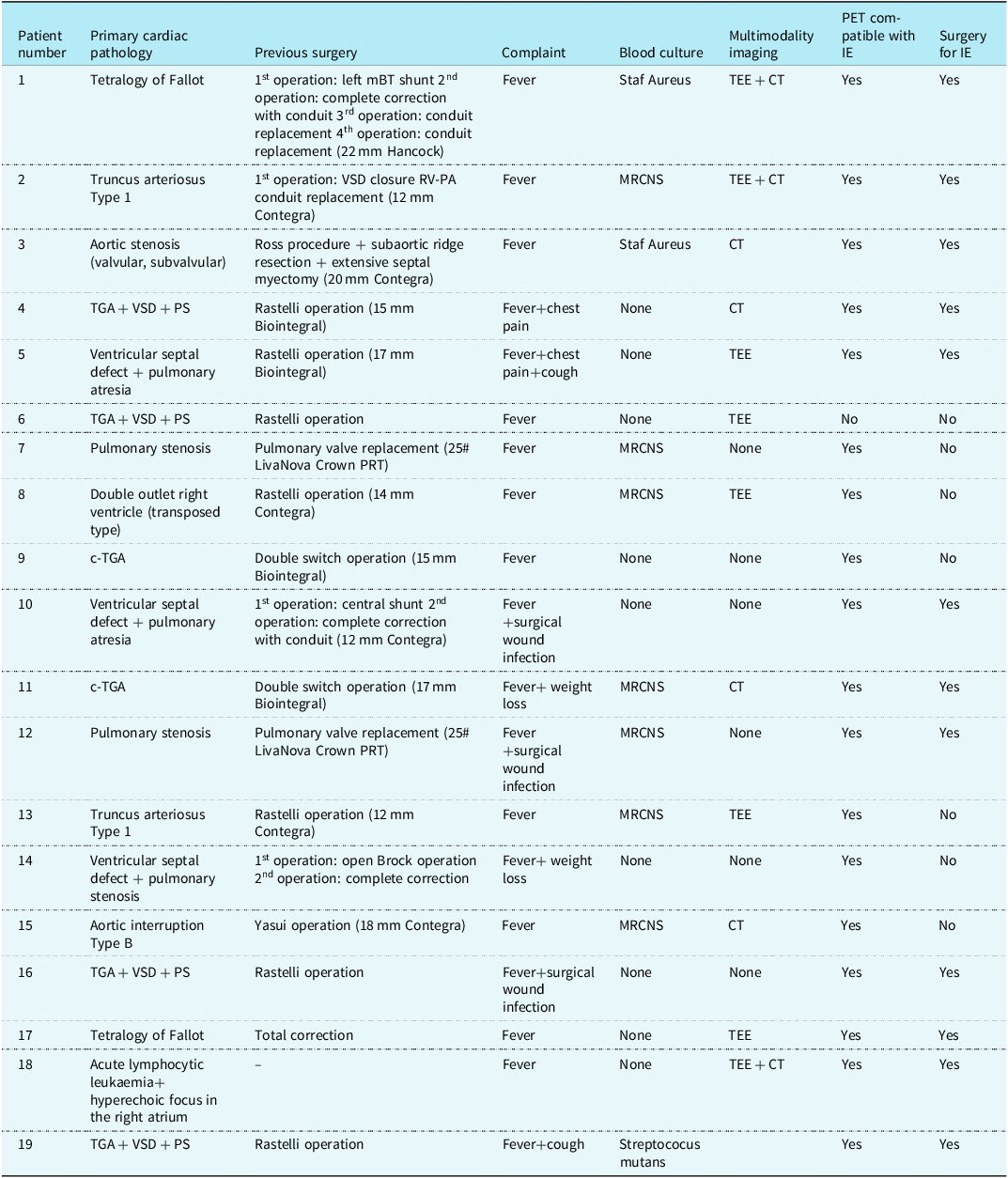
Between 2018 and 2023, our paediatric cardiology inpatient clinic admitted 54 patients with infectious endocarditis. Among them, 29 patients underwent 18F-fluorodeoxyglucose positron emission tomography/CT studies, with 19 of these patients ultimately diagnosed with right-sided infective endocarditis. Table 1 presents the demographic characteristics and clinical presentations of these 19 patients. The median age of the patients was 11 years (range: 1–22 years), comprising 11 males and eight females. Two of these patients had DiGeorge syndrome, and one was undergoing treatment for acute lymphocytic leukaemia. Among the patients, 14 had conduit replacement, and two had pulmonary valve replacement (Table 2).
Demonstrates the demographic characteristics of the patients. The duration between the complaints started and hospitalisation, initial operation and hospitalization, antibiotic therapy administration, and positron emission tomography-CT imaging were also presented

Demonstrates the primary cardiac pathology, previous surgery, initial complaints, and used additional multimodality imaging techniques. Blood culture results and whether surgery was performed were also presented in this table

CT = computed tomography; IE = infectious endocarditis; MRCNS = methicillin-resistant coagulase-negative staphylococci; PS = pulmonary stenosi; TEE = transesophageal echocardiography; TGA = transposition of the great arteries; VSD = ventricular septal defect.
All patients presented with fever, and additionally, two patients reported chest pain, two had cough, three had surgical wound infections, and two experienced more than 10% weight loss. Upon admission to our clinic, patients had been experiencing symptoms for an average of 9.4 ± 2.5 days. Physical examinations and abdominal ultrasounds revealed splenomegaly in eight (52%) patients. Among the cases, 18 had prior cardiac surgeries, with all but one of these initial surgeries performed at our centre. The mean interval between the first operation and hospitalisation was 66.8 ± 11.2 months (Table 1).
Transthoracic echocardiography was conducted for all patients during their initial hospitalisation. In three (15.8%) patients, no pathological findings were identified in transthoracic echocardiography, while vegetation was suspected but not confirmed in six (31.6%) patients, and vegetation was detected in 10 (52.6%) patients. Transesophageal echocardiography was performed in eight patients, with no pathological findings detected in three of them, while vegetation was observed in four patients.
Cardiac CT was carried out in seven patients, revealing paravalvular abscesses in three and vegetation in two patients. Haematuria was detected in three (15.7%) patients, proteinuria in 4 (21%) patients, and rheumatoid factor positivity in three (15.7%) patients. C3 values were low in six (31.5%) patients, and C4 values were low in five (26.3%) patients. In one (5.2%) patient, acute glomerulonephritis developed, characterised by elevated creatinine levels and reduced urine output. According to the Duke criteria, blood cultures were positive in 10 (52.6%) patients. Methicillin-resistant coagulase-negative staphylococci were isolated in nine cases, while streptococcus mutans was identified in one case. Nine patients had two of the major criteria of the Duke criteria, while eight patients had one major criterion, and two patients had no major criterion. Among the patients, 12 had two of the minor criteria, three patients had three criteria, and four patients had one criterion of the Duke criteria.
Fluorodeoxyglucose-positron emission tomography-CT was conducted approximately 5.2 ± 2.3 days after the initiation of infective endocarditis treatment. Patients followed an appropriate diet for an average of 3.3 days before fluorodeoxyglucose-positron emission tomography-CT, with two patients requiring two positron emission tomography-CT scans due to non-compliance with dietary instructions. In one patient, a second fluorodeoxyglucose-positron emission tomography-CT was performed to evaluate the progress of infectious endocarditis treatment, which revealed a regression of infection. Among the patients who underwent fluorodeoxyglucose-positron emission tomography-CT, 18 (94.7%) showed findings consistent with infective endocarditis (Figure 1). The average maximum standardised uptake value (SUV max) was 4.8. Pulmonary septic embolism was observed in 15 (78.9%) cases, and spleen involvement was identified in 12 (57.8%) cases.
Red arrows: Septic embolism, which is an evidence for disseminated infection is shown. Blue arrow: Infection of the conduit extending from the right ventricle to the pulmonary artery and the fistula of this infection focus to the skin are seen in this image.

Fluorodeoxyglucose-positron emission tomography-CT yielded negative results in one patient for whom no vegetations were observed on transthoracic/transesophageal echocardiography. Conversely, fluorodeoxyglucose-positron emission tomography-CT showed positive results in one patient, despite the absence of vegetations on transthoracic echocardiography, and blood culture results were negative. This patient was not diagnosed with infective endocarditis and experienced a favourable outcome during the follow-up period.
In six patients, transthoracic/transesophageal echocardiography either failed to detect vegetation or provided uncertain results. In these cases, the involvement of infective endocarditis was subsequently diagnosed based on fluorodeoxyglucose-positron emission tomography-CT findings, which were confirmed following surgical intervention. The mean hospital stay for our patients was 38.1 ± 5.6 days. Among them, 11 patients underwent surgery for infective endocarditis, while two patients tragically succumbed to the condition.
Discussion
Guidelines for the use of positron emission tomography-CT in inflammation and infection have been published by the European Association of Nuclear Medicine and the Society of Nuclear Medicine and Molecular Imaging. Reference Casali, Lauri and Altini6,Reference Pijl, Kwee and Legger7 These guidelines primarily highlight the major indications for positron emission tomography-CT, including sarcoidosis, peripheral bone osteomyelitis, suspected spinal infection, evaluation of fever of unknown origin, and the primary assessment of vasculitis. However, the guidelines currently state that it remains unclear whether positron emission tomography-CT offers advantages over other imaging techniques for diagnosing endocarditis. In an effort to address this issue, we share our experience with 18F-fluorodeoxyglucose positron emission tomography/CT in diagnosing right-sided endocarditis in paediatric patients.
Infectious endocarditis is a diagnostic challenge due to its highly variable clinical presentation. Reference Chau, Renella and Arrieta8,Reference Eleyan, Khan and Musollari9 Many patients with CHDs now possess surgical implants such as stents, mechanical valves, conduits, and assist devices that are exceptionally difficult to assess using transthoracic/transesophageal echocardiography, MRI, or even conventional CT due to imaging artefacts. Therefore, a more precise diagnostic tool is essential when there is suspicion of infection. In our study, most of the cases included had conduits or biological valves that are challenging to visualise via echocardiography. These patients often undergo recurrent surgeries and reinterventions, such as conduit replacement, which can carry a high risk of mortality and morbidity. However, when suspicious lesions arise from the right heart structures, inadequate echocardiographic views and difficulties in imaging conduits and biologic valves complicate decision-making. In our experience, transthoracic/transesophageal echocardiography yielded uncertain results in six patients, while 18F-fluorodeoxyglucose positron emission tomography/CT revealed involvement in right-sided structures. The diagnosis of infective endocarditis was subsequently confirmed following surgical intervention based on the fluorodeoxyglucose-positron emission tomography-CT findings. We conclude that 18F-fluorodeoxyglucose positron emission tomography/CT may offer high diagnostic accuracy in confirming or excluding infectious endocarditis in right-sided heart structures, aiding decision-making in paediatric cases.
18F-fluorodeoxyglucose positron emission tomography/CT can be used for two primary purposes when there is suspicion of infectious endocarditis: it can directly establish the presence of an infection in the endocardium or be employed to uncover evidence of disseminated infection, such as septic pulmonary embolism. Reference Orvin, Goldberg and Bernstine10,Reference Ten Hove, Slart, Sinha, Glaudemans and Budde11 According to data from the European Infective Endocarditis Registry, extracardiac uptake was observed in 38.8% of patients, with the most frequent uptake being in the lungs (27.1% of cases), spine (21.8%), spleen (19.6%), liver (5.0%), and bowel (18.9%). Reference Habib, Erba and Iung12 Similar to this comprehensive study, we also identified septic pulmonary embolism and 18F-fluorodeoxyglucose uptake in the spleen in most of our patients. Consequently, the identification of extracardiac foci of infection can aid in establishing the diagnosis and significantly impact treatment decisions.
In 18F-fluorodeoxyglucose positron emission tomography/CT, several pathological conditions can mimic the pattern of focally increased 18F-fluorodeoxyglucose uptake typically seen in infectious endocarditis, including active thrombi, soft atherosclerotic plaques, postsurgical inflammation, and more. Reference Shmueli, Thomas, Flint, Setia, Janjic and Siegel5 In one of our patients who had undergone a double switch operation, fluorodeoxyglucose-positron emission tomography/CT revealed 18F-fluorodeoxyglucose uptake in the conduit. However, the blood culture was negative, and no evidence of vegetation was observed on transthoracic echocardiography. Following discussions among the paediatric cardiology and cardiac surgery teams, the decision was made to monitor the patient without surgery, and the patient showed improvement during one year of follow-up.
The information provided by 18F-fluorodeoxyglucose positron emission tomography/CT can not only be used for diagnosing infectious endocarditis but also for monitoring the effectiveness of treatment, guiding therapeutic decision-making. In one of our patients who had undergone surgery five times before the infectious endocarditis episode, the sixth surgery posed a significant risk. 18F-fluorodeoxyglucose positron emission tomography/CT helped us assess the efficacy of the antibiotic treatment we administered and aided in deciding whether the patient needed surgery again despite antibiotic therapy. Since we observed a reduction in the extent of involvement in the follow-up 18F-fluorodeoxyglucose positron emission tomography/CT, the patient did not undergo additional surgery, thereby avoiding a potentially life-threatening recurrent surgery. This experience underscores the importance of considering 18F-fluorodeoxyglucose positron emission tomography/CT for evaluating the efficacy of infectious endocarditis treatment.
Given that fluorodeoxyglucose is a glucose analogue, it is typically taken up by myocardial cells. Therefore, patient preparation for infectious endocarditis involves limiting metabolic activity in the myocardium. This is achieved through dietary modifications, such as a high-fat, low-carbohydrate diet, before the 18F-fluorodeoxyglucose positron emission tomography/CT procedure. Reference Jiménez-Ballvé, Pérez-Castejón and Delgado-Bolton13 However, compliance with this diet can be challenging, especially in young children and those with chronic diseases. In our initial experience, despite diet education, we had to terminate the positron emission tomography study due to widespread fluorodeoxyglucose uptake caused by diet incompatibility in two patients. This prompted us to emphasise the importance of diet compliance during positron emission tomography procedures to both the families and nurses of the patients.
In our study, 18F-fluorodeoxyglucose positron emission tomography-CT was performed at a mean of 5.2 ± 2.3 days after the initiation of infective endocarditis treatment. In comparison, data from the European Infective Endocarditis Registry reveal that 18F-fluorodeoxyglucose positron emission tomography/CT was conducted at a mean of 8 (IQR 4.0–15.0) days after inclusion, with 46.7% of cases having the procedure within the first week of hospitalisation. Reference Habib, Erba and Iung12 The shorter time frame for conducting 18F-fluorodeoxyglucose positron emission tomography-CT in our study can be attributed to close communication with the nuclear medicine team and our proactive approach in seeking alternative imaging methods when echocardiographic findings were uncertain. This underscores the importance of having experienced infective endocarditis teams in centres specialising in structural heart diseases to facilitate prompt diagnosis and early treatment initiation.
In conclusion, based on our experience, we suggest that positron emission tomography/CT could be a valuable tool for making well-informed decisions in patients with right-sided endocarditis.
Acknowledgements
We extend our gratitude to Bekir Yükcü for his contributions to this study.
Financial support
The clinicians received no grants from any funding agency and no support from any commercial or not-for-profit sector.
Competing ınterests
None.
Ethical standards
The authors assert that this work complies with the ethical standards of the relevant national guidelines on human experimentation and with the Helsinki Declaration of 1975, as revised in 2008.