Bodily isomerism, often referred to as heterotaxy, is the clinical entity in which the thoracic organs are themselves mirror-imaged in the same individual.Reference Jacobs, Anderson and Weinberg 1 , Reference Loomba, Hlavacek, Spicer and Anderson 2 The syndrome has historically been segregated on the basis of splenic anatomy into subsets of asplenia and polysplenia.Reference Ivemark 3 , Reference Van Mierop, Gessner and Schliebler 4 Some authorities have continued to doubt the presence of isomerism within the heart.Reference Van Praagh and Van Praagh 5 When assessed on the extent of the pectinate muscles, however, it is suggested that the morphology of the atrial appendages serves to distinguish right as opposed to left isomerism.Reference Loomba, Hlavacek, Spicer and Anderson 2 , Reference Uemura, Ho, Devine, Kilpatrick and Anderson 6 Subdivision of the syndromes is important, as various findings are associated with the two subsets. Right thoracic isomerism is associated with eparterial bronchuses and trilobed lungs bilaterally, absence of a spleen, and congenital malformations of the heart requiring functionally univentricular repair. Left thoracic isomerism, in contrast, is associated with hyparterial bronchuses and bilobed lungs bilaterally, multiple spleens, and less-severe congenital cardiac malformations.Reference Jacobs, Anderson and Weinberg 1 , Reference Uemura, Ho, Devine, Kilpatrick and Anderson 6 – Reference Loomba, Shah and Anderson 10
These considerations go beyond simply being anatomical curiosities, having important functional implications. It is intuitive to understand that congenital malformations of the heart can have haemodynamic consequences. Splenic anatomy also has functional implications, with decreased or absent splenic function being present even in the setting of a single spleen, which can rarely be found in both subsets, or multiple spleens in the setting of left isomerism.Reference Nagel, Williams, Stewart, Paul and Stumper 11 Arrhythmias are also frequently noted, with different examples being found in those with right as opposed to left isomerism, likely due to known variability in the cardiac conduction system.Reference Loomba, Willes, Kovach and Anderson 12 – Reference Smith, Ho and Anderson 14 Identification of the type of isomerism within the heart, should it truly exist, therefore, can allow for appropriate anticipation and surveillance of associated findings. Echocardiography remains the current standard modality for imaging congenital malformations of the heart; therefore, it follows that it is also important to identify structures that might be identified echocardiographically so as to segregate the subsets of isomerism.Reference Teele, Jacobs, Border and Chanani 15 A recent review suggested that the atrial appendages could be distinguished echocardiographically.Reference Loomba, Aggarwal and Gupta 13 This fact, however, is not universally appreciated. In a previous study, we examined specimens contained within a single archive, confirming the morphology of the appendages served to distinguish right from left isomerism within the heart.Reference Loomba, Ahmed, Spicer, Backer and Anderson 16 We pointed at that time to the need for examination of a larger series of archived specimens. We have, therefore, now extended our previous study, examining specimens from several archives, with concentration on structures that might be imaged echocardiographically to determine the presence of right as opposed to left isomerism.
Methods
We used sequential segmental analysis to analyse hearts from patients known on the basis of previous postmortem examination to have heterotaxy.Reference Anderson, Becker and Freedom 17 , Reference Anderson and Shirali 18 The hearts were part of the Farouk S. Idriss Cardiac Registry at the Ann & Robert H. Lurie Children’s Hospital in Chicago, Illinois, the Van Mierop Archive at University of Florida, Gainesville, Florida, the archive at Johns Hopkins All Children’s Heart Institute in St. Petersburg, Florida, and the Robert M. Freedom archive held at the Hospital for Sick Children, Toronto, Canada. When available, we also examined the thoracic and abdominal organs. In this regard, bronchial morphology was assessed using the length of the bronchuses, as well as the branching pattern of the bronchial tree relative to the pulmonary arteries. Short, eparterial bronchuses were considered to be morphologically right, whereas long, hyparterial bronchuses were considered to be morphologically left. Trilobed lungs were considered to be morphologically right, whereas bilobed lungs were considered to be morphologically left.
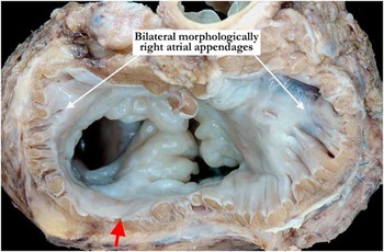
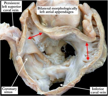
The atrial appendages were classified as either being right or left on the basis of their shape, but more importantly according to the extent of the pectinate muscles.Reference Uemura, Ho, Devine, Kilpatrick and Anderson 6 Broad and pyramidal appendages, with pectinate muscles surrounding the entirety of the adjacent atrioventricular junction, were classified as being morphologically right (Fig 1), whereas finger-like appendages, with pectinate muscles confined to the appendage, with smooth vestibules interposing between their orifice and the cardiac crux, were classified as being morphologically left (Fig 2).
This heart with isomerism of the right atrial appendages is viewed from the base. The atrial appendages are bilaterally morphologically right. Note the pectinate muscles extending around the atrioventricular junction to the crux of the heart.

A heart from a patient with left isomerism viewed from the base. Note the atrial appendages are morphologically left bilaterally. The appendages are finger-like with a narrow attachment to the venous component (double-headed red arrows). The pectinate muscles are confined to the appendages and do not extend around the atrioventricular junction with a smooth vestibule on both sides. No Eustachian valve is appreciated in this heart. A persistent left superior caval vein drains to the coronary sinus. There is also a common atrioventricular junction with tissue connecting the superior and inferior bridging leaflets across the crest of the ventricular septum, resulting in two orifices.

The superior caval veins were analysed with respect to their sidedness and drainage, as was the inferior caval vein, taking particular note of hepatic venous drainage and interruption of the return from the abdominal organs. The pulmonary veins were identified, taking particular note of non-cardiac connections. In hearts that had undergone surgical repair of the pulmonary venous connection, we consulted the original autopsy reports when available to identify the initial pulmonary venous connections. Having ascertained overall veno-atrial connections, we categorised the venous returns as being mixed, quasi-normal, or quasi-mirror-imaged. The quasi-normal arrangement refers to the situation where all of the systemic venous return was to the right-sided atrial chamber, with the pulmonary venous return entirely to the left-sided chamber. The quasi-mirror-imaged arrangement describes the reversed situation.
Within the atrial chambers, we took note of the morphology of the interatrial communication, and assessed the presence or absence of terminal crests and valves guarding the venous orifices. We also assessed the atrioventricular junctions for presence of the coronary sinus. The atrioventricular junctions were defined as being separate or common, with particular attention directed to the number of orifices within the atrioventricular valves. The atrioventricular connections themselves were classified as being biventricular and mixed or double-inlet ventricle. None of our hearts had absence of an atrioventricular connection. The ventricular mass was assessed to determine the number of discrete ventricles present, as well as the topological arrangement. We described the latter feature on the basis of right or left handedness, according to the way the palms of the hands could be laid on the septal surface of the morphologically right ventricle with the thumb in the inlet and the fingers in the outlet. Coarse apical trabeculations were used to identify the morphologically right ventricle, whereas fine and criss-crossing apical trabeculations were used to identify the morphologically left ventricle.
The ventriculo-arterial connections were classified as being concordant, discordant, double outlet, or single outlet, specifying the ventricular origin of the arterial trunks in the latter situations, with single outlet being present in the presence of aortic or pulmonary atresia. In specimens with a solitary and indeterminate ventricle, the ventriculo-arterial connections, of necessity, could only be double outlet or single outlet from the solitary ventricle. The relationships between the arterial trunks were assessed on the basis of aortic position relative to the pulmonary trunk. We described the arrangement of the arterial trunks, as they exited from the heart and entered the mediastinum, as being normally arranged and spiralling, mirror-imaged and spiralling, or parallel. We recorded the sidedness of the aortic arch depending on whether it crossed the left or the right bronchus. The aortic arch was further inspected for evidence of coarctation or interruption. The aortic valvar sinuses were inspected for the orifices of the coronary arteries, the coronary arteries themselves being assessed by following their epicardial course.
Data regarding splenic anatomy, the location of the abdominal organs, and presence of malrotation and other non-cardiac anomalies were obtained by direct examination when possible, but also by using reports from the original postmortem examination.
The frequencies of specific findings were compared between hearts found to have isomeric right as opposed to left atrial appendages. Each characteristic could not be assessed for each specimen, and hence there were missing data. The number of specimens for which data were unavailable is included for each characteristic. Percentages assigned to findings are based on a denominator consisting only of specimens for which data were available. Thus, the denominator used for each characteristic may differ. χ2 Analysis was carried out to compare categorical variables using SPSS Version 20.0 (SPSS, Chicago, Illinois, United States of America). Frequencies are reported as percentages.
Results
We assessed a total of 188 hearts obtained from patients known on the basis of the initial postmortem reports to have heterotaxy. Of these, as assessed on the basis of the atrial pectinate muscles, 131 (70%) cases had right isomerism, whereas 57 (30%) cases had left isomerism.
Right isomerism
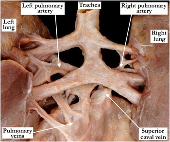
All findings are summarised in Table 1. Most (94%) of the patients with isomeric right atrial appendages also had morphologically right bronchuses bilaterally. Usual arrangement of the bronchuses was found in 4%, with mirror-imaged arrangement in 1%. A single patient had an unusual bronchial pattern, in which the trachea branched into two bronchuses, with continuation of the trachea beyond this bifurcation to produce a second bifurcation (Fig 3). In this patient, therefore, there were four bronchuses, two on each side. The lungs were morphologically right bilaterally in 89% of patients but morphologically left bilaterally in 3% of patients.
Bronchuses from a patient with right isomerism viewed from the posterior aspect. The bronchuses demonstrate an unusual branching pattern. There are four bronchuses that arise separately, two on each side.

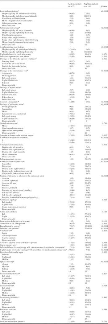
Summary of necroscopy findings

* Data were unavailable for some specimens, and thus the percentage is based on the percentage for which data were available. The denominator does not include number of specimens for which data were unavailable, thus denominators for different characteristics do differ slightly. p-Values are based on calculation excluding specimens for which there were no data
The atrial appendages were morphologically right in all cases, with concordance in all between the shape and the extent of the pectinate muscles (Fig 1). In all right isomeric hearts, we also found evidence of terminal crests, with presence of a valve guarding the venous orifice noted in 43 patients. A right-sided superior caval vein was present in 83% of patients, which always drained directly into the roof of the right-sided atrium. A left-sided superior caval vein was present in 68%, again draining directly into the roof of the left-sided atrium. The coronary sinus was universally absent. The inferior caval vein drained into the right-sided atrium in 73% of patients, into the left-sided atrium in 33%, and was divided to drain into the atrial chambers bilaterally in 4% of cases. In 66% of hearts, the hepatic veins drained directly into the inferior caval vein, but drained separately into the left-sided atrium in 2% and directly into the right-sided atrium in 28%. The pulmonary veins connected directly to the right-sided atrium in 20% of patients and to the left-sided atrium in 19%. Totally anomalous infradiaphragmatic connection was present in 23%, supracardiac connection in 28%, and mixed connections in 7%. Taken together, the veno-atrial connections were mixed in 90%, quasi-usual in 7%, and quasi-mirror-imaged in 3%. The cardiac apex was leftward in 60% and rightward in 40%.
The atrioventricular connections were biventricular and mixed in 82%, with 3% showing double inlet into a solitary and indeterminate ventricle, 10% showing double-inlet right ventricle, and 5% showing double-inlet left ventricle. A common atrioventricular junction was noted in 79%. In 109 of these hearts, there was also a common atrioventricular valvar orifice (Fig 1). There was right-handed ventricular topology in 61%, left-handed topology in 36%, and a single indeterminate ventricle in 3%. Concordant ventriculo-arterial connections were found in 6% of hearts with right-handed ventricular topology and in 4% of hearts with left-handed topology. The ventriculo-arterial connections were discordant in 53%, double outlet from the right ventricle in 34%, single outlet from an indeterminate ventricle with pulmonary atresia in 2%, and double outlet from an indeterminate ventricle in 1%. Pulmonary atresia was present in 58 (45%) of hearts, with coarctation noted in only 11%, with no patient having interruption of the aortic arch. The aorta was most commonly anterior and rightward in relation to the pulmonary trunk (33%). A left-sided aortic arch was present in 63% and a right-sided arch in 37%. Abnormal coronary arterial distribution was identified in 66% of the hearts. The abnormal patterns noted included a single coronary artery arising from sinus 1, a single coronary artery arising from sinus 2, two coronary arteries arising separately from sinus 1 with a third arising from sinus 2, and two coronary arteries arising separately from sinus 2, with a third arising from sinus 1. In one of the patients with three coronary arterial orifices, evidence of multiple myocardial infarctions had been noted during the initial autopsy, none of which were deemed to be acute. Thrombi occluding portions of the vessels were present in multiple coronary arteries.
In patients with available information, we ascertained that 87% had absence of spleen, with 4% having multiple spleens, and 9% having a solitary left-sided spleen. The stomach was left sided in 64% of patients in whom this information had been recorded. Nearly two-thirds (64%) of all patients had a midline liver, with 28% described as having a right-sided liver and 8% a left-sided liver.
Left isomerism
All findings are detailed in Table 1. All except two patients with left isomerism had morphologically left bronchuses, with the remaining two patients having usual bronchial arrangement. Most (80%) had morphologically left lungs bilaterally. In 4%, we found single-lobed lungs bilaterally, whereas 8% had usual pulmonary arrangement. In 4% of patients, we found morphologically right lungs bilaterally, whereas the remaining 4% of patients had mirror-imaged pulmonary arrangement.
Both atrial appendages were morphologically left in all hearts (Fig 2), with the anticipated shape of the appendages being concordant with the extent of the pectinate muscles, even in hearts obtained from patients known to have had significant haemodynamic disturbances as outlined in the reports of postmortem examinations. A terminal crest could not be identified in any patient with isomeric left appendages, but 17% had valvar structures guarding the veno-atrial junctions.
The atrioventricular connections were biventricular and mixed in 91% of patients. In five patients, there was a double inlet, with four of these patients having a double-inlet right ventricle and one having a double-inlet single ventricle. There was a common atrioventricular junction in 65% of hearts, with 14 of these hearts having separate atrioventricular orifices for the right and left ventricles (Figs 1 and 3). Left-handed ventricular topology was found in 34% and right-handed ventricular topology in 66% patients. The cardiac apex was pointed leftward in 57%, rightward in 41%, and was midline in the remainder. The ventriculo-arterial connections were concordant in 58% and discordant in 13%, and double-outlet right ventricle was found in 28% and double outlet from an indeterminate ventricle in 1%. All hearts with double-outlet right ventricles had subaortic interventricular communications. Pulmonary atresia was present in 9%. Coarctation of the aorta was present in 16%, and interruption of the aortic arch in 2%. The aortic arch was left sided in 75%. Abnormal coronary arterial distribution was noted in 66% of patients, with a single coronary arising either from sinus 1 or from sinus 2 in five of these patients (Fig 6).
A right-sided superior caval vein was present in 82% of hearts, with the vein always entering the roof of the right atrium. A left-sided superior caval vein was present in 64%, draining through the coronary sinus to the right-sided atrium in 17% of these patients and to the atrial roof in the remainder (Fig 4). The inferior caval vein was most often interrupted, with this feature being found in 78% of hearts. When interrupted, the abdominal venous return reached the superior caval vein through the azygos venous system. In 6%, the inferior caval vein drained directly into the left-sided atrium, and in 16% it drained directly into the right-sided atrium (Fig 4). The hepatic veins drained into the inferior caval vein in 69%. In 14%, the hepatic veins drained directly into the right-sided atrium, and in another 7% the hepatic veins drained into the left-sided atrium. A coronary sinus within the atrioventricular groove was identified in 80% of hearts, with its mouth identified in the right-sided atrium (Fig 5). The pulmonary veins connected symmetrically in 36%, to the right-sided atrium in 39%, and to the left-sided atrium in 25% (Fig 6). Assessed overall, the veno-atrial connections were mixed in 83%, showed quasi-normal arrangement in 7%, and were quasi-mirror-imaged in 10% (Figs 7 and 8).
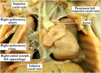
A heart from a patient with left isomerism viewed from the anterior aspect. A right-sided morphologically left appendage is visible. A right-sided superior caval vein is connecting to the right-sided atrium with a left-sided superior caval vein returning to the left-sided atrium. The inferior caval vein is also connecting to the right-sided atrium.

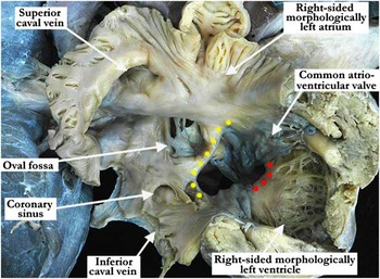
A heart from a patient with left isomerism viewed from the right-sided superior aspect. The right-sided atrium and ventricle are opened, revealing a morphologically left appendage. Although the atrium is dilated, it retains the finger-like shape and has pectinate muscles that are mostly confined to the appendage and do not extend around the entirety of the atrioventricular junction. A superior caval vein is connecting to the right-sided atrium as is an inferior caval vein. There is a fenestrated oval fossa defect and a well-formed coronary sinus. A common atrioventricular junction is present with a single orifice, with the yellow dots marking the leading edge of the atrial septum and the red dots the crest of the ventricular septum. Note the right-sided, morphologically left ventricle.

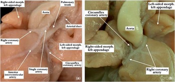
A heart from a fetus at 15 weeks of gestation with left isomerism. ( a ) The heart from the anterior and leftward aspect. There is a single coronary artery that is arising from the leftward-facing sinus (sinus 1). The anterior interventricular artery and the right coronary artery are arising from this single coronary artery. The right coronary artery later gives rise to the circumflex coronary artery and it extends posterior to the arterial pedicle to resume its usual position in the left atrioventricular groove. ( b ) Same heart from the anterior and rightward aspect. The right coronary artery gives rise to the circumflex artery, which extends posterior to the arterial pedicle.

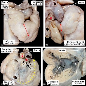
( a ) The left-sided, morphologically left atrial appendage, with a left superior caval vein. The appendage is tubular with a narrow attachment (double-headed red arrow) to the atrial vestibule. ( b ) The right-sided, morphologically left atrial appendage that is tubular and has a narrow attachment (double-headed red arrow) to the atrial vestibule. There is a persistent, right superior caval vein that drains to the coronary sinus within the left-sided atrium. Note the dilated appearance of the left-sided atrium in a (red star – right pulmonary artery). ( c ) The posterior aspect of the atria, with the superior and inferior caval veins draining into the left-sided atrium and the pulmonary veins (black square and arrows) draining into the right-sided atrium. The persistent right superior caval vein (yellow arrow) drains postero-inferiorly (yellow dots) into the left-sided coronary sinus (red stars – right and left pulmonary arteries). ( d ) The apex of the heart towards the right and the right ventricle is opened in clam-shell manner. There is a common atrioventricular (AV) valve, concordant ventriculo-arterial connections, and left-hand topology. Note the increased ventricular trabeculations consistent with so-called non-compaction.

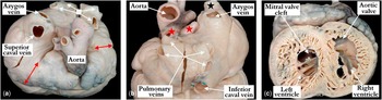
( a ) Superior view of the base of the heart demonstrating bilateral, morphologically left atrial appendages. Both appendages are tubular in nature with a narrow attachment with the atrial vestibule (double-headed red arrows). The superior caval vein drains to the right-sided atrium, and there is a large azygos vein in this patient with azygos continuation of the inferior caval vein. The pulmonary veins (white square and arrows) drain to the left-sided atrium. ( b ) Posterior view of the venous drainage with the superior (black star) and inferior caval veins draining to the right-sided atrium. The pulmonary veins (white arrows) drain to the left-sided atrium. The right and left pulmonary arteries are marked with red stars. ( c ) A short-axis apical view demonstrating the so-called non-compaction of both right and left ventricles. The right ventricle is left-sided and there is transposition. There is a ventricular septal defect (not seen) and a true cleft of the right-sided mitral valve.

In patients in whom it was possible to determine the location of the abdominal organs, we ascertained that multiple spleens were present in 88%, with 2% noted to have absence of spleen, and 10% patients noted to have solitary spleens. The stomach was known to be left sided in the majority (57%). The liver was right-sided in 41% of patients with available data, midline in 38%, and left-sided in 21%. The gallbladder was left sided in 21% of the patients in whom this feature had been described.
Differences between right and left isomerism
We have summarised the significant differences between patients having isomeric right as opposed to left atrial appendages in Table 1. The bronchial morphology is almost always consistent with the type of appendage isomerism (p<0.0001). The same is noted for pulmonary morphology (p<0.0001). A valvar structure guarding a ven-oatrial connection was frequently noted with right isomerism (p<0.0001), with a terminal crest always noted with right isomerism, but never with left isomerism (p<0.0001). The drainage of the left-sided superior caval vein differed significantly between the subsets (p<0.0001), driven by the fact that the coronary sinus was found only in those with isomeric left appendages (p<0.0001). The hepatic veins more commonly drained directly into the inferior caval vein in those with right isomerism and directly into the right-sided atrium in those with left isomerism (p<0.0001). Pulmonary venous connections also differed between the two subsets, with totally anomalous pulmonary venous connection being the rule in presence of isomeric right appendages, even when the pulmonary veins connect directly to either the right-sided or the left-sided atrial chamber (p<0.0001). No difference was noted, however, in the overall patterns of venous return.
The presence of a common atrioventricular junction was more frequent in right isomerism (p=0.035), although the presence of a common atrioventricular junction divided into a valve with separate atrioventricular orifices was more frequently found in the setting of left isomerism (p<0.0001). Pulmonary atresia was more frequent in the setting of right isomerism (p<0.0001). The ventriculo-arterial connections also differed between the two subsets, with right isomerism being associated with discordant ventriculo-arterial connections and double-outlet right ventricle more commonly than left isomerism, which was more commonly associated with concordant ventriculo-arterial connections (p<0.0001). The aortic arch was more commonly right-sided in those with right isomerism (p=0.034). Left-handed ventricular topology with concordant ventriculo-arterial connections was more frequently found in those with left isomerism when compared with those with right isomerism. Right-handed ventricular topology with concordant ventriculo-arterial connections was also more frequently found in those with left isomerism when compared with those with right isomerism (p<0.0001).
Discussion
Our study has served to confirm the notion that, when assessing cardiac morphology, heterotaxy can reliably be differentiated into isomeric subsets on the basis of the morphology of the atrial appendages, with this feature being distinguished according to the extent of the pectinate muscles relative to the atrioventricular junction.Reference Uemura, Ho, Devine, Kilpatrick and Anderson 6 The atrial appendage is known to be the most consistent feature of the atrial chambers, and is now confirmed by our study to demonstrate isomerism. Our study also confirms that the terminal crest is found only in the presence of isomeric right atrial appendages, whereas the coronary sinus is universally absent in this setting. We have also shown that, as might be expected, there is good concordance between the bronchial morphology and the arrangement of the atrial appendages. Splenic morphology, in contrast, although indicative of the type of isomerism, was more frequently discordant. This is again not unexpected, as the abdominal organs, although usually being abnormally located in the setting of heterotaxy, do not show evidence of isomerism.
When considered from the stance of cardiac morphology, the distinction between right and left isomerism is important, as each subset is associated with other anatomical and functional disturbances. This finding disputes the notion that isomerism within the heart is no more than a “useful mnemonic”.Reference Van Praagh and Van Praagh 5 Our investigation endorses the concept established by Uemura and his colleagues, namely that it is the appendages that are isomeric, and that their morphology can be distinguished according to the extent of their contained pectinate muscles. Classification of isomerism on this basis, therefore, can allow for anticipation of, surveillance for, and counselling with regard to associated findings. This is particularly important with respect to cardiac arrhythmias, as well as splenic function and subsequent risk of infection.
In a minority of our cases, the venous returns were such as to produce “normal” drainage, with all the systemic venous return draining to the right-sided atrium, and all the pulmonary veins draining to the left-sided atrium. If assessed in terms of haemodynamics, these arrangements could be considered to represent “situs solitus”. Other hearts in our cohort showed the venous returns to have mirror-imaged drainage. In both these situations, however, the atrial arrangement, as based on the morphology of the appendages, was isomeric. It is this latter feature that determines the location of the sinus node, and hence paramount when defining atrial arrangement. It is also important, nonetheless, to describe the venous drainage, which we achieved by accounting for the quasi-normal or quasi-mirror-imaged arrangements. The significance of these arrangements has recently been emphasised in the clinical setting. They are of particular significance when assessed in the context of the ventriculo-arterial connections present – for example, if patients with quasi-mirror-imaged venous drainage have left-handed ventricular topology, with concordant ventriculo-arterial connections, this will produce the haemodynamic situation synonymous to that of transposition. Quasi-usual venous drainage, with left-handed topology and concordant ventriculo-arterial connections, will resemble congenitally corrected transposition. Such arrangements were found in the cited, recent clinical study.Reference Laux, Houyel, Bajolle, Raimondi, Boudjemline and Bonnet 19 Some might describe these rare combinations as “isolated ventricular discordance” if found with usual venous drainage, or even as “isolated ventricular non-inversion” if found with quasi-mirror-imaged venous drainage. Our preference in such situations is to provide a full segmental description, but by placing emphasis on the isomeric nature of the atrial appendages.
Our study is exclusively morphological. This is relevant to the fact that echocardiography remains the gold standard for clinical assessment of cardiac anatomy and function in the setting of congenital cardiac malformations. It would be of great value, therefore, if it were shown to be possible to differentiate between right and left isomerism by echocardiographic interrogation of the appendages. Although ideal in this setting, this process is difficult and time-consuming. Evidence has recently emerged, nonetheless, to show that distinction is feasible.Reference Teele, Jacobs, Border and Chanani 15 These investigators showed that other distinguishing features, as identified by our study, are readily identified by echocardiography. Thus, the presence or absence of the coronary sinus can readily be established, as can the presence of the terminal crest. In the setting of a clinical diagnosis of heterotaxy, if the coronary sinus is present, then it can reliably be inferred that there is left isomerism. Absence of the coronary sinus, however, does not rule out left isomerism. If present, the body of the coronary sinus can readily be visualised running in the left atrioventricular groove along the posterior aspect of the heart in the parasternal long-axis plane. It is particularly prominent from this echocardiographic plane in the setting of a left-sided superior caval vein, which drains through the coronary sinus. The four-chamber plane also allows for visualisation of the body of the coronary sinus, although it can also be imaged echocardiographically using a subcostal imaging plane through the most posterior aspect of the heart.Reference Eidem, Cetta and O’Leary 20 Echocardiogarphic imaging of atrial morphology, therefore, should now make it feasible to segregate heterotaxy into its right and left isomeric subtypes.Reference Teele, Jacobs, Border and Chanani 15 Bronchial morphology is usually concordant with atrial appendage morphology, but not perfect. The concepts of asplenia and polysplenia syndromes, in contrast, are now well established, from the cardiac stance, as being inadequate in appropriately segregating heterotaxy.Reference Uemura, Ho, Devine, Kilpatrick and Anderson 6 , Reference Teele, Jacobs, Border and Chanani 15 Although they may be of historical significance, they should no longer be used when trying to infer the likely cardiac associations.
The strengths of our study include the ability to thoroughly assess cardiac structures, as postmortem specimens were being analysed. In addition, ours is one of the largest investigations of autopsied hearts from patients known to have heterotaxy. The availability of original postmortem reports also allowed collection of additional clinical data. Limitations of the study are the lack of availability of clinical and functional data for all patients. We have shown, nonetheless, that so-called heterotaxy can reliably be segregated into the subsets of left and right isomerism on the basis of the morphology of the atrial appendages, defining this feature according to the extent of the pectinate muscles. Identification of isomerism in this way allows for anticipation and surveillance of associated findings.
Conclusion
Isomerism is uniformly segregated on the basis of the morphology of the atrial appendages, itself defined by the extent of the pectinate muscles. Other features such as the presence of a coronary sinus and the systemic venous return can further help with such segregation of isomerism.