


Highlights
-
There are ethnic phenotypic and genotypic differences in PLA2G6-associated neurodegeneration among the Indian, Asian, Middle East and European cohorts.
-
The variant c.2222G > A,p.R741Q was common in the Indian and Middle East cohorts.
-
The c.2222G > A,p.R741Q variant had a relatively severe phenotype, whereas the c.991G > T,p.D331Y variant had a milder phenotype.
Introduction
PLA2G6-associated neurodegeneration (PLAN) (OMIM: 610217) is a heterogeneous group of rare autosomal recessive neurodegenerative disorders caused by variants in the PLA2G6 gene. Reference Guo, Tang and Guo1 PLAN is the second most common type of neurodegeneration with brain iron accumulation (NBIA) spectrum disorders and is characterised by abnormal iron accumulation in the basal ganglia, substantia nigra, thalamus and cerebellum, axonal swelling and presence of the spheroid bodies throughout the peripheral and central nervous systems including the cerebellar cortex, globus pallidus (GP), brainstem and dorsal column of the spinal cord. Reference Balsinde and Balboa2 This gene encodes for the enzyme iPLA2β/iPLA2-VI, a group VIA calcium-independent A2 phospholipase, which is involved in the hydrolysis stage of the ester bonds in fatty acyl groups of phospholipids, producing free fatty acids and lysophospholipids. It is involved in the phospholipids metabolism essential for maintaining cell membrane integrity as well as in oxidative stress because the hydrolytic activity of iPLA2-VI on the membrane phospholipids releases arachidonic acid, whose metabolism generates reactive oxygen species (ROS). This gene is located on the long arm of chromosome 22 (22q13.1) and contains 17 exons spanning more than 69 k base pairs. Reference Holla, Samim and Kumari3 It contains three domains: the conserved GXSXG catalytic site (517–521), the calmodulin binding region (747–759) and the ankyrin repeat region (150–381). The phenotypic presentation is varied based on the age at onset, clinical features and disease progression and can be classified as infantile neuroaxonal dystrophy (INAD, NBIA2A; MIM 256600), atypical neuroaxonal dystrophy (aNAD, NBIA2B; MIM 610217) and adult-onset dystonia-parkinsonism (DP, PARK14; MIM 612953) and autosomal recessive early-onset parkinsonism (AREP). INAD and aNAD present during childhood, whereas DP and AREP are reported to occur in adults. The other phenotypic presentation of adult-onset PLAN is ataxia, hereditary spastic paraplegia (HSP), complex dystonia and dystonia-parkinsonism-myoclonus (DPM). Reference Dehnavi, Bemanalizadeh and Kahani4,Reference Bohlega, Al-Mubarak and Alyemni5 Brain MRI shows mineralisation in the basal ganglia, substantia nigra and cerebellar, basal ganglia and cerebral atrophy. The adult-onset PLAN shows phenotypic variability as different variants cause a diversity in the enzymatic activity. Reference Engel, Jing, O’Brien, Sun and Kotzbauer6,Reference Gui, Xu, Wen, Liu, Zhao and Hu7 There are reported cases of adult-onset PLAN mainly from China, Taiwan and Japan. The clinical, imaging and genetic profile from the Indian cohort is lacking. The objective of this study was to describe the clinical, imaging and genetic profile of our patients with adult-onset PLAN and compare the Indian cohort with the Asian, Middle East and European cohorts, with a focus on genotype–phenotype correlations, and to study ethnic phenotypic and genotypic differences of adult-onset PLAN.
Methods
Patients and methods
This was a retrospective, cross-sectional, descriptive analysis of patients with adult-onset PLAN conducted at a quaternary care hospital in India. Hospital records of patients with genetic confirmation of adult-onset PLAN (onset age older than 18 years) between 2015 and 2023 were searched and included. Reference Chu, Lin, Chen and Lin8 The following clinical data were extracted, which include age at clinical presentation, age at onset of symptoms (age at which the first neurological sign had been noted), duration of disease (onset of first symptom to the time of presentation), sex, types of symptoms (e.g. dystonia, myoclonus, parkinsonism, spasticity, cerebellar ataxia, psychiatric symptoms, cognitive symptoms, etc.), birth history, consanguinity and family history. The clinical examination findings including fundus, cranial nerves, motor, sensory, cerebellar, extrapyramidal examination findings were recorded. A detailed description of dystonia – including the initial site of onset (cranial/oro-mandibular, cervical or brachial/crural), progression to segmental/generalised dystonia, and time from onset to generalisation and ambulatory status – was collected. Details about site and side of onset of parkinsonian symptoms, levodopa response, levodopa-induced dyskinesia, motor fluctuations, disease severity and staging were collected. The phenotypic classifications included were DP, DPM, AREP, cerebellar ataxia, HSP and isolated or complex dystonia. Brain MRI was analysed to look for abnormalities that include atrophy in the cortical, basal ganglia, cerebellar and brainstem regions; signal changes on T1-weighted imaging, T2-weighted imaging, fluid-attenuated inversion recovery, diffusion-weighted imaging and apparent diffusion coefficient image; and iron deposition on susceptibility-weighted imaging. Information about the PLA2G6 variants – missense, frameshift, stop-gain and splice-site loss – and their zygosity (homozygous or compound heterozygous) was collected. The study was conducted after approval by the ethics committee (No: NIMH/DO/DEAN [Basic Science]/2020–21). Informed written consent was obtained from the patients for publication. The study was conducted in accordance with the Declaration of Helsinki (revised in 2013).
Genetic analysis
Genomic DNA extraction using QIAamp DNA Blood Mini Kit (Qiagen Germany, #51104), was performed on the blood samples of the recruited patients. Identification of variants employed the Genome Analysis Toolkit framework (Broad Institute, Cambridge, MA, USA). The variants were annotated utilising the online platform ANNOVAR (http://www.openbioinformatics.org/annovar/ [34791404]). The common variants with a minor allele frequency >0.01 were not considered. Comparison was made with Exome Aggregation Consortium, 1000 Genome project and gnomAD database (https://gnomad.broadinstitute.org/). The individual sequence variants were interpreted using PolyPhen-2, Sorting Intolerant from Tolerant (SIFT) webserver and MutationTaster. The variants were classified in accordance with American College of Medical Genetics and Genomics standards and guidelines into benign, likely pathogenic or pathogenic. The variants were checked for their novelty by analysis in mutation databases (ClinVar).
Review of literature
We conducted a comprehensive search across the publicly accessible medical database of PubMed, employing specific medical subject headings: “adult-onset,” “PLA2G6-associated neurodegeneration” and “PLAN” to identify relevant studies. Initially, studies underwent a rigorous title and abstract screening process. Subsequently, studies focusing on the Asian and European populations were included for further analysis. We included our current study data and other cases published in India and compared them with the Asian and European cohorts that were last updated in 2023. The data extraction process encompassed the retrieval of critical information, including demographic parameters, clinical phenotype descriptions and variant details, to facilitate a comprehensive evaluation of the existing literature on this topic. Studies not presented in English or lacking patient details were excluded from consideration. We did shadow search of the reference articles to avoid missing key articles.
Statistical analysis
Summary statistics were used to describe the clinical, imaging and genetic characteristics that were reported. The continuous variables were expressed as means with standard deviation/median and categorical variables as frequencies/percentages. Summary statistics was used for the clinical, genetic and imaging characteristics of patients based on the genotype and phenotype. The comparison of means among the groups was performedusing ANOVA and the frequencies using the chi-square test.
Results
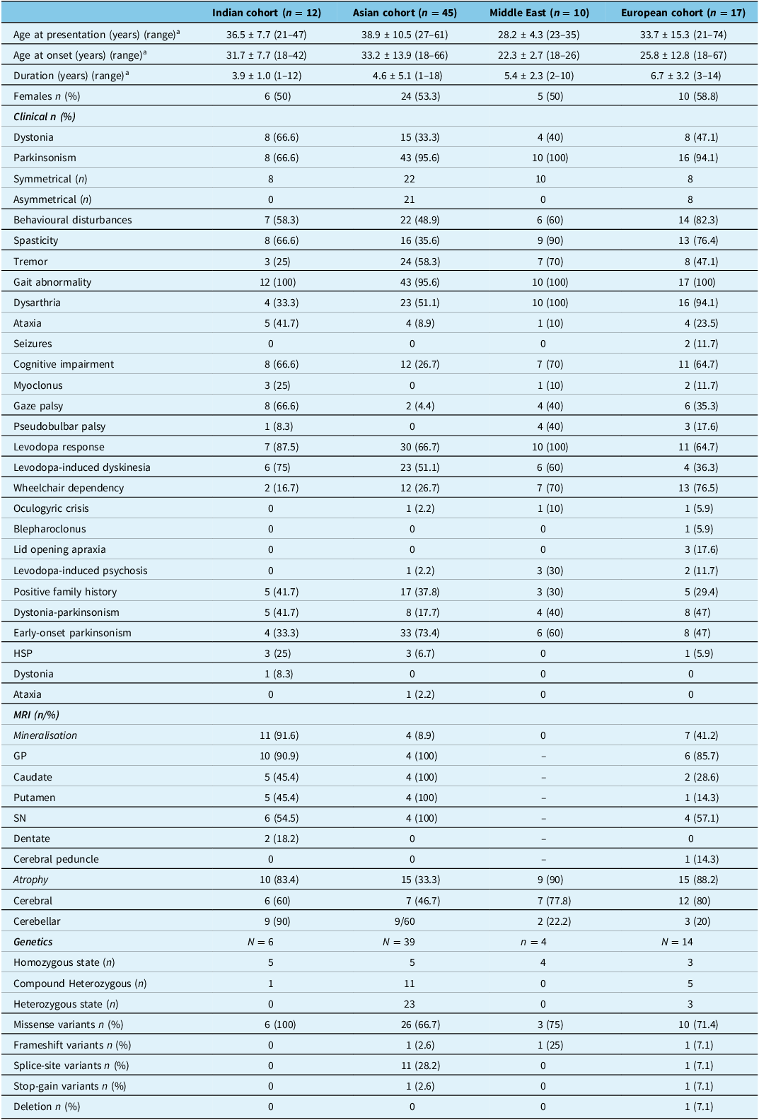
We had two patients with adult-onset PLAN available for assessment in our cohort. Ten cases of adult-onset PLAN were already published from our institute. The Indian cohort of 12 patients was compared with the Asian, Middle East and the European cohorts. Two patients included in this study were both males aged 37 and 47 years and had a median onset at 37 years (33–42 years) of age. The median duration of symptoms was 4.5 years (4–5 years).
Clinical findings (n = 2)
Case 1
The patient was born of consanguineous parentage and had initial symptoms of slowness in walking and slowness in daily activities of 3 years duration. He had depression 1 year before the onset of parkinsonism. There was no anxiety, tremors, myoclonus or seizures. Family history was negative. Examination showed affected cognitive domains involving attention, execution and memory, vertical gaze palsy, slow saccades, pseudobulbar palsy, brisk deep tendon reflexes with extensor plantar responses, symmetrical bradykinesia and rigidity (axial and appendicular), postural instability and bibrachial dystonia. The patient was treated with 800 mg of levodopa, but there was no response. The patient was wheelchair bound within 4 years of onset. The overall phenotype was early-onset parkinsonism-dystonia with dementia and rapid progression.
Case 2
The patient was born out of non-consanguineous parentage and presented with slowness in walking and activities of daily life of 2 years duration. The parkinsonism was symmetrical in onset without tremors, associated with postural instability. Patient had freezing of gait (FOG) with start and turn hesitation. There was no dystonia, myoclonus, seizures or psychiatric symptoms. There were no affected family members. Examination showed impaired attention and execution, vertical gaze palsy, slow saccades, symmetrical bradykinesia and rigidity (axial and appendicular), postural instability and FOG. The patient was treated with 600 mg of levodopa (tolerated dose) and showed no response. The patient was ambulant at 1-year follow-up. The overall phenotype was early-onset parkinsonism (EOP) with FOG.
Imaging profile
Brain MRI showed diffuse cerebral and cerebellar atrophy with mineralisation in bilateral GP and dentate in cases 1 and 2.
Genetic profile
Case 1
A homozygous missense variant in exon 16 of the PLA2G6 gene (c.2264G > A) causing amino acid substitution of glutamine for arginine at codon 755 (p.Arg755Gln; ENST00000668949.1) was detected. The p.Arg755Gln variant has not been reported in the 1000 genomes and gnomAD databases. The in silico predictions of the variant are possibly damaging by PolyPhen-2(HumDiv) and damaging by SIFT, LRT and MutationTaster2.
Case 2
A homozygous missense variant in exon 16 of the PLA2G6 gene (c.2222G>A) causing amino acid substitution of glutamine for arginine at codon 741 (p.Arg741Gln) was detected. The p.Arg741Gln variant has not been reported in the 1000 genomes and gnomAD databases. The in silico predictions of the variant are possibly damaging by PolyPhen-2(HumDiv) and damaging by SIFT, LRT and MutationTaster2.
Other cases from the Indian cohort (n = 10) Reference Holla, Samim and Kumari3
A review of 10 reported cases from India showed male predominance (60%), median age of onset of 23 years (range: 18–28 years) and median duration of 3 years. The predominant symptoms were gait abnormalities, dystonia and spasticity, followed by parkinsonism, ataxia, psychiatric symptoms and cognitive impairment. Tremor, myoclonus and pseudobulbar palsy were less common symptoms. Family history was positive in five patients. Levodopa response was seen in seven patients with levodopa-induced dyskinesia in six patients. Nine patients were ambulant at the time of presentation. The phenotypic presentations were DP in two, DPM in two, complicated HSP in three, complex dystonia in one and EOP in two patients. Brain MRI showed mineralisation in nine patients, with GP mineralisation in eight patients. Cerebellar and cerebral atrophy was seen in seven and four patients, respectively. Genetic analysis showed four homozygous and one compound heterozygous variants, and all were missense variants. Six patients had homozygous missense variant (c.2222G > A p.R741Q) (Table 1).
Comparison of clinical features, imaging and genetic findings among the cohorts

Table 1 Long description
The table compares clinical features, imaging, and genetic findings across four cohorts: Indian, Asian, Middle East, and European. It has 12 rows and 17 columns. The columns are labeled as Indian cohort (n = 12), Asian cohort (n = 45), Middle East (n = 10), and European cohort (n = 17). The rows are labeled with various clinical and genetic parameters. Row 1: Age at presentation (years) (range), Row 2: Age at onset (years) (range), Row 3: Duration (years) (range), Row 4: Females n (%), Row 5: Dystonia, Row 6: Parkinsonism, Row 7: Symmetrical (n), Row 8: Asymmetrical (n), Row 9: Behavioural disturbances, Row 10: Spasticity, Row 11: Tremor, Row 12: Gait abnormality, Row 13: Dysarthria, Row 14: Ataxia, Row 15: Seizures, Row 16: Cognitive impairment, Row 17: Myoclonus, Row 18: Gaze palsy, Row 19: Pseudobulbar palsy, Row 20: Levodopa response, Row 21: Levodopa-induced dyskinesia, Row 22: Wheelchair dependency, Row 23: Oculogyric crisis, Row 24: Blepharospasm, Row 25: Lid opening apraxia, Row 26: Levodopa-induced psychosis, Row 27: Positive family history, Row 28: Dystonia-parkinsonism, Row 29: Early-onset parkinsonism, Row 30: HSP, Row 31: Dystonia, Row 32: Ataxia, Row 33: MRI (n/%): Mineralisation, Row 34: GP, Row 35: Caudate, Row 36: Putamen, Row 37: SN, Row 38: Dentate, Row 39: Cerebral peduncle, Row 40: Atrophy: Atrophy, Row 41: Cerebellar, Row 42: Cerebral, Row 43: Genetics: Homozygous state (n), Row 44: Compound Heterozygous (n), Row 45: Heterozygous state (n), Row 46: Missense variants n (%), Row 47: Frameshift variants n (%), Row 48: Splice-site variants n (%), Row 49: Stop-gain variants n (%), Row 50: Deletion n (%). Each cell contains specific data points relevant to the respective cohort and parameter.
HSP = hereditary spastic paraplegia; GP = globus pallidus; SN = substantia nigra.
a Mean ± standard deviation.
Asian cohort (n = 45) Reference Gui, Xu, Wen, Liu, Zhao and Hu7–Reference Kim, Lyoo, Hong, Kim and Lee22
Clinical findings
The Asian cohort had 13 patients reported from China, 21 patients from Japan, 10 patients from Taiwan and 1 patient from South Korea. The median age of presentation of the reported patients from China was 39 years (range: 27–61 years), median age at onset was 36 years (range: 23–59 years), median duration of illness of 2 years (range: 1–12 years) and male preponderance (69.2%). Family history was reported in 15.4%. The median age of presentation of the reported patients from Japan was 38 years (range: 34–53 years), median age at onset was 27.5 years (range: 18–66 years), median duration of illness of 11.5 years (range: 8–16 years) and slight female preponderance (52.4%). Family history was positive in 57.1%. The median age of presentation of the reported patients from Taiwan was 36 years (range: 31–48 years), median age at onset was 29 years (range:19–31 years), median duration of illness was 9 years (range: 5–18 years) and female preponderance (80%). Family history was seen in 20%. Parkinsonism and tremor were the most common symptoms in the reported cases from China and Japan, and parkinsonism and psychiatric symptoms were commonly reported from Taiwan. One patient had an oculogyric crisis from Taiwan. The parkinsonism was levodopa responsive in 50%–81% of patients, and levodopa-induced dyskinesias occurred in 55%–100% of patients. The phenotypic presentations were parkinsonism in 33 patients, DP in 8 patients, HSP in 3 patients and cerebellar ataxia in 1 patient. Wheelchair dependency was reported in 12 patients. Brain MRI showed mineralisation in three patients with cerebellar and cerebral atrophy in six and two patients, respectively, from China. Mineralisation was not reported from Taiwan and Japan, and two patients had cerebellar and cerebral atrophy from Taiwan and three patients from Japan had only cerebral atrophy. Heterozygous variants were common from China and Japan and compound heterozygous variants from Taiwan. Missense variants were commonly reported in all these countries. The homozygous/compound heterozygous variant (c.991G > T, p.D331Y) was the most common variant in the Asian cohort (33.3%) (Table 1).
Middle East cohort (n = 10) Reference Dehnavi, Bemanalizadeh and Kahani4,Reference Bohlega, Al-Mubarak and Alyemni5,Reference Sina, Shojaee, Elahi and Paisán-Ruiz23
The Middle East cohort had 10 cases reported with a median age of presentation of 29 years (range: 23–35 years), median age of onset of 22.5 years (range: 18–26 years) and median duration of illness of 6 years (range: 2–10 years), with an equal gender ratio. Parkinsonism, tremor and cognitive impairment were the common symptoms. Parkinsonism was levodopa responsive with levodopa-induced dyskinesia in 60% of patients with parkinsonism. One patient had an oculogyric crisis. Three patients had a positive family history. The phenotypic presentation was parkinsonism in six and DP in four patients. Seven patients were wheelchair dependent. Cerebral and cerebellar atrophy was seen in seven and two patients, respectively, with none of the patients having mineralisation on MRI. Three homozygous variants were reported. Two were missense and one frameshift variant (Table 1).
European cohort (n = 17) Reference Agarwal, Hogarth and Hayflick24 –Reference Giri, Guven and Hanagasi34
There were 17 reported cases from Europe with a median age of presentation of 32 years (range: 21–74 years), median age at onset of 25 years (range: 18–67 years) and median duration of illness of 7.5 years (range: 3–14 years), with female preponderance (58.8%). Parkinsonism, spasticity, psychiatric symptoms and cognitive impairment were the common symptoms. The phenotypic presentation was parkinsonism in eight, DP in eight and HSP in one patient. Levodopa response was seen in 68% of patients with levodopa-induced dyskinesia in 36% of patients. Lid opening apraxia, levodopa-induced psychosis, seizures and blepharoclonus were additional findings reported in this cohort. Family history was positive in five patients. Wheelchair dependency was seen in 13 patients. Cerebral and cerebellar atrophy was seen in 12 and 3 patients, respectively, with mineralisation in 7 patients on MRI. There were three homozygous, three heterozygous and five compound heterozygous variants. The majority of the variants were missense (Table 1). The genetic variants of all cohorts are summarised in Supplementary Table 1.
Comparison of patients with different phenotypic presentations of PLAN
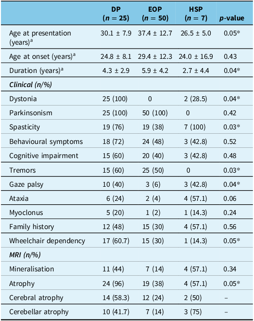
Patients with EOP had significantly older age at presentation with longer disease duration, but the difference in the age at onset among the three phenotypes was not significant. Gaze palsy was significantly less common in EOP than in DP and HSP phenotypes. Tremor was common in DP and EOP. Wheelchair dependency was common in DP. There was no difference in the frequency of mineralisation among the phenotypes, but cerebral and cerebellar atrophy was significantly frequent in DP (Table 2).
Comparison of patients with different phenotypic presentations of PLA2G6-associated neurodegeneration

Table 2 Long description
The table compares patients with different phenotypic presentations of PLA2G6-associated neurodegeneration across three groups: DP (n = 25), EOP (n = 50), and HSP (n = 7). It includes age at presentation, age at onset, and duration in years, along with clinical and MRI findings. Row 1: Age at presentation (years), DP: 30.1 ± 7.9, EOP: 37.4 ± 12.7, HSP: 26.5 ± 5.0, p-value: 0.05*. Row 2: Age at onset (years), DP: 24.8 ± 8.1, EOP: 29.4 ± 12.3, HSP: 24.0 ± 16.9, p-value: 0.43. Row 3: Duration (years), DP: 4.3 ± 2.9, EOP: 5.9 ± 4.2, HSP: 2.7 ± 4.4, p-value: 0.04*. Clinical findings include dystonia, parkinsonism, spasticity, behavioral symptoms, cognitive impairment, tremors, gaze palsy, ataxia, myoclonus, family history, and wheelchair dependency. MRI findings include mineralisation, atrophy, cerebral atrophy, and cerebellar atrophy. Notable trends include significant differences in age at presentation, duration, dystonia, spasticity, tremors, gaze palsy, wheelchair dependency, and atrophy among the groups.
DP = dystonia-parkinsonism; EOP = early-onset parkinsonism; HSP = hereditary spastic paraplegia.
* p-value ≤ 0.05;
a mean ± standard deviation.
Comparison of patients with different PLAN-related genotypes
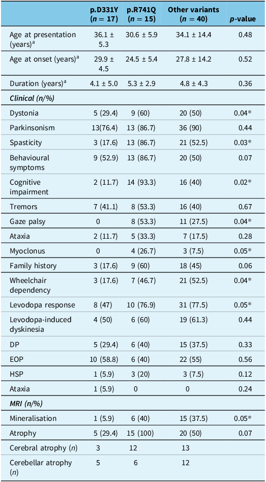
We compared the common variant in the Asian cohort (p.D331Y) with p.R741Q, a common variant in the Indian and Middle East cohorts and other variants to look for phenotypic variability based on the genotypes. There was no difference among the groups with respect to age at presentation, age at onset and duration of disease. Dystonia, spasticity, psychiatric symptoms, cognitive impairment, myoclonus and gaze palsy were significantly common in the p.R741Q variant. Wheelchair dependency, levodopa responsiveness of parkinsonism, mineralisation and atrophy on MRI were less frequent in p.D331Y variant (Table 3).
Comparison of patients with different PLA2G6-associated neurodegeneration-related genotypes

Table 3 Long description
The table compares clinical and MRI characteristics of patients with different PLA2G6-associated neurodegeneration genotypes. It has 20 rows and 6 columns. The columns are labeled p.D331Y (n = 17), p.R741Q (n = 15), Other variants (n = 40), and p-value. The rows are grouped into categories: Age at presentation (years), Age at onset (years), Duration (years), Clinical (n/%): Dystonia, Parkinsonism, Spasticity, Behavioural symptoms, Cognitive impairment, Tremors, Gaze palsy, Ataxia, Myoclonus, Family history, Wheelchair dependency, Levodopa response, Levodopa-induced dyskinesia, DP, EOP, HSP, Ataxia, MRI (n/%): Mineralisation, Atrophy, Cerebral atrophy (n), Cerebellar atrophy (n). Each row provides specific values for each genotype and p-value. Notable trends include significant differences in the frequency of dystonia, spasticity, cognitive impairment, gaze palsy, wheelchair dependency, levodopa response, mineralisation, and atrophy among the genotypes.
DP = dystonia-parkinsonism; EOP = early-onset parkinsonism; HSP = hereditary spastic paraplegia; n = number.
* p-value < 0.05;
a mean ± standard deviation.
Discussion
The study aimed to describe the clinical, imaging and genetic profile of reported patients with adult-onset PLAN from India and compare the same with the Asian, Middle East and European cohorts of patients. There were 12 cases reported in the Indian cohort. The Asian cohort had 45, the Middle East cohort had 10 and the European cohort had 17 patients. The Indian cohort and Asian cohorts had a median age of onset of 33 years and 30 years, respectively. Patients of Middle East and European cohorts had a younger age at onset of 22 and 25 years, respectively. There was female predominance in the Asian and European cohorts, with equal sex distribution in the Middle East cohort and male predominance in the Indian cohort. The duration of symptoms was lower in the Indian cohort (3 years) as compared to the Asian (7 years), Middle East (6 years) and European (7.5 years) cohorts. Dystonia, parkinsonism and spasticity were common symptoms in the Indian cohort, whereas parkinsonism, tremors and psychiatric symptoms were reported from the Asian cohort. Parkinsonism, spasticity, psychiatric symptoms and cognitive impairment were common in Middle East and European cohorts. DP, EOP and HSP were the common phenotypic presentations in the Indian, Asian and European cohorts, whereas EOP was common in the Middle East cohort. Myoclonus was reported from the Indian, Middle East and European cohorts, but none from the Asian cohort. Levodopa-induced dyskinesia was common in the Asian and Indian cohorts as compared to the Middle East and European cohorts. Gaze palsy was less commonly reported from the Asian cohort. Levodopa-induced psychosis was reported from the Middle East cohort. Patients with DP phenotype had more frequent spasticity, behavioural and cognitive disturbances, gaze palsy, wheelchair dependency and cerebral atrophy on MRI. The HSP phenotype had more frequent ataxia and gaze palsy as compared to other phenotypes. Parkinsonism is due to the role of the enzyme iPLA2-VI in ROS generation and lipid peroxidation and the demonstration of Lewy bodies in the neocortex in adult-onset dystonia-parkinsonism cases with PLA2G6 mutations.
Brain MRI showed abnormalities in the form of mineralisation and the presence of cerebral and cerebellar atrophy. Both mineralisation and cerebral/cerebellar atrophy were reported in the Indian cohort and European cohort, with the majority of mineralisation noted in the GP, followed by the substantia nigra, caudate and putamen. Mineralisation was not reported in the Middle East cohort and had mainly cerebral rather than cerebellar atrophy, whereas in the Asian cohort, the frequency of mineralisation was low (8.9%), and cerebellar atrophy was more frequently reported than cerebral atrophy. Missense variants were reported from all cohorts. The homozygous missense variant (c.2222G > A,p.R741Q) was common in the Indian and Middle East cohorts, whereas the homozygous/compound heterozygous variant (c.991G > T, p.D331Y) was the most common variant in the Asian cohort. The c.991G > T (p.D331Y) variant was found in Chinese and Taiwanese patients, suggesting a common founder effect of this variant in Chinese populations. Reference Chu, Lin, Chen and Lin8 A homozygous c.991G > T (p.D331Y) knock-in mouse model showed dopaminergic neuronal degeneration in the substantia nigra and had 30% enzymatic activity remaining in the PLA2G6 protein. Reference Shi, Tang and Wang11 This explains the relatively milder clinical and neuroimaging phenotypes of patients with c.991G > T (p.D331Y) variant. The c.2222G > A,p.R741Q variant had a relatively severe phenotype of dystonia, parkinsonism, spasticity, behavioural and cognitive symptoms, gaze palsy, ataxia, wheelchair dependency and cerebral/cerebellar atrophy on MRI. Engel et al. (2010) reported that the variants R741Q, R747W and R632W did not impair phospholipase catalytic activity in vitro. The exact mechanism of the R741Q variant causing clinical manifestations remains to be elucidated. Reference Engel, Jing, O’Brien, Sun and Kotzbauer6
The strength of the study was the detailed clinical, radiological and genetic description of cases from the Indian, Asian, Middle East and European cohorts and the genotype–phenotype correlations. The limitations of the study are the small sample size of the Indian cohort, variability in the number of reported cases with variable clinical assessments among the groups, lack of long-term follow-up and prognosis of the Indian cohort.
Conclusion
Adult-onset PLAN has three distinct phenotypes of DP, EOP and HSP. There was an ethnic phenotypic difference among the cohorts as dystonia, myoclonus and gaze palsy were common in the Indian cohort; parkinsonism and tremor in the Asian cohort; parkinsonism, tremor, spasticity and cognitive impairment in the Middle East cohort; and parkinsonism and behavioural disturbances in the European cohort. Mineralisation on MRI was commonly reported in the Indian cohort and none from the Middle East cohort. Cerebral/cerebellar atrophy was less frequent in the Asian cohort. The c.991G > T (p.D331Y) was reported mainly in the Asian cohort, and c.2222G > A,p.R741Q variant was mainly from the Indian and Middle East cohorts. PLAN should be suspected in patients presenting with parkinsonism or DP or HSO with early cognitive decline, psychiatric symptoms, spasticity and gaze palsy even if there is no mineralisation. Dopaminergic treatment should be titrated carefully as there is a risk of levodopa-induced psychosis.
Supplementary material
The supplementary material for this article can be found at https://doi.org/10.1017/cjn.2025.10352.
Acknowledgements
Nil.
Author contributions
-
1. Research project: A. Conception, B. Organisation, C. Execution;
-
2. Statistical Analysis: A. Design, B. Execution, C. Review and critique;
-
3. Manuscript Preparation: A. Writing of the first draft, B. Review and critique;
RRM: 1A, 1B, 1C, 2A, 2B, 3B.
HP: 1A, 2C, 3B.
PM: 1A, 2C, 3B.
Funding statement
There is no funding received for this study.
Competing interests
There are no competing interests.




Target article
Adult-Onset PLA2G6-Associated Neurodegeneration (PLAN): A Clinical, Imaging and Genetic Profile Review
Related commentaries (1)
Reviewer Comment on Mahale et al. “Adult-onset PLA2G6-Associated Neurodegeneration (PLAN): A clinical, imaging and genetic profile review”